
Quantitation of thiol metabolites from mammalian cells using fluorous tagging and HILIC-MS†
Abstract
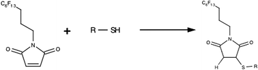
Oxidative stress diminishes reduced thiols by decreasing their production or by forming disulfide bonds. This instigates an equilibrium shift in further downstream metabolic pathways. The goal of this work is to develop a rapid and selective fluorous labeling strategy for improved sensitivity in LC-MS analysis of reduced thiols in biological samples. Fluorous tagging is used to augment the MS signal and has limited stationary phase interactions. Thiol standards were reacted with a fluorous maleimide at a ratio of 100 : 1 in an 80% acetonitrile buffer containing 5 mM ammonium formate at pH 8. The reaction time and temperature were optimized. The mixture of thiol metabolites was then separated using a cyano HILIC column with a linear gradient from 90% to 70% acetonitrile. Signal intensity from fluorous-tagged thiols was improved over the untagged thiols by at least four fold. This demonstrates a quick method that can be used to compare levels of reduced thiols in diabetic and normal mammalian endothelial cells. Human tissue was also analyzed using this tagging method. This method will further elucidate the impact of oxidative stress on thiol metabolism and therefore help identify therapeutic targets for the treatment of diabetic complications.
- This article is part of the themed collection: Emerging Investigators
 Please wait while we load your content...

