Analysis of affinities between specific biological ligands using atomic force microscopy
Abstract
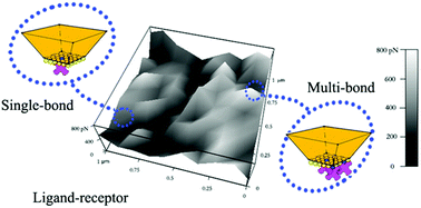
In the cell, protein–ligand recognition involves association and dissociation processes controlled by the affinity of the two binding partners and chemical harvesting of adenosine triphosphate energy. Fundamental knowledge of selected recognition events is currently translated in a synthetic environment for biosensors, immunoassays and diagnosis applications, or for pharmaceutical development. However, in order to advance such fields, one needs to determine the lifetime and binding efficiency of the two partners, as well as the complex energy landscape parameters. We employed contact mode atomic force microscopy to evaluate the association and dissociation events between streptavidin protein and its anti-streptavidin antibody ligand currently used for nucleotide array, ELISA, and flow cytometry applications, just to name a few. Using biotin as the control, our analysis helped characterize and differentiate multi- or single bonds of different strengths as well as associated energy landscapes to determine the protein–ligand structural arrangement at nanointerfaces and how these depend on the specificity of the ligand-recognition reaction. Our results suggest that understanding the importance of the rupture forces between a protein and its ligand could serve as the first step to protect on–off switches for biomedical research applications where specificity and selectivity are foremost sought.


 Please wait while we load your content...