Multimodal chemical imaging of molecular messengers in emerging Pseudomonas aeruginosa bacterial communities†
Abstract
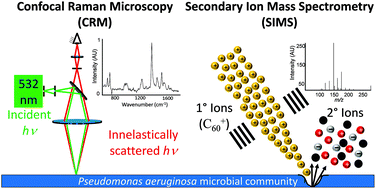
Two label-free molecular imaging techniques, confocal Raman microscopy (CRM) and secondary ion mass spectrometry (SIMS), are combined for in situ characterization of the spatiotemporal distributions of quinolone metabolites and signaling molecules in communities of the pathogenic bacterium Pseudomonas aeruginosa. Dramatic molecular differences are observed between planktonic and biofilm modes of growth for these bacteria. We observe patterned aggregation and a high abundance of N-oxide quinolines in early biofilms and swarm zones of P. aeruginosa, while the concentrations of these secreted components in planktonic cells and agar plate colonies are below CRM and SIMS detection limits. CRM, in conjunction with principal component analysis (PCA) is used to distinguish between the two co-localized isomeric analyte pairs 4-hydroxy-2-heptylquinoline-N-oxide (HQNO)/2-heptyl-3-hydroxyquinolone (PQS) and 4-hydroxy-2-nonylquinoline-N-oxide (NQNO)/2-nonyl-hydroxyquinolone (C9-PQS) based on differences in their vibrational fingerprints, illustrating how the technique can be used to guide tandem-MS and tandem-MS imaging analysis. Because N-oxide quinolines are ubiquitous and expressed early in biofilms, these analytes may be fundamentally important for early biofilm formation and the growth and organization of P. aeruginosa microbial communities. This study underscores the advantages of using multimodal molecular imaging to study complex biological systems.


 Please wait while we load your content...