Contribution of Raman spectroscopy in nephrology: a candidate technique to detect hydroxyethyl starch of third generation in osmotic renal lesions†
Abstract
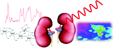
Background and objectives: HydroxyEthyl Starch (HES) has been one of the most commonly used colloid volume expanders in intensive care units for over 50 years. The first and second generation HES, with a high molecular weight (≥200 kD) and a high degree of substitution (≥0.5), has been associated with both renal dysfunction and osmotic nephrosis-like lesions in histological studies. Recently, third generation HES (130 kD/<0.5) has also been shown to impair renal function in critically ill adult patients although tubular accumulation of HES has never been proven in the human kidney. Our objective was to demonstrate the potential of Raman micro-imaging to bring out the presence of third generation-HES in the kidney of patients having received the volume expander. Design: Four biopsies presenting osmotic nephrosis-like lesions originated from HES-administrated patients with impaired renal function were compared with HES-negative biopsies (n = 10) by Raman microspectroscopy. Results: The first step was dedicated to the identification of a specific vibration of HES permitting the detection of the cellular and tissue accumulation of the product. This specific vibration at 480 cm−1 is assigned to a collective mode of the macromolecule; it is located in a spectral region with a limited contribution from biological materials. Based on this finding, HES distribution within tissue sections was investigated using Raman micro-imaging. Determination of HES positive pixels permitted us to clearly distinguish positive cases from HES-free biopsies (proportions of positive pixels from the total number of pixels: 23.48% ± 28 vs. 0.87% ± 1.2; p = 0.004). Conclusions: This study shows that Raman spectroscopy is a candidate technique to detect HES in kidney tissue samples currently manipulated in nephrology departments. In addition, on the clinical aspect, our approach suggests that renal impairment related to third generation HES administration is associated with osmotic nephrosis-like lesions and HES accumulation in the kidney.


 Please wait while we load your content...