Issue 10, 2015

3D elemental sensitive imaging by full-field XFCT

Abstract

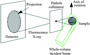
X-ray fluorescence computed tomography (XFCT) is a stimulated emission tomography modality that maps the three-dimensional (3D) distribution of elements. Generally, XFCT is done by scanning a pencil-beam across the sample. This paper presents a feasibility study of full-field XFCT (FF-XFCT) for 3D elemental imaging. The FF-XFCT consists of a pinhole collimator and X-ray imaging detector with no energy resolution. A prototype imaging system was set up at the Shanghai Synchrotron Radiation Facility (SSRF) for imaging the phantom. The first FF-XFCT experimental results are presented. The cadmium (Cd) and iodine (I) distributions were reconstructed. The results demonstrate FF-XFCT is fit for 3D elemental imaging and the sensitivity of FF-XFCT is higher than a conventional CT system.

Graphical abstract: 3D elemental sensitive imaging by full-field XFCT

Article information

Article type
Paper
Submitted
04 Jan 2015
Accepted
19 Mar 2015
First published
19 Mar 2015

Analyst, 2015,140, 3521-3525

Author version available

3D elemental sensitive imaging by full-field XFCT

B. Deng, G. Du, G. Zhou, Y. Wang, Y. Ren, R. Chen, P. Sun, H. Xie and T. Xiao, Analyst, 2015, 140, 3521 DOI: 10.1039/C4AN02401J

To request permission to reproduce material from this article, please go to the Copyright Clearance Center request page.

If you are an author contributing to an RSC publication, you do not need to request permission provided correct acknowledgement is given.

If you are the author of this article, you do not need to request permission to reproduce figures and diagrams provided correct acknowledgement is given. If you want to reproduce the whole article in a third-party publication (excluding your thesis/dissertation for which permission is not required) please go to the Copyright Clearance Center request page.

Read more about how to correctly acknowledge RSC content.

Social activity

Spotlight

Advertisements