Differentiation of prostate cancer from normal tissue in radical prostatectomy specimens by desorption electrospray ionization and touch spray ionization mass spectrometry†
Abstract
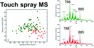
Radical prostatectomy is a common treatment option for prostate cancer before it has spread beyond the prostate. Examination for surgical margins is performed post-operatively with positive margins reported to occur in 6.5–32% of cases. Rapid identification of cancerous tissue during surgery could improve surgical resection. Desorption electrospray ionization (DESI) is an ambient ionization method which produces mass spectra dominated by lipid signals directly from prostate tissue. With the use of multivariate statistics, these mass spectra can be used to differentiate cancerous and normal tissue. The method was applied to 100 samples from 12 human patients to create a training set of MS data. The quality of the discrimination achieved was evaluated using principal component analysis – linear discriminant analysis (PCA-LDA) and confirmed by histopathology. Cross validation (PCA-LDA) showed >95% accuracy. An even faster and more convenient method, touch spray (TS) mass spectrometry, not previously tested to differentiate diseased tissue, was also evaluated by building a similar MS data base characteristic of tumor and normal tissue. An independent set of 70 non-targeted biopsies from six patients was then used to record lipid profile data resulting in 110 data points for an evaluation dataset for TS-MS. This method gave prediction success rates measured against histopathology of 93%. These results suggest that DESI and TS could be useful in differentiating tumor and normal prostate tissue at surgical margins and that these methods should be evaluated intra-operatively.


 Please wait while we load your content...