
Rapid biodiagnostic ex vivo imaging at 1 μm pixel resolution with thermal source FTIR FPA
Abstract
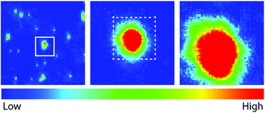
A recent upgrade to the optics configuration of a thermal source FTIR microscope equipped with a focal plane array detector has enabled rapid acquisition of high magnification spectrochemical images, in transmission, with an effective geometric pixel size of ∼1 × 1 μm2 at the sample plane. Examples, including standard imaging targets for scale and accuracy, as well as biomedical tissues and microorganisms, have been imaged with the new system and contrasted with data acquired at normal magnification and with a high magnification multi-beam synchrotron instrument. With this optics upgrade, one can now conduct rapid biodiagnostic ex vivo tissue imaging in-house, with images collected over larger areas, in less time (minutes) and with comparable quality and resolution to the best synchrotron source FTIR imaging capabilities.
- This article is part of the themed collections: Alzheimer's Research Month 2016 and Optical Diagnosis (2014)
 Please wait while we load your content...

