
Probe spectrum measurements of Eu3+ ions as a relevant tool for monitoring in vitro hydroxyapatite formation in a new borate biomaterial
Abstract
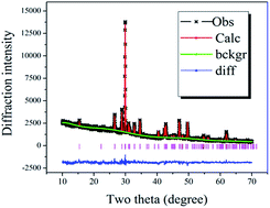
CaB2O4 powders and ceramics were prepared by the conventional solid-state reaction. In vitro hydroxyapatite (HA) mineralization was investigated by soaking the samples in simulated body fluid (SBF) for various time periods. X-ray diffraction and structural refinements, scanning electron microscopy and X-ray energy-dispersive spectra measurements were applied to investigate apatite formation before and after immersion in SBF. HA can easily form flower-like nanostructures with nano-needles even when soaked in SBF for several hours. The in vitro bioactivity of CaB2O4 was attributed to easy formation of B–OH groups in the CaB2O4 structure when soaked in SBF solutions. In the process of mineralization, the luminescence evolution of Eu3+ ions, a well-known structural probe, was detected by photoluminescence spectra and photoluminescence decay curves. This suggested that the process of mineralization can be monitored by the luminescence intensity of Eu3+ ions in the mineralization products. The current study will open up a new and simple in vivo avenue for in situ monitoring of hydroxyapatite conversion using a fiber luminescence spectrometer.
 Please wait while we load your content...

