
The ultrastructure of type I collagen at nanoscale: large or small D-spacing distribution?†
Abstract
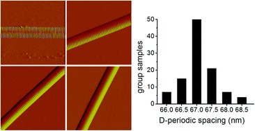
D-Spacing is the most significant topographic feature of type I collagen fibril, and it is important for our understanding of the structure and function in collagens. Traditionally, the D-spacing of type I collagen fibril was shown to have a singular value of 67 nm, but recent works indicated that the D-spacing values have a large distribution of up to 10 nm when measured by atomic force microscopy. We found that this large distribution of D-spacing values mainly resulted from image drift during measurement. Note that the D-spacing was homogeneous in a single type I collagen fibril. Our statistical analysis indicated that the D-spacing values of type I collagen fibrils exhibited only a narrow distribution of 2.5 nm around the value of 67 nm. In addition, the D-spacing values of the collagen fibrils were nearly identical not only within a single fibril bundle, but also in different fibril bundles. The measurement of the D-spacing values of collagen may provide important structural information in many research areas such as collagen related diseases, construction of molecular model of collagen, and collagen fibrogenesis.
 Please wait while we load your content...

