ElectroTaxis-on-a-Chip (ETC): an integrated quantitative high-throughput screening platform for electrical field-directed cell migration†
Abstract
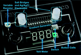
Both endogenous and externally applied electrical stimulation can affect a wide range of cellular functions, including growth, migration, differentiation and division. Among those effects, the electrical field (EF)-directed cell migration, also known as electrotaxis, has received broad attention because it holds great potential in facilitating clinical wound healing. Electrotaxis experiment is conventionally conducted in centimetre-sized flow chambers built in Petri dishes. Despite the recent efforts to adapt microfluidics for electrotaxis studies, the current electrotaxis experimental setup is still cumbersome due to the needs of an external power supply and EF controlling/monitoring systems. There is also a lack of parallel experimental systems for high-throughput electrotaxis studies. In this paper, we present a first independently operable microfluidic platform for high-throughput electrotaxis studies, integrating all functional components for cell migration under EF stimulation (except microscopy) on a compact footprint (the same as a credit card), referred to as ElectroTaxis-on-a-Chip (ETC). Inspired by the R–2R resistor ladder topology in digital signal processing, we develop a systematic approach to design an infinitely expandable microfluidic generator of EF gradients for high-throughput and quantitative studies of EF-directed cell migration. Furthermore, a vacuum-assisted assembly method is utilized to allow direct and reversible attachment of our device to existing cell culture media on biological surfaces, which separates the cell culture and device preparation/fabrication steps. We have demonstrated that our ETC platform is capable of screening human cornea epithelial cell migration under the stimulation of an EF gradient spanning over three orders of magnitude. The screening results lead to the identification of the EF-sensitive range of that cell type, which can provide valuable guidance to the clinical application of EF-facilitated wound healing.


 Please wait while we load your content...