
A light-sheet microscope compatible with mobile devices for label-free intracellular imaging and biosensing†
Abstract
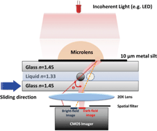
The inner structure, especially the nuclear structure, of cells carries valuable information about disease and health conditions of a person. Here we demonstrate a label-free technique to enable direct observations and measurements of the size, shape and morphology of the cell nucleus. With a microfabricated lens and a commercial CMOS imager, we form a scanning light-sheet microscope to produce a dark-field optical scattering image of the cell nucleus that overlays with the bright-field image produced in a separate regime of the same CMOS sensor. We have used the device to detect nuclear features that characterize the life cycle of cells and have used the nucleus volume as a new parameter for cell classification. The device can be developed into a portable, low-cost, point-of-care device leveraging the capabilities of the CMOS imagers to be pervasive in mobile electronics.
 Please wait while we load your content...

