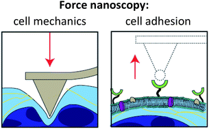
Cells are constantly exposed to mechanical stimuli in their environment and have several evolved mechanisms to sense and respond to these cues. It is becoming increasingly recognized that many cell types, from bacteria to mammalian cells, possess a diverse set of proteins to translate mechanical cues into biochemical signalling and to mediate cell surface interactions such as cell adhesion. Moreover, the mechanical properties of cells are involved in regulating cell function as well as serving as indicators of disease states. Importantly, the recent development of biophysical tools and nanoscale methods has facilitated a deeper understanding of the role that physical forces play in modulating cell mechanics and cell adhesion. Here, we discuss how atomic force microscopy (AFM) has recently been used to investigate cell mechanics and cell adhesion at the single-cell and single-molecule levels. This knowledge is critical to our understanding of the molecular mechanisms that govern mechanosensing, mechanotransduction, and mechanoresponse in living cells. While pushing living cells with the AFM tip provides a means to quantify their mechanical properties and examine their response to nanoscale forces, pulling single surface proteins with a functionalized tip allows one to understand their role in sensing and adhesion. The combination of these nanoscale techniques with modern molecular biology approaches, genetic engineering and optical microscopies provides a powerful platform for understanding the sophisticated functions of the cell surface machinery, and its role in the onset and progression of complex diseases.
You have access to this article
 Please wait while we load your content...
Something went wrong. Try again?
Please wait while we load your content...
Something went wrong. Try again?