
Comparative proteome profiling of breast tumor cell lines by gel electrophoresis and mass spectrometry reveals an epithelial mesenchymal transition associated protein signature†
Abstract
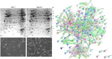
The epithelial to mesenchymal transition (EMT) is a cellular program associated with the organ morphogenesis but also with the disease progression. EMT in the cancer field fuels neoplastic progression promoting the resistance to cell death, the resistance to chemotherapy and the acquisition of stem cell properties. Considering the crucial role of EMT in breast cancer metastasis, a better understanding of this process may provide new therapeutic options. Here, by using a proteomic approach we identified a set of proteins differentially expressed between an epithelial and a mesenchymal breast cancer cell line. The protein–protein network of these identified proteins was determined by an in silico analysis highlighting, in the EMT program, the role of proteins involved in cell adhesion, migration, and invasion, together with protein kinases involved in proliferation and survival, with many of these emerging as possible targets of novel biological agents. Finally, the pharmacological inhibition of some of these kinases was able to reverse the mesenchymal phenotype to an epithelial phenotype.
- This article is part of the themed collection: Molecular BioSystems 2013 Proteomics
 Please wait while we load your content...