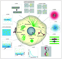
A cell can be viewed as a dynamic puzzle, where single pieces shuffle in space, change their conformation to fit different partners, and new pieces are generated while old ones are destroyed. Microscopy has become capable of directly observing the pieces of the puzzle, which are single molecules. Single-molecule microscopy in vivo provides new insights into the molecular processes underlying the physiology of a cell, allowing not only for visualizing how molecules distribute with nanometer resolution in the cellular environment, but also for characterizing their movement with high temporal precision. This approach reveals molecular behaviors normally invisible in ensemble measurements. Depending on the molecule, the process, and the cellular region studied, single molecules can be followed by conventional epifluorescence microscopy, or by illuminating only a thin region of the cell, as in Total Internal Reflection Fluorescence (TIRF) and Selective Plane Illumination Microscopy (SPIM), and by limiting the amount of detectable molecules, as in Fluorescence Speckle Microscopy (FSM) and Photo-Activation (PA). High spatial resolution can be obtained by imaging only a fraction of the molecules at a time, as in Photo-Activated Localization Microscopy (PALM) and Stochastic Optical Reconstruction Microscopy (STORM), or by de-exciting molecules in the periphery of the detection region as in Stimulated Emission-Depletion (STED) microscopy. Single-molecule techniques in vivo are becoming widespread; however, it is important to choose the most suited technique for each biological question or sample. Here we review single-molecule microscopy techniques, describe their basic principles, advantages for in vivo application, and discuss the lessons that can be learned from live single-molecule imaging.


 Please wait while we load your content...