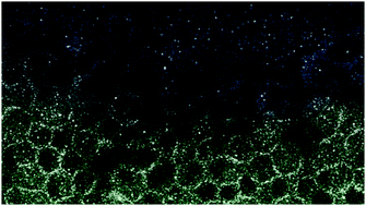
The morphology of ionic clusters that form in polyelectrolyte membranes has a strong effect on transport and electrical properties. In spite of considerable research effort the link between morphology and properties has not been clearly established, mainly due to difficulties in assessing nanoscale morphology. Electron microscopy (EM) has the potential to visualize morphology. However success in visualization has so far been moderate. In this review we focus on the potential of EM techniques to characterize the ionic domains. We use both experimental data and models to compare the capabilities of several EM techniques: BF TEM, HAADF, core-loss EELS, and low-loss EELS in projection imaging and STEM modes. The main problems common for all these EM modes are radiation damage and overlap of features in projection. Our models show that core loss EELS with exposures that are below the typical damage threshold is incapable of resolving 2 nm diameter sulfur-rich clusters in PEMs. While low loss EELS requires lower exposure, the insight it can provide is quite limited. HAADF and BF TEM present the most effective modes for imaging the sulfur clusters in PEMs. While BF TEM uses scattered electrons more efficiently, HAADF using slightly higher doses can provide unique information due to in-focus imaging and transparent interpretation of the images. Fortunately, in at least some interesting cases the clusters themselves are much more radiation resistant than the polymer and can be studied at exposures high enough to obtain clear images. Our simulations also show that tomographic 3D reconstruction provides the best approach for solving the overlap problem. In spite of the abilities of electron tomography, data obtained from all EM techniques improve if thin sections are studied. We briefly discuss methods for obtaining such sections.

You have access to this article
 Please wait while we load your content...
Something went wrong. Try again?


 Please wait while we load your content...