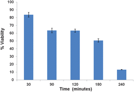
The effects of simulated solar irradiation of an artificial skin model have been examined using Raman spectroscopy and the results are compared with cytotoxicological and histological profiling. Samples exposed for times varying between 30 minutes and 240 minutes were incubated post exposure for a period of 96 hours. The cytotoxicological response as measured by the MTT [3-(4,5-dimethylthiazol-2-yl)-2,5-diphenyl tetrazolium bromide] assay demonstrated a ∼50% loss of viability of the artificial tissue after 120 minutes exposure. Histological staining of tissue sections showed considerable loss of cellular content in the epidermal layer at this endpoint. Raman spectroscopic mapping of tissue sections, coupled with K-means cluster analysis (KMCA) clearly identified the dermal and stratum corneum layers and differentiated further substructures of the epidermis. Post irradiation, a significant loss of DNA features in the basal layer was apparent in the results of the KMCA. Principal Components Analysis (PCA) of layers identified by the KMCA post exposure compared with controls indicated a significant increase in the lipidic signatures of the stratum corneum. In the dermal layer, little photodamage was observed, but a similar increase in lipidic signatures in the basal layer was accompanied by a decrease in DNA and protein contributions. The spectral profiles of the photodamage to the basal layer as identified by PCA are consistent over the exposure periods of 30–240 minutes, but an examination of the evolution of features associated with specific biochemical components indicated DNA damage and loss of lipidic signatures at the early exposure times, whereas changes in protein signatures appeared to evolve over longer periods. In comparison to the cytotoxicological responses, the study demonstrates that Raman spectroscopy can identify biochemical changes as a result of solar exposure at time points significantly earlier than changes in tissue viability are observed.

You have access to this article
 Please wait while we load your content...
Something went wrong. Try again?


 Please wait while we load your content...