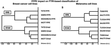
Over the past few decades, Fourier transform infrared (FTIR) spectroscopy coupled to microscopy has been recognized as an emerging and potentially powerful tool in cancer research and diagnosis. For this purpose, histological analyses performed by pathologists are mostly carried out on biopsied tissue that undergoes the formalin-fixation and paraffin-embedding (FFPE) procedure. This processing method ensures an optimal and permanent preservation of the samples, making FFPE-archived tissue an extremely valuable source for retrospective studies. Nevertheless, as highlighted by previous studies, this fixation procedure significantly changes the principal constituents of cells, resulting in important effects on their infrared (IR) spectrum. Despite the chemical and spectral influence of FFPE processing, some studies demonstrate that FTIR imaging allows precise identification of the different cell types present in biopsied tissue, indicating that the FFPE process preserves spectral differences between distinct cell types. In this study, we investigated whether this is also the case for closely related cell lines. We analyzed spectra from 8 cancerous epithelial cell lines: 4 breast cancer cell lines and 4 melanoma cell lines. For each cell line, we harvested cells at subconfluence and divided them into two sets. We first tested the “original” capability of FTIR imaging to identify these closely related cell lines on cells just dried on BaF2 slides. We then repeated the test after submitting the cells to the FFPE procedure. Our results show that the IR spectra of FFPE processed cancerous cell lines undergo small but significant changes due to the treatment. The spectral modifications were interpreted as a potential decrease in the phospholipid content and protein denaturation, in line with the scientific literature on the topic. Nevertheless, unsupervised analyses showed that spectral proximities and distances between closely related cell lines were mostly, but not entirely, conserved after FFPE processing. Finally, PLS-DA statistical analyses highlighted that closely related cell lines are still successfully identified and efficiently distinguished by FTIR spectroscopy after FFPE treatment. This last result paves the way towards identification and characterization of cellular subtypes on FFPE tissue sections by FTIR imaging, indicating that this analysis technique could become a potential useful tool in cancer research.

 Please wait while we load your content...

