Issue 3, 2012

Lens proteomics: analysis of rat crystallins when lenses are exposed to dexamethasone

Abstract

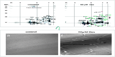
To identify glucocorticoid induced cataract (GIC)-specific modified crystallins and related changes, we analyzed rat crystallins and related changes in lenses exposed to dexamethasone (Dex). To carry out proteomics analyses, we separated soluble lens proteins with two-dimensional electrophoresis (2-DE) and modified crystallins were analyzed with matrix assisted laser desorption/ionization time-of-flight tandem mass spectrometry (MALDI-TOF-MS/MS). Related changes in mRNA, protein levels and morphological and functional changes of modified crystallins were also determined. Measured masses (except for γD-crystallin as the larger and cross-link form), the isoelectric points (PIs; except for βB3-crystallin as the alkalinization form) and amino acid sequences of all known rat crystallins matched previously reported data. Analysis by 2-DE indicated that αA, αB, βB3 and γD increased when lenses were exposed to 5 μM Dex; βA4 increased when lenses were exposed to 1 μM Dex and the five proteins that had the highest expressional trend were identical with the results of Q-PCR. βA3/A1 crystallin (expressional trend identical with results of Q-PCR) and the serum albumin precursor gradually disappeared when exposed to 1–50 μM Dex. Results of Western blotting, immunohistochemistry or fluorescence analysis showed that αA and αB increased most when exposed to 5 μM Dex and βA1/A3 and KI-67 decreased obviously when exposed to 1–50 μM Dex. Electron microscopy showed that the condition of the lens was better when lenses were exposed to 5 μM Dex than at other levels and cracks between the fiber cells became larger when lenses were exposed to 1–50 μM Dex. A chaperone role of α-crystallin protecting heated catalase (CAT) and the activity of superoxide dismutase (SOD), glutathione (GSH), and caspase-3 were highest when exposed to 5 μM Dex. Moreover, αA-crystallins were associated with increased phosphorylation (PI decreased). In conclusion, the proteomics analysis and related changes of rat crystallins when lenses were exposed to Dex in this study will be useful for comparison with normal lens proteins and GIC. We also provided a mechanism for GIC from a proteomics aspect based on the in vitro model.

Graphical abstract: Lens proteomics: analysis of rat crystallins when lenses are exposed to dexamethasone

Supplementary files

Article information

Article type
Paper
Submitted
09 Nov 2011
Accepted
13 Dec 2011
First published
23 Jan 2012

Mol. BioSyst., 2012,8, 888-901

Lens proteomics: analysis of rat crystallins when lenses are exposed to dexamethasone

L. Wang, W. C. Zhao, X. L. Yin, J. Y. Ge, Z. G. Bu, H. Y. Ge, Q. F. Meng and P. Liu, Mol. BioSyst., 2012, 8, 888 DOI: 10.1039/C2MB05463A

To request permission to reproduce material from this article, please go to the Copyright Clearance Center request page.

If you are an author contributing to an RSC publication, you do not need to request permission provided correct acknowledgement is given.

If you are the author of this article, you do not need to request permission to reproduce figures and diagrams provided correct acknowledgement is given. If you want to reproduce the whole article in a third-party publication (excluding your thesis/dissertation for which permission is not required) please go to the Copyright Clearance Center request page.

Read more about how to correctly acknowledge RSC content.

Spotlight

Advertisements