Issue 16, 2012

Gelatin methacrylate as a promising hydrogel for 3D microscale organization and proliferation of dielectrophoretically patterned cells

Abstract

Establishing the 3D microscale organization of cells has numerous practical applications, such as in determining cell fate (e.g., proliferation, migration, differentiation, and apoptosis) and in making functional tissue constructs. One approach to spatially pattern cells is by dielectrophoresis (DEP). DEP has characteristics that are important for cell manipulation, such as high accuracy, speed, scalability, and the ability to handle both adherent and non-adherent cells. However, widespread application of this method is largely restricted because there is a limited number of suitable hydrogels for cell encapsulation. To date, polyethylene glycol-diacrylate (PEG-DA) and agarose have been used extensively for dielectric patterning of cells. In this study, we propose gelatin methacrylate (GelMA) as a promising hydrogel for use in cell dielectropatterning because of its biocompatibility and low viscosity. Compared to PEG hydrogels, GelMA hydrogels showed superior performance when making cell patterns for myoblast (C2C12) and endothelial (HUVEC) cells as well as in maintaining cell viability and growth. We also developed a simple and robust protocol for co-culture of these cells. Combined application of the GelMA hydrogels and the DEP technique is suitable for creating highly complex microscale tissues with important applications in fundamental cell biology and regenerative medicine in a rapid, accurate, and scalable manner.

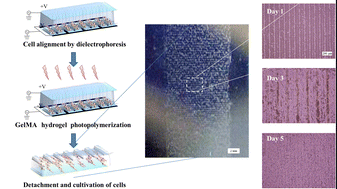
Graphical abstract: Gelatin methacrylate as a promising hydrogel for 3D microscale organization and proliferation of dielectrophoretically patterned cells

Supplementary files

Article information

Article type
Paper
Submitted
28 Feb 2012
Accepted
14 May 2012
First published
15 May 2012

Lab Chip, 2012,12, 2959-2969

Gelatin methacrylate as a promising hydrogel for 3D microscale organization and proliferation of dielectrophoretically patterned cells

J. Ramón-Azcón, S. Ahadian, R. Obregón, G. Camci-Unal, S. Ostrovidov, V. Hosseini, H. Kaji, K. Ino, H. Shiku, A. Khademhosseini and T. Matsue, Lab Chip, 2012, 12, 2959 DOI: 10.1039/C2LC40213K

To request permission to reproduce material from this article, please go to the Copyright Clearance Center request page.

If you are an author contributing to an RSC publication, you do not need to request permission provided correct acknowledgement is given.

If you are the author of this article, you do not need to request permission to reproduce figures and diagrams provided correct acknowledgement is given. If you want to reproduce the whole article in a third-party publication (excluding your thesis/dissertation for which permission is not required) please go to the Copyright Clearance Center request page.

Read more about how to correctly acknowledge RSC content.

Social activity

Spotlight

Advertisements