Issue 1, 2012

Resonant Mie scattering (RMieS) correction applied to FTIR images of biological tissue samples

Abstract

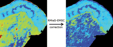
Recently a resonant Mie scattering (RMieS) correction approach has been developed and demonstrated to be effective for removing the baseline distortions that compromise the raw data in individual spectra. In this paper RMieS correction is extended to FTIR images of a tissue section from biopsy of the human cervical transformation zone and a coronal tissue section of a Wistar rat brain and compared to the uncorrected images. It is shown that applying RMieS correction to FTIR images a) removes baseline distortions from the image spectra and thus reveals previously hidden information on spatial variation of chemical contents within the tissue and b) can lead to improved automatic tissue feature classification through multivariate cluster analysis.

Graphical abstract: Resonant Mie scattering (RMieS) correction applied to FTIR images of biological tissue samples

Article information

Article type
Paper
Submitted
21 Jul 2011
Accepted
21 Oct 2011
First published
10 Nov 2011

Analyst, 2012,137, 126-132

Resonant Mie scattering (RMieS) correction applied to FTIR images of biological tissue samples

K. R. Bambery, B. R. Wood and D. McNaughton, Analyst, 2012, 137, 126 DOI: 10.1039/C1AN15628D

To request permission to reproduce material from this article, please go to the Copyright Clearance Center request page.

If you are an author contributing to an RSC publication, you do not need to request permission provided correct acknowledgement is given.

If you are the author of this article, you do not need to request permission to reproduce figures and diagrams provided correct acknowledgement is given. If you want to reproduce the whole article in a third-party publication (excluding your thesis/dissertation for which permission is not required) please go to the Copyright Clearance Center request page.

Read more about how to correctly acknowledge RSC content.

Social activity

Spotlight

Advertisements