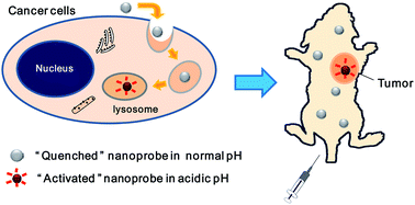
Optical imaging is promising in tumor diagnosis due to its high sensitivity, no radioactive irradiation and low running cost. Compared to small molecular probes, fluorescence nanoprobes demonstrate a tunable circulation lifetime, an up-regulated intratumoral accumulation and enhanced sensitivity by labeling multiple imaging reporters on a single nanoparticle. Acidic extracellular fluid is a universal phenomenon of solid tumors. Therefore, a nanoprobe that responds to the acidic microenvironment is promising in visualizing a tumor in vivo, regardless of the tumor type or even developmental stage. Moreover, the fluorescence “activation” in the tumor but not the normal tissues will greatly increase the “target to background” signal ratio, which benefits the visualization of small volume tumors with high sensitivity. In this review, we summarize the recent developments in pH responsive fluorescence nanoprobes for tumor visualization.
You have access to this article
 Please wait while we load your content...
Something went wrong. Try again?
Please wait while we load your content...
Something went wrong. Try again?