Issue 1, 2011

Quantitative determination of intact free cisplatin in cell models by LC-ICP-MS

Abstract

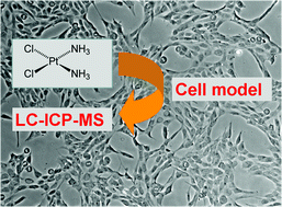
For the first time cellular uptake studies of cisplatin were addressed by elemental speciation analysis at biological relevant concentration levels, i.e. drug exposure concentration ranging at 5 µM. The quantification of intact, free cisplatin in cell models was investigated by two complementary LC-ICP-MS methods, using chromatographic separations based on pentafluorophenylpropyl siloxane bonded stationary phases (Discovery HS F5) and on porous graphitized carbon (Hypercarb). Limits of detection for cisplatin were 0.013 and 0.11 µg L−1 (given as total drug), respectively. Cisplatin—once entering the cancer cell—is known to undergo reactions with proteins and peptides in the cytosol by forming adducts. Hence, due to the limited selectivity of one-dimensional LC separation, efficient protein removal was a prerequisite for accurate quantification in such complex biological matrix as cell lysate. Centrifugal filtration (cut-off of 10 kDa) was the method of choice. Exposure of two different cell models to 5 µM cisplatin for 24 hours resulted in cisplatin concentration levels ranging between 0.2 and 1.5 µg g−1 protein. Despite the poor recovery of the columns regarding total Pt in filtrated samples, the accuracy of cisplatin quantification was given, which was shown via species specific IDMS and standard addition. Using the HS F5 column, the selection of the cut-off filter was crucial, otherwise cytosolic samples upon 24 hours of drug exposure contained Pt species which could not be separated from cisplatin.

Graphical abstract: Quantitative determination of intact free cisplatin in cell models by LC-ICP-MS

Supplementary files

Article information

Article type
Paper
Submitted
16 Jun 2010
Accepted
22 Sep 2010
First published
14 Oct 2010

J. Anal. At. Spectrom., 2011,26, 109-115

Quantitative determination of intact free cisplatin in cell models by LC-ICP-MS

T. Falta, P. Heffeter, A. Mohamed, W. Berger, S. Hann and G. Koellensperger, J. Anal. At. Spectrom., 2011, 26, 109 DOI: 10.1039/C0JA00047G

To request permission to reproduce material from this article, please go to the Copyright Clearance Center request page.

If you are an author contributing to an RSC publication, you do not need to request permission provided correct acknowledgement is given.

If you are the author of this article, you do not need to request permission to reproduce figures and diagrams provided correct acknowledgement is given. If you want to reproduce the whole article in a third-party publication (excluding your thesis/dissertation for which permission is not required) please go to the Copyright Clearance Center request page.

Read more about how to correctly acknowledge RSC content.

Social activity

Spotlight

Advertisements