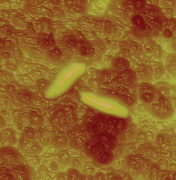
The nanoscale surface analysis of microbial cells represents a significant challenge of current microbiology and is critical for developing new biotechnological and biomedical applications. Using atomic force microscopy (AFM) topographic imaging, researchers can visualize the ultrastructure of live cells under physiological conditions and their subtle modifications upon cell growth or treatment with drugs. Chemical force microscopy, in which AFM tips are modified with specific functional groups, allows investigators to measure molecular forces and chemical properties of cell surfaces on a scale of only 25 functional groups. Molecular recognition imaging using AFM offers a means to localize specific receptors on cells, such as cell adhesionproteins or antibiotic binding sites. With this Highlight on AFM, it is hoped that more and more microbiologists and biophysicists will take advantage of this powerful, multifunctional nanotechnique.
You have access to this article
 Please wait while we load your content...
Something went wrong. Try again?


 Please wait while we load your content...