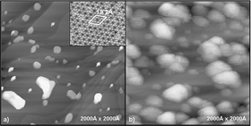
Epitaxial TiO2nanoparticles on Pt(111): a structural study by photoelectron diffraction and scanning tunneling microscopy
Abstract
Angle-scanned

* Corresponding authors
a
Dipartimento di Scienze Chimiche and Unità di Ricerca INFM, Università di Padova, Via Marzolo, Padova, Italy
E-mail:
gaetano.granozzi@unipd.it
Fax: +39 049 827 5161
Tel: +39 049 827 5722
b Institut für Physik der kondensierten Materie, Heinrich-Heine Universität Düsseldorf, Universitätsstrasse 1, Düsseldorf, Germany
Angle-scanned

 Please wait while we load your content...
Something went wrong. Try again?
F. Sedona, M. Eusebio, G. Andrea Rizzi, G. Granozzi, D. Ostermann and K. Schierbaum, Phys. Chem. Chem. Phys., 2005, 7, 697 DOI: 10.1039/B415402A
To request permission to reproduce material from this article, please go to the Copyright Clearance Center request page.
If you are an author contributing to an RSC publication, you do not need to request permission provided correct acknowledgement is given.
If you are the author of this article, you do not need to request permission to reproduce figures and diagrams provided correct acknowledgement is given. If you want to reproduce the whole article in a third-party publication (excluding your thesis/dissertation for which permission is not required) please go to the Copyright Clearance Center request page.
Read more about how to correctly acknowledge RSC content.
 Fetching data from CrossRef.
This may take some time to load.
Loading related content
