
Anisotropy in magnetic materials for sensors and actuators in soft robotic systems
Hyeokju
Kwon
 ,
Yeonhee
Yang
,
Geonsu
Kim
,
Dongyeong
Gim
and
Minjeong
Ha
*
,
Yeonhee
Yang
,
Geonsu
Kim
,
Dongyeong
Gim
and
Minjeong
Ha
*
School of Materials Science and Engineering, Gwangju Institute of Science and Technology (GIST), Gwangju 61005, Republic of Korea. E-mail: minjeongha@gist.ac.kr
First published on 2nd March 2024
Abstract
The field of soft intelligent robots has rapidly developed, revealing extensive potential of these robots for real-world applications. By mimicking the dexterities of organisms, robots can handle delicate objects, access remote areas, and provide valuable feedback on their interactions with different environments. For autonomous manipulation of soft robots, which exhibit nonlinear behaviors and infinite degrees of freedom in transformation, innovative control systems integrating flexible and highly compliant sensors should be developed. Accordingly, sensor–actuator feedback systems are a key strategy for precisely controlling robotic motions. The introduction of material magnetism into soft robotics offers significant advantages in the remote manipulation of robotic operations, including touch or touchless detection of dynamically changing shapes and positions resulting from the actuations of robots. Notably, the anisotropies in the magnetic nanomaterials facilitate the perception and response with highly selective, directional, and efficient ways used for both sensors and actuators. Accordingly, this review provides a comprehensive understanding of the origins of magnetic anisotropy from both intrinsic and extrinsic factors and summarizes diverse magnetic materials with enhanced anisotropy. Recent developments in the design of flexible sensors and soft actuators based on the principle of magnetic anisotropy are outlined, specifically focusing on their applicabilities in soft robotic systems. Finally, this review addresses current challenges in the integration of sensors and actuators into soft robots and offers promising solutions that will enable the advancement of intelligent soft robots capable of efficiently executing complex tasks relevant to our daily lives.
 Hyeokju Kwon | Hyeokju Kwon received his BS from the Department of Organic Materials Science and Engineering, Pusan National University, South Korea, in 2023. He is currently pursuing his Combined Master's–Doctoral Program at the School of Materials Science and Engineering, Gwangju Institute of Science and Technology (GIST), South Korea, under the supervision of Prof. Minjeong Ha. His research interests are in the development of magnetic soft composites and applications for soft robotics. |
 Yeonhee Yang | Yeonhee Yang received her MS from Materials Science and Engineering, Gwangju Institute of Science and Technology (GIST), South Korea, in 2022 and BS from the Department of Display and Semiconductor Physics, Korea University, Sejong, South Korea in 2020. She is currently pursuing her Doctoral program at the School of Materials Science and Engineering, GIST, under the supervision of Prof. Minjeong Ha. Her research interests are in the development of magnetic field sensors for flexible electronics and soft robotics. |
 Geonsu Kim | Geonsu Kim received his MS from the the Department of Carbon Composites Convergence Materials Engineering, Jeonbuk National University, South Korea, in 2023 and BS in the Department of Organic Materials and Textile Engineering from Jeonbuk National University in 2021. He is currently pursuing his Doctoral Program at the School of Materials Science and Engineering, Gwangju Institute of Science and Technology (GIST), under the supervision of Prof. Minjeong Ha. His research interests are in the development of flexible multifunctional sensing devices for human health monitoring and soft robotics. |
 Dongyeong Gim | Dongyeong Gim is currently pursuing his BS program at the Department of Chemistry, Gwangju Institute of Science and Technology (GIST), South Korea. His research interests are in the development of magnetic soft composites and applications for EMI shielding. |
 Minjeong Ha | Minjeong Ha is currently an assistant professor in the School of Materials Science and Engineering at Gwangju Institute of Science and Technology (GIST), Republic of Korea. She received her PhD and MS from the School of Energy and Chemical Engineering at Ulsan National Institute of Science and Technology (UNIST) in 2019 and her BS from the School of Nano-Bioscience and Chemical Engineering at UNIST in 2013. She worked as a postdoctoral fellow at Helmholtz-Zentrum Dresden-Rossendorfe (HZDR) in Dresden, Germany, from 2019 to 2020 and as a researcher at the Electronics and Telecommunications Research Institute (ETRI) in the Republic of Korea from 2020 to 2021. Her research interests include magnetic nanomaterials and stimulus-responsive soft composites for flexible sensors, soft actuators, and energy-harvesting devices. |
1. Introduction
Soft robots, known for their flexible designs and interactive features, introduce a paradigm shift in robotics by offering unique attributes for compliant, continuum, and configurable behaviors.1 These complete soft-bodied systems demonstrate seamless adaptation to irregular surfaces and high degrees of freedom (DoFs) of transformation while exhibiting mechanical resilience.2–4 This capability is achieved using intrinsically deformable and stretchable yet mechanically robust materials such as silicone elastomers, tough gels, functionalized polymers, and polymer composites.5 These characteristics render soft robots superior in areas where conventional rigid robots struggle, playing roles in safe human interactions, handling of delicate objects, navigation of soft robots via confined spaces, and execution of intricate motions of these robots. However, the control of soft robots is more complex than that of rigid robots, which relies on well-defined kinematics of pre-formed joints. Soft bodies exhibit nonlinear viscoelastic behavior with significant hysteresis and different degrees of transformation based on their designs and material compositions. Therefore, predicting their responses and changes in their shapes and positions in a three-dimensional (3D) space is challenging.6The locomotion of soft robots depends on adjustments of the dimension and stiffness of the materials constituting the robot bodies. Stimuli-responsive materials demonstrate notable actuating mechanisms, including energy-efficient and precise control of motion in response to external triggers such as heat, light, humidity, and electric and magnetic fields.7 During the operation of soft robots in untethered states and dynamic environments as illustrated in Fig. 1, magnetic field-responsive materials allow robot bodies to undergo immediate transformation owing to a relatively fast response time of these materials as compared with those of other stimuli-responsive materials.8 Additionally, magnetic field-driven actuation facilitates remote operation of untethered soft robots because magnetic fields can penetrate via various media while decoupling from other stimuli, for example, mechanical stress, radiation, illumination, and humidity.
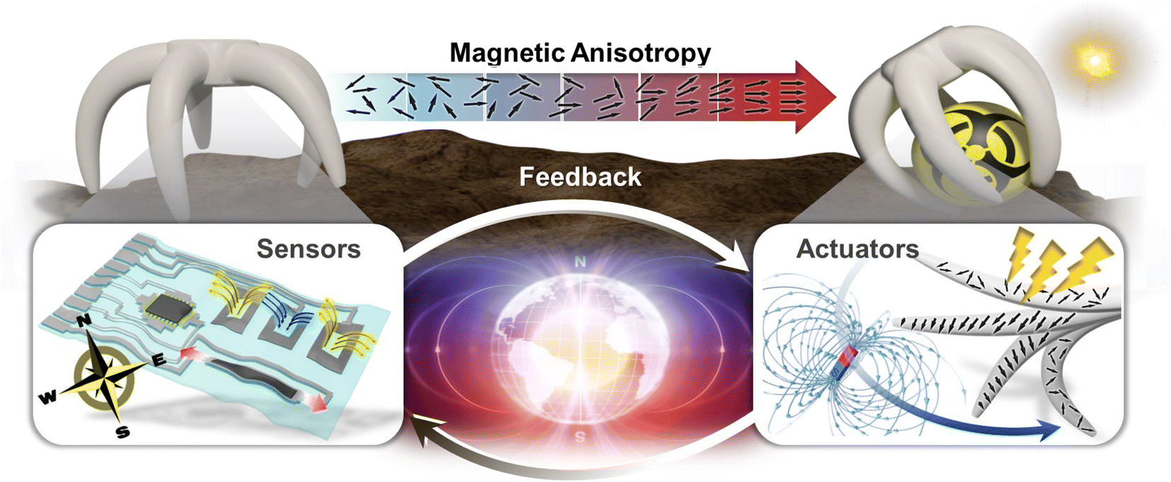 | ||
| Fig. 1 Magnetic soft robot autonomously operating in dynamic environments. The schematic illustration depicts that the magnetic anisotropy facilitates the sensing capabilities for both robotic motions and the surrounding changes as well as the precise manipulation of actuation for the applications of soft robots. | ||
For delicate control of a soft robot, the capability for directional actuation has attracted significant attention.9,10 However, achieving a directional response to external stimuli often requires complicated designs. By employing materials with inherent anisotropic characteristics, it becomes possible to implement directional actuation without the design of complex structures.11 For example, Kim et al. demonstrated an electrothermal soft actuator utilizing the anisotropic thermal expansion of low-density polyethylene (LDPE).12 They designed a bilayer structure comprising LDPE, which has a large anisotropic thermal expansion, and polyvinyl chloride, which exhibits a small isotropic thermal expansion. The mismatch in thermal expansion between the layers results in directional bending in response to electrical stimuli. The significance of magnetism in manipulating robotics is attributed to the selective and directional response capabilities of magnetic materials induced by magnetic anisotropy, which can be obtained by either localized magnetization or the strategical distribution of micro/nano-scale magnets at desired spots in the soft matrix. Magnetic anisotropy with a preferred pole enables the generation of strong torques or programmable actuations along the magnetic easy axes without pre-defined structures.
Soft robotic systems mimicking the sensorimotor functions of biological organisms exhibit adaptabilities to variable and uncertain environments because of the integration of magnetic sensors into their actuating bodies. A closed-loop control system with magnetic sensors and actuators allows the robot to autonomously adjust its activities based on sensory feedback.13–15 Without sensory feedback, even minor variations in material properties or environmental factors can cause errors that disrupt the sequential actuation and hinder task completion. Nevertheless, traditional rigid, chip-type magnetic sensors encounter difficulties in delivering high-quality signals due to their inferior mechanical compliance with soft bodies.16 Fortunately, progress in materials science and flexible electronics has led to the development of stretchable, highly deformable, and conformable magnetic sensors that can be integrated into soft robots without disturbing their motion and degrading the softness of the robot bodies. To monitor both the movements of a robot and its surroundings, two distinct sensing modalities are necessary: proprioception and exteroception.16–18 Proprioceptive sensors provide information about the internal states of the robot bodies during actuation,18 whereas exteroceptive sensors detect changes in the surroundings to identify the location of the robot.16 Magnetic sensors are versatile for these sensing modalities because they not only can detect the variations in stray fields caused by the actuations of magnetic robot bodies, but can also determine the proximity of external magnetic field sources. Particularly, magnetic anisotropy demonstrates strong responses to certain axial directions of the magnetic field. Sensors with magnetic anisotropy offer exceptional sensitivity, accuracy, and selectivity for recognizing the shape and position of soft robots while minimizing interference and crosstalk resulting from varying magnetic field orientations or the integration of multiple magnetic sensors into the robot bodies. Despite the challenges associated with integrating sensors and actuators due to the potential for interference among the components, this integration remains essential for the advancement of soft robotics with seamless operation. Thus, anisotropy in magnetic nanomaterials is indispensable for high compatibility of both sensors and actuators in terms of controlling actuation and tracking changes in the internal and external states of magnetic soft robots. The anisotropy guarantees the successful execution of various missions and tasks.
This review aims to investigate magnetic nanomaterials with a particular focus on magnetic anisotropy and applications of these nanomaterials in sensors and actuators, which are key components in soft robotics (Fig. 1). First, we discuss the origins of magnetic anisotropy and underlying mechanisms that drive anisotropy in magnetism considering the energy in the system. Then, a variety of magnetic nanomaterials with enhanced magnetic anisotropy through alignment, shape control, interlayer coupling, and external energy source (e.g. mechanical and magnetic energy) are highlighted. These magnetic nanomaterials are categorized based on their dimensions and dominant magnetic anisotropy, which will be discussed in section 2. Furthermore, we present the compatibility of each magnetic nanomaterial with distinct magnetic anisotropy and discuss how these materials and corresponding magnetisms satisfy the specific requirements of sensors, actuators, or both in soft robotic applications. As these magnetic nanomaterials are sufficiently compliant with the pliable bodies of soft robots, we investigate their design, fabrication, and manufacturing processes aimed at preserving the anisotropic properties of these materials and maintaining the requisite softness of the corresponding soft robots. Finally, we examine how magnetic anisotropy contributes to enhancing the performances of magnetic sensors and actuators with a particular emphasis on the crucial roles of magnetic sensors and actuators in enabling various functions in soft robots.
2. Fundamentals of magnetic anisotropy
Magnetic anisotropy denotes the pinning of magnetic moments in a specific orientation, leading to directional dependence of the magnetization, which is observed in ferromagnetic (FM) materials. To understand this directional dependence, anisotropy energy needs to be considered. In other words, a preferred direction of magnetization, namely, the magnetic easy axis, arises to minimize the anisotropy energy. Various anisotropies originate from different mechanisms and the total energy of the system is determined by the interactions between them, rather than a single mechanism. The total energy in magnetic materials is expressed as follows:| Etotal = EZeeman + Ecrys + Esh + Eme + Eex | (1) |
Note that the E terms in this review denote the energy density caused by the dominant mechanism of anisotropy while the EZeeman is associated with the interaction between the magnetic moments and external magnetic field (Hext). Specifically, the magnetic anisotropy energy density originates from several key factors: crystal orientation (Ecrys), dimension and shape (Esh), magnetoelasticity (Eme), and interfacial exchange coupling (Eex) of magnetic materials. The complex interplay of these energies elucidates how FM materials exhibit a magnetic easy axis resulting in directional responses under Hext (Fig. 2). Therefore, we will discuss how the dominant energy term varies with respect to material size, dimension, shape, surface, and other relevant variables in this section (Fig. 2a). Subsequently, we discuss how the anisotropy energy influences the directional behavior and magnetization state (Fig. 2b), considering both intrinsic and extrinsic properties of magnetic materials.
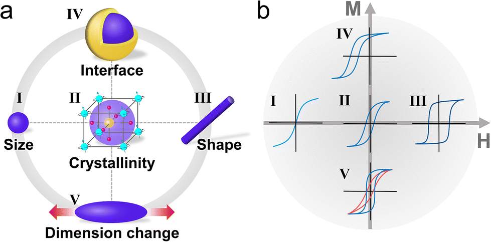 | ||
| Fig. 2 Factors influencing the magnetization states of nanomaterials. (a) Magnetic properties induced by (I) size, (II) crystallinity, (III) shape, (IV) interfacial coupling, and (V) dynamic changes in the dimensions of magnetic nanomaterials. (b) Magnetization state verified by magnetization hysteresis loops based on the factors in (a): (I) a decrease of size led to superparamagnetism, (II) the crystal structure determined the inherent magnetizations of materials and hysteresis curve shapes, (III) shape anisotropy resulted in increasing coercivity and remanent magnetization, (IV) exchange coupling at the interface between different magnetic materials caused curve shifts, and (V) there were dynamic changes in the magnetization states according to the magnetostrictive properties of the materials. | ||
2.1. Intrinsic magnetocrystalline anisotropy
Crystallographic orientations in magnetic materials yield preferential directions of magnetization for minimizing anisotropy energy known as magnetocrystalline anisotropy. This fundamental mechanism of magnetocrystalline anisotropy originates from spin–orbit coupling along with the interaction between the orbital motion of electrons and the crystal field of the lattice.19,20 Assuming that the orbital contributions to magnetic moments are quenched, magnetic properties are primarily determined by the spin of the electron. Notably, the orientations of the crystal structures do not strongly affect the electron spin. However, spin–orbit coupling can bridge the gap between the spin and crystal lattice as the orbital orientation becomes firmly fixed to the lattice.21 This interaction leads to an anisotropy energy that defines the preferred orientation of magnetization.
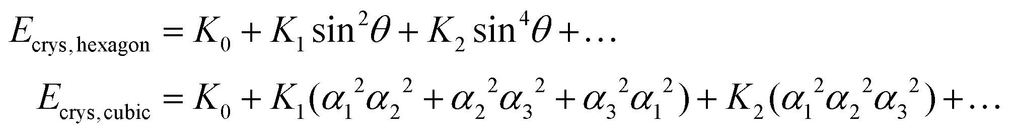
Fig. 3 illustrates the magnetocrystalline anisotropy of magnetic materials, particularly for the simplest cases of hexagonal close-packed cobalt (hcp-Co), body-centered cubic iron (bcc-Fe), and face-centered cubic nickel (fcc-Ni). For cobalt, which has a hexagonal crystal structure, the easy axis is along the [0001] direction, while the hard axis is in the 〈10![[1 with combining macron]](https://www.rsc.org/images/entities/char_0031_0304.gif) 0〉 directions (Fig. 3a). The hard axis is defined as an unfavored direction of magnetization, requiring a higher field to be saturated in that direction. The behavior of hcp-Co can be explained by the energy of the system, which is described in the following equation:
0〉 directions (Fig. 3a). The hard axis is defined as an unfavored direction of magnetization, requiring a higher field to be saturated in that direction. The behavior of hcp-Co can be explained by the energy of the system, which is described in the following equation:
Ecrys, hexagon = K0 + K1![[thin space (1/6-em)]](https://www.rsc.org/images/entities/char_2009.gif) sin2θ + K2sin4θ + … sin2θ + K2sin4θ + … | (2) |
0〉 direction. Since hcp-Co has a single easy axis along the c-axis, it exhibits uniaxial anisotropy.
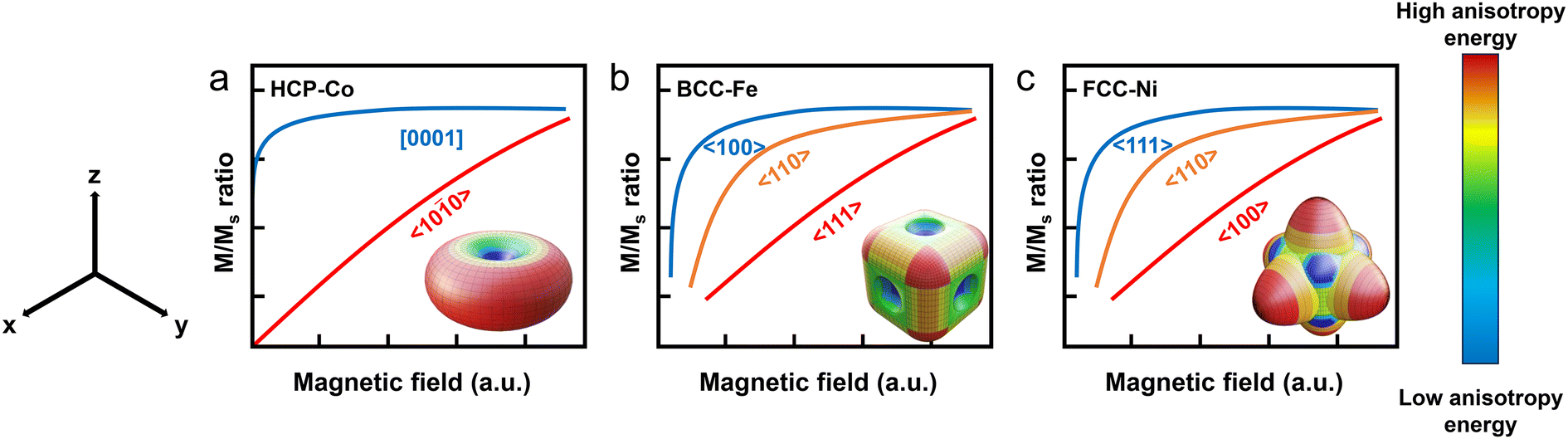 | ||
| Fig. 3 Magnetocrystalline anisotropy of the simplest magnetic materials such as (a) hcp-Co, (b) bcc-Fe, and (c) fcc-Ni. The inset images in each figure represents magnetocrysalline ansisotropy energy. The red color represents high magnetic anisotropy energy resulting in a hard axis along that direction while the blue color represents low magnetic anisotropy energy forming an easy axis in the direction. | ||
The situation for bcc-Fe and fcc-Ni is quite different compared with hcp-Co (Fig. 3b and c). They have multiple easy axes, which can be also explained by the energy of the system. For cubic symmetry,
 | (3) |
2.2. Shape anisotropy and finite size effect
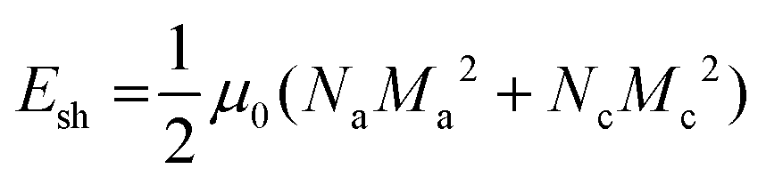
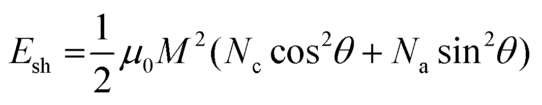
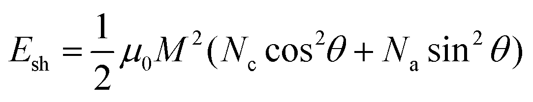
The preferential magnetization direction induced by magnetic anisotropy energy is influenced by not solely the intrinsic crystallographic geometry, but also the shapes and sizes of the magnetic materials. Unlike the case of magnetocrystalline anisotropy, the energy derived from shape anisotropy can be deliberately adjusted by tailoring the designs of magnetic materials with specific structures and dimensions. All magnetic materials inherently possess a demagnetizing field (Hd) resulting from the magnetostatic interaction between the north and south poles in materials when they are subjected to Hext (Fig. 4a).25 Moreover, Hd is directly proportional to −NdM, where Nd indicates the demagnetizing factor and M corresponds to the magnetization of the materials. Nd is a shape-dependent parameter, which provides an opportunity to modulate the magnitude of Hd in magnetic materials. For example, in the cases of isotropic and spherical magnetic particles, Nd is equal to 1/3. However, for an ellipsoidal magnetic particle, Nd varies depending on the axis of the particle (Fig. 4b). Along the longest axis, denoted as “c”, Nc is less than 1/3 as the magnetostatic forces decrease with respect to the pole distance (r), demonstrating an inverse square relationship with r (1/r2). Simultaneously, along the shortest axis, denoted as “a”, Na is larger than 1/3 because the sum of the demagnetizing factors is equal to 1 for each axis.25,26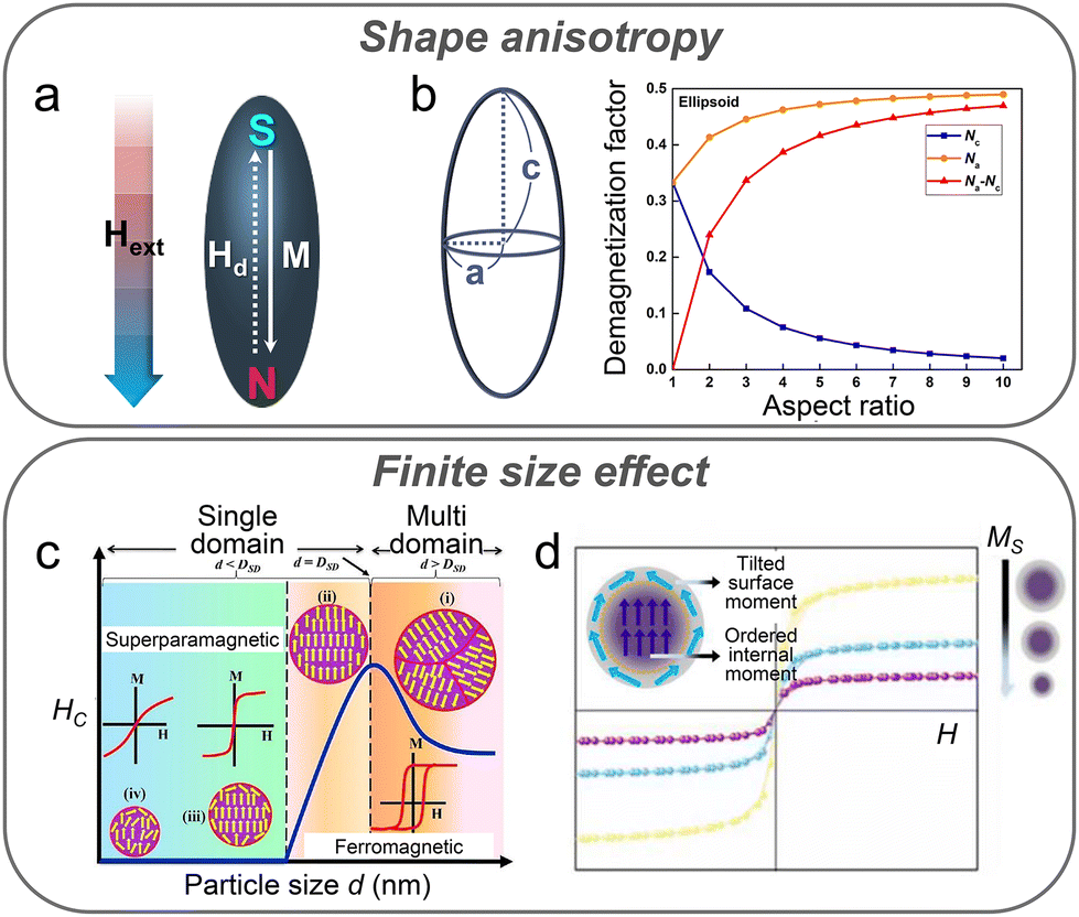 | ||
| Fig. 4 Magnetism based on the shape and size of magnetic materials. Under external magnetic fields, (a) a demagnetizing field generated along the long-axis direction due to shape anisotropy. (b) Relationship between demagnetizing factors and the aspect ratio of a prolate ellipsoid. Reproduced with permission.26 Copyright, the Creative Commons CC BY License. (c) Size-dependent behavior of coercivity in magnetic nanomaterials. Reproduced with permission.28 Copyright, the Creative Commons CC BY License. (d) Decrease in saturation magnetization owing to magnetic dead layers with a decrease in the size of the magnetic nanomaterials. Reproduced with permission.36 Copyright, the Creative Commons CC BY License. | ||
The magnitude of the demagnetizing field resulting from the shape-dependent differences in Nd defines the shape anisotropy energy Esh, which can be expressed as follows:
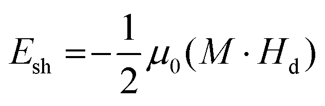 | (4) |
 | (5) |
 | (6) |
The size of the magnetic materials is another key factor affecting the magnetic properties and even contributes to magnetic anisotropy, specifically with a decrease in dimensions (Fig. 4c and d).28 Bulk magnets naturally form a multi-domain state to minimize the magnetostatic energy instead of involving all spins aligned in parallel.29 However, when the size of a magnetic particle is reduced to the nanoscale, it transitions into a single-domain state upon reaching a specific diameter (Dsd). Assuming an uniaxial magnetic material for simplification, Dsd can be calculated as follows:
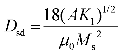 | (7) |
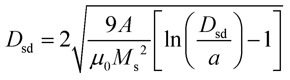 | (8) |
In the single-domain state, the magnetization is uniformly aligned along a particular direction, and the magnetization reversal is caused by the coherent rotation of the magnetic moment which is described by the Stoner–Wohlfarth model, leading to higher coercivity than that in the multi-domain state.30 With a decrease in the diameter of a magnetic particle to below the sub-nanometer scale, Ecrys reduces across the entire volume of the particle. When Ecrys falls below the thermal energy threshold, the magnetic orientation within the particle experiences thermal fluctuations, which are commonly observed in paramagnetic materials. This state is referred to as superparamagnetism.31 The critical diameter (Dsp) at which a particle exhibits superparamagnetic properties can be defined as follows:
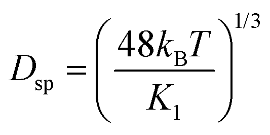 | (9) |
2.3. Exchange anisotropy
Interfacial coupling between magnetic bilayers or multilayers can be observed by an extrinsic factor, namely, exchange anisotropy. This phenomenon occurs at the interface between an FM material and an adjacent antiFM (AFM) material, resulting in a unidirectional magnetization orientation in the FM material.37,38 This interfacial coupling is not limited to FM/AFM bilayers but extends to other magnetic configurations, such as FM/AFM superlattices,39 ferrimagnet (FI)/AFM,40 FI/FM,41 soft FM/hard FM,42 and FM/spin glass systems.43 Unidirectional pinning of the magnetization notably alters the hysteresis loop of the FM/AFM coupled layers when compared with that of a standalone FM layer, where the center of the hysteresis loop shifts from the zero magnetic field position (Fig. 5a). This shift in the magnetic hysteresis loop is termed bias, which is a behavior associated with exchange anisotropy and is thereby called exchange bias. During magnetic-thermal treatment (Fig. 5a), the exchange bias appears after cooling the FM/AFM coupled system. Typically, critical temperature limits exist for both FM and AFM materials, denoted as the Curie temperature (TC) and Néel temperature (TN), at which magnetic materials exhibit phase transitions and subsequently change or lose their magnetism. Within the temperature range of TN < T < TC, the spins in the FM layer can align in the direction of Hext, while the spins in the AFM layer randomly orient. When the temperature decreases below TN, the spins in the AFM layer adjacent to the FM layer are oriented to the spin configurations of the FM layer. The other spin planes in the AFM layer follow the rule of antiparallel alignment for a zero net magnetization. If the magnetic field is reversed, the spins in the FM layer rotate, whereas those in the AFM layer remain unchanged. The stationary spins exert a torque that resists the spin rotation in the FM layer, holding the spins in their initial orientation. Thus, the complete reversal of the spins in the FM layer coupled with an AFM layer requires a larger field to overcome the torque. Owing to this interfacial coupling, the magnetic hysteresis loop shifts and coercivity changes.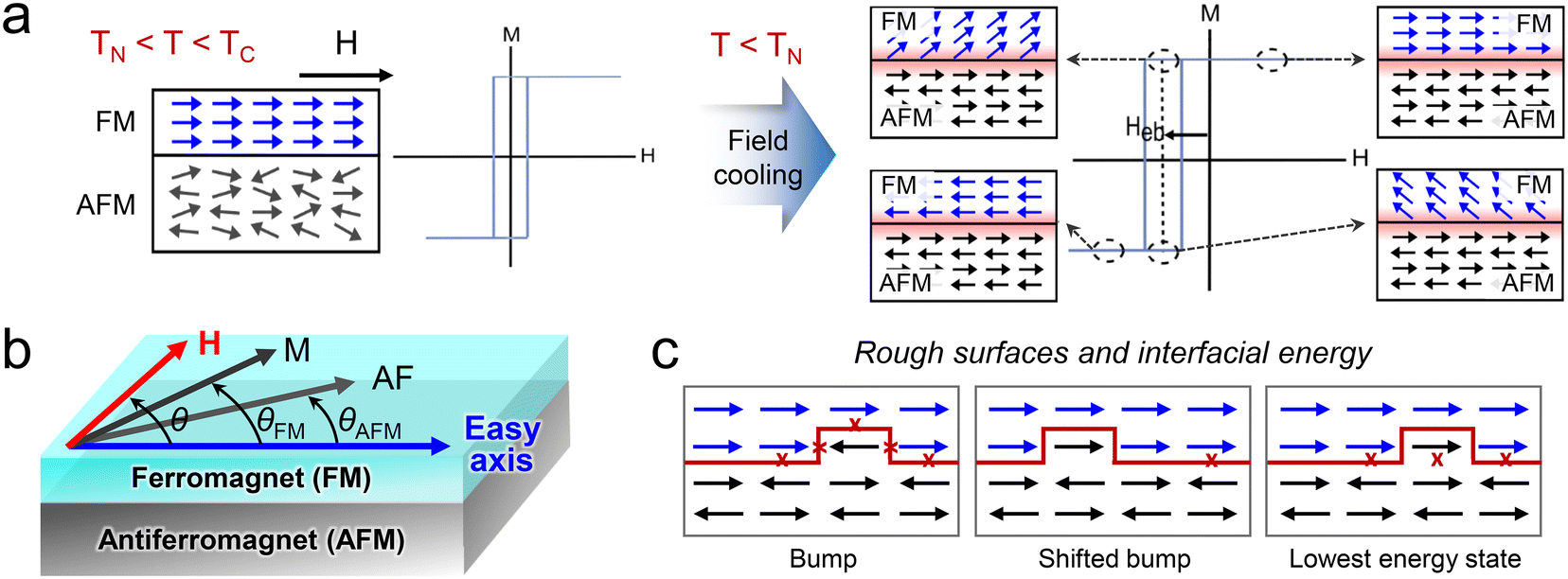 | ||
| Fig. 5 Exchange bias in magnetic interlayers. (a) Spin configurations and magnetization curves of the FM/AFM bilayer. At TN < T < Tc, the centered magnetization curve and the paramagnetic state of the AFM layer are alongside the aligned spin state in the FM layer (left). A field cooling process induces a shift in the magnetization curve, and this shift is known as exchange bias. Reproduced with permission.37 Copyright 1999, Elsevier Science B.V. (b) Magnetic easy axis based on exchange anisotropy arising from FM/AFM interlayer coupling and angular relationships of the magnetization of the FM layer with respect to the external field direction. (c) Magnetic moment configuration at the FM/AFM interface with a bump. The cross symbol represents frustrated bonds. Starting from the fully compensated state of the AFM layer, the introduction of a bump generates AFM deviations. A shifted bump induces FM deviations resulting in a net interfacial energy difference. When the lowest energy state is achieved, the interfacial energy difference is reduced. Reproduced with permission.46 Copyright 1987, American Physical Society. | ||
Numerous models have been proposed to explain exchange bias in different systems. An early simple model was suggested by Meiklejohn-Bean to describe oxidized FM particles (Co/CoO).44 This model assumed an atomically smooth FM/AFM interface, where both magnetic materials existed in a single domain. In the AFM material, the uncompensated spins are aligned in the same crystallographic plane and direction at the interface of FM/AFM, due to the aforementioned cooling step. Then, the exchange anisotropy energy required to overcome the resistance of spin rotation in the coupled FM material was calculated. This exchange bias model considered the total energy (E) originating from the coherent rotation of FM magnetization as follows:37
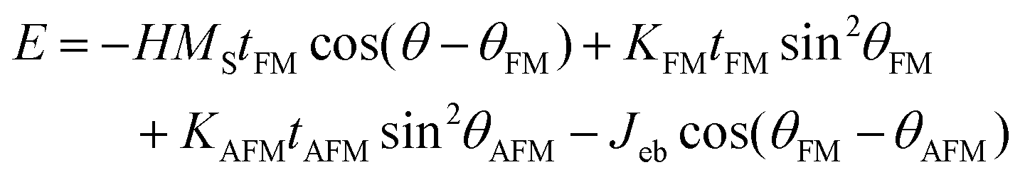 | (10) |
The sequence in eqn (10) presents the uniaxial anisotropy of FM and AFM materials, and exchange anisotropy energy. Here, H is the applied field, MS is the saturation magnetization of the FM layer, and tFM and tAFM denote the thickness of the FM and AFM layers, respectively. KFM and KAFM represent the uniaxial anisotropy constants of the FM and AFM layers. As KAFM is typically larger than KFM, KFM can be ignored. Jeb is the interlayer exchange anisotropy constant. In addition, θ, θFM, and θAFM denote the angles of the applied magnetic field, magnetization of the FM layer, and sublattice magnetization of the AFM layer with respect to the predetermined easy axis of the FM and AFM layers (Fig. 5b). Then, the exchange anisotropy energy was defined as follows:
| Eex = −Jebcos(θFM − θAFM) | (11) |
The exchange anisotropy is often regarded as unidirectional anisotropy rather than uniaxial, since it is proportional to the first power of the cosine. When the spin alignment of the AFM layer matches the easy axis, that is, θAFM ≈ 0, the field required to switch the magnetization of the FM layer is defined as the exchange bias field, Heb. Considering the energy stability condition (∂E/∂θ = 0), Heb can be obtained as follows:
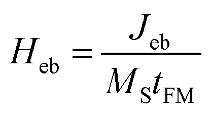 | (12) |
According to eqn (12), the exchange bias field is inversely proportional to the thickness of the FM layer because of its association with interfacial characteristics.37 However, in this model, the assumption regarding the presence of fully uncompensated spins at the interface causes differences in Heb with experimental results.45 In the uncompensated case, all AFM spins at the FM/AFM interface are aligned in the same direction and the Jeb is directly proportional to the FM exchange constant Ji. However, the actual spin configuration of the AFM surface is considerably complex due to a non-ideal FM/AFM layer.
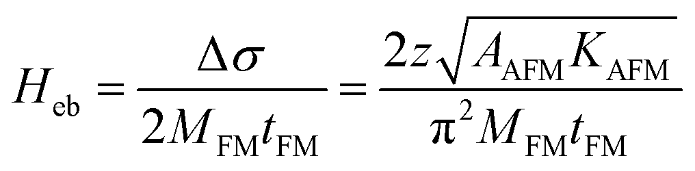
Several models have been proposed to address this gap and elucidate the mechanism of exchange bias at the FM/AFM interface. According to the random field model proposed by Malozemoff, the dynamics of interfacial conditions, including defects at the interface between the FM and AFM layers, produces a randomness in the Heb of the system.46 In the presence of local bumps (Fig. 5c), the net interfacial energy difference decreases as the AFM spin in the bumps is inverted at the lowest energy state, thus affecting the total exchange anisotropy constants. For example, the net interfacial energy difference, which is calculated as 6 × 2Ji = 12Ji, is reduced by 5 × 2Ji owing to the existence of the bump when the system reaches its lowest energy state. The energy difference 4J results from the summation of the FM exchange constant (2Ji) and AFM constant (2JA), assuming J ≈ Ji ≈ JA. Eventually, a perpendicular domain-like region is formed to minimize the net random unidirectional interfacial anisotropy. In this model, Heb is acquired as follows:
 | (13) |
Average interfacial energy is defined as, Δσ = 4zJ/πaL, where z, J, a, and L represent the number of antiparallel pairs, exchange coupling constant, cubic lattice parameter, and domain size, respectively. Furthermore, AAFM is the exchange stiffness of the AFM layer, also represented as AAFM ≡ J/a. The inherent randomness influences the behavior of the domain wall in the AFM layer, which weakens the Heb strength.
Domain wall formation in the AFM layer bridges the theoretical and experimental differences in exchange bias. The random field model associates the exchange bias with a domain wall perpendicular to the FM/AFM interface, while Mauri's model assumes that the domain wall in the AFM layer is parallel to this interface. Thus, the calculated Heb is lower than what is predicted by the Meiklejohn–Bean model.47 Nevertheless, when tAFM reduces below a critical level, the domain wall is not formed, leading to the disappearance of Heb. As an ultra-thin AFM layer forms island-like grains rather than a continuous film, this layer is insufficient for coupling with the FM layer.48 Moreover, the domain state model indicates that a domain is formed in the bulk AFM layer during magnetic thermal treatment.49 In this process, the AFM layer experiences magnetic dilution due to non-magnetic (NM) defects. Domain wall formation is energetically favorable as this wall passes through the NM defects, causing its pinning. Subsequently, this pinned domain wall lowers Heb by reducing the number of uncompensated spins at the FM/AFM interface. Consequently, tAFM is a critical factor in determining the potential of the defects that may impact exchange anisotropy.
Although the interfacial conditions of the FM/AFM layers affect the exchange bias, the crystallographic orientation of the AFM material should also be considered for explaining exchange anisotropy because of the relationship between the bulk crystallinity and spin configurations at the interface of the FM/AFM layers.50,51 Assuming that the AFM spins at the FM/AFM interface are similar to those in the bulk material, which are significantly affected by the crystallographic structure, an angle is inevitably formed between the AFM and FM spins. Then, the exchange bias depends on the angle between the AFM and FM spins. According to the Hamiltonian equation for exchange bias, Jint|SAFM||SFM|cosα, where α = 0° results in maximum Heb and α = 90° indicates no exchange bias.52 The crystal orientation of the AFM layer at the interface determines whether this layer is in a compensated or an uncompensated state. Consequently, the exchange anisotropy is not only evidently determined by the interfacial condition between the FM and AFM layers, but also significantly influenced by the thickness and crystal structure of these layers. By considering the factors associated with exchange bias, the strength and stability of Heb can be controlled. This capability is of considerable importance as it tunes the behavior of the magnetic material based on exchange anisotropy according to the specific requirements of magnetic field sensors. For example, exchange bias is applied to anisotropic magnetoresistance (AMR)-based sensors to adjust the magnetization direction.53,54 Furthermore, exchange bias is applied to spin valve55 and magnetic tunnel junction devices56 to appropriately pin the magnetization of the FM layer.
2.4. Stress-induced magnetic anisotropy
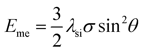
In the previous sections, the different parameters, including both intrinsic magnetocrystalline anisotropy and extrinsic influences such as adjusting the dimensions, shapes, and interlayer coupling of the magnetic materials, governing magnetic anisotropy and its associated energy are comprehensively discussed. Magnetic anisotropy has been explored in a static state without considering dynamic conditions, for instance, applied mechanical stress, temperature changes, and the different strengths and directions of stray fields. In this section, we investigate magnetic materials exhibiting dynamic behaviors in which anisotropy can be induced by mechanical stress.Magnetostriction is a phenomenon where the dimensions of a material vary in response to alterations in its magnetization orientation, accompanied by domain wall motion under Hext.57,58 Magnetic materials exhibit strain of the saturation magnetostriction coefficient (λsi) in the direction of the magnetic field during magnetization.59 Thus, the presence of magnetostriction implies that mechanical stress can vary the magnetic domain and can be a new source of magnetic anisotropy.60–62 This variation of magnetization under stress is known as inverse magnetostriction or, more commonly, the magnetoelastic effect.62,63 The magnetoelastic effect is correlated with λsi and the magnetic behaviour of a material under stress (σ). λsi can be positive or negative depending on the crystal structure of the materials and composition of the alloys. For example, bcc-Fe exhibits a positive λsi, whereas fcc-Ni shows a negative λsi.64 Conversely, Fe20Co80 alloys are in the fcc phase for a positive λsi or bcc phase for a negative λsi depending on the fabrication methods.65 If a magnetic material has a positive λsi, it will elongate during magnetization. This implies that applying a tensile stress to lengthen the material (λsiσ > 0) increases the magnetization of the material, which facilitates the formation of a preferential orientation of the easy axis.58,66 However, magnetization in a material may not always be parallel to stress, and the resulting Ms can be understood by considering the magnetic anisotropy energy of the system. Assuming a simple cubic crystal, the overall magnetic anisotropy energy can be expressed as
 | (14) |
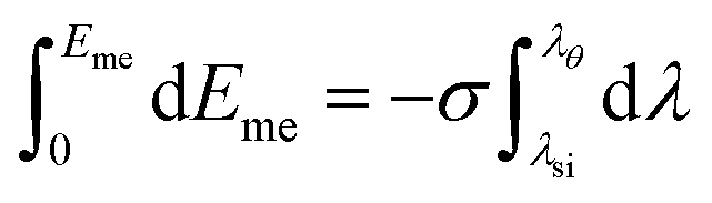
| dEme = −σdλ | (15) |
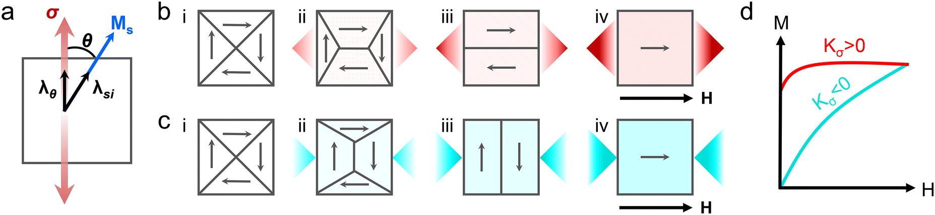 | ||
| Fig. 6 Magnetic anisotropy induced by external stress. (a) The angle θ of the resultant saturation magnetization in magnetic materials involving magnetostriction coefficient (λ) under tensile stress (σ). Domain wall motion of a polycrystalline magnetic material under tensile stress with (b) positive Kσ (λsi > 0, σ > 0) and (c) negative Kσ (λsi > 0, σ < 0). (d) Corresponding magnetization curves of (b) iv (red) and (c) iv (blue) under an external magnetic field. | ||
Therefore, the total Eme can be derived by using a simple term:
 | (16) |
 | (17) |
 | (18) |
| Eme = Kσsin2θ | (19) |
According to eqn (19), when a material has a positive Kσ, Eme is minimum at θ = 0° and maximum at θ = 90°.61 Thus, the magnetic easy axis is formed along the direction of stress to minimize the energy of the system. For a negative Kσ, the situation is reversed, as the magnetic easy axis is perpendicular to the system. Since external stress induces a single easy axis even in cubic materials, stress-induced anisotropy can be treated as uniaxial anisotropy.
A polycrystalline material typically demonstrates a weak crystal anisotropy without a preferential orientation, often existing in a demagnetized state (Fig. 6b-i and c-i).69 An applied tensile stress initiates domain wall motion, increasing the volume of the domains magnetized in a stress-induced easy axis, thereby exerting the direction of Ms (Fig. 6b-ii and c-ii).61 As all domains are aligned with the easy axis, Eme is minimized (Fig. 6b-iii and c-iii).70,71 Upon applying Hext (Fig. 6b-iv and c-iv), the magnetization curve of a positive Kσ would appear as the case of easy-axis magnetization of the uniaxial magnetic material, whereas for a negative Kσ, the magnetization curve would be similar to that of hard-axis magnetization (Fig. 6d).58
Representative materials with a magnetostriction effect include Co, Ni, Fe, permalloy (Ni–Fe alloys), Terfenol-D (TbxDy1−xFe2), and Galfenol (Fe–Ga alloys).59,72–74 However, the levels of magnetoelasticity in these materials may vary. Considering the total energy in eqn (14), materials with a relatively larger value of K1 than λsi are primarily influenced by crystal anisotropy rather than stress anisotropy. Modulation of the magnetic anisotropy in this crystal system via applied stress requires an energy contribution from stress anisotropy that is at least equal to that of the crystal anisotropy energy. Therefore, materials with a low λsi value need significantly higher stress levels to achieve a high Kσ, as expressed by the relationship, Kσ = 3/2λsiσ. This may exceed the yield stress of the materials, potentially causing failure. Thus, magnetic anisotropy is not determined by a single mechanism, but is affected by complicated relationships between various mechanisms. A summary of magnetic anisotropies ranging from intrinsic magnetocrystalline anisotropy to extrinsic magnetic anisotropies is presented at Table 1.
| Anisotropies in magnetic materials | Influencing factors | Origin of anisotropies | Energy equations | |
|---|---|---|---|---|
| Intrinsic | Magnetocrystalline anisotropy | Crystal structures | Spin–orbit coupling |
|
| Extrinsic | Shape anisotropy | Overall shape and aspect ratio | Demagnetizing field |
|
| Exchange anisotropy | Interfacial coupling between magnetic materials | Exchange interaction |
E
ex = −Jebcos(θFM − θAFM) |
|
| Stress-induced anisotropy | External mechanical stress | Magnetoelastic effect |
E
me = Kσsin2θ |
3. Synthesis and design strategies for anisotropy in magnetic materials
3.1. Anisotropic assemblies of zero-dimensional magnetic nanomaterials
Magnetic behaviors of spherical MNPs, characterized by isotropic morphologies and properties in all directions, exhibit size dependence instead of directional dependence. When reduced to the single-domain size, typically, below tens of nanometers, these MNPs commonly demonstrate superparamagnetism. This phenomenon is attributed to the fact that the thermal energy-induced transition of spin direction drives magnetic fluctuations, resulting in decayed remanence and coercivity, as discussed in section 2.2. Therefore, the single-domain spin confinement of spherical MNPs presents challenges in maintaining ferromagnetism and achieving magnetic anisotropy. However, monodisperse MNPs with high magnetization values can achieve long-range ordering and transform into anisotropic MNPs via magnetic dipole–dipole interactions.75 In addition to the magnetic dipole–dipole interactions, colloidal systems induce random agglomeration or clustering of monodisperse MNPs via physical interactions.76 Magnetic anisotropy can considerably vary depending on the assembly processes and resulting chain orientations of MNPs. This variability in magnetic anisotropy expands the applicability for magnetic soft robots. Therefore, highly stabilized monodisperse MNPs are necessarily prepared because their arrangements can be appropriately controlled to prevent their random agglomeration.Wet chemical synthesis of monodisperse MNPs provides many advantages such as controllability over the desired shape, size, and dimension of NPs and obtaining highly stabilized MNP dispersion by tailoring the solvents, additives, and capping surfactant. A variety of solution processing methods have been extensively employed for the synthesis of MNPs, including co-precipitation,77 thermal decomposition,78,79 and the solvothermal approach.80 These well-established synthesis methods reliably produce high-quality and monodisperse MNPs. Nevertheless, the anisotropic assembly of MNPs has proved challenging owing to the weaker dipolar attractions when compared with thermal fluctuations or isotropic van der Waals interactions between MNPs.76 Klokkenburg et al. discussed the potential for chain conformations in monodisperse MNPs, suggesting the requirement for magnetic dipole–dipole interactions more than that for thermal fluctuations.75 As-synthesized magnetite NPs with a size of 21 nm obtained by thermal decomposition were successfully assembled into chain conformations in a ferrofluid. However, these assembled MNPs with the anisotropic geometry exhibited instability and disassociation, requiring further efforts to achieve prolonged stability in anisotropic chain conformations from the isotropic MNPs.
A straightforward approach to obtain sustainable and stable anisotropic configurations of isotropic MNPs involves one-dimensional (1D) template-assisted assembly. Correa-Duarte et al. used functionalized multi-wall carbon nanotubes (MWCNTs) as templates.81 Layer-by-layer coating of poly(sodium 4-styrene sulfonate) (PSS) and poly(dimethyldiallylammonium chloride) (PDDA) on the MWCNT surface facilitated electrostatic interactions of positively charged MWCNTs with negatively charged γ-Fe2O3/Fe3O4 NPs (Fig. 7a). Since 1D MWCNTs served as templates for the anisotropic assembly of MNPs, γ-Fe2O3/Fe3O4 NP-decorated MWCNTs exhibited magnetic anisotropy and aligned in response to an external magnetic field (0.2 T, Fig. 7b). Fan et al. proposed an in situ attachment of MNPs to the surface of MWCNTs during the synthesis of MNPs via thermal decomposition.82 Vacuum pumping during the thermal decomposition of the Fe(CO)5 precursor afforded precise control over the size of the MNPs as well as ensured a uniform attachment of MNPs to the MWCNT surfaces. Unlike the aforementioned methods based on the surface treatment of a template, a recent template-assisted assembly included the decoration of Fe3O4 NPs on MWCNTs without acid treatment. The mussel-inspired catechol chemistry enabled in situ attachment of Fe3O4 NPs to pristine MWCNTs via a co-precipitation process.83
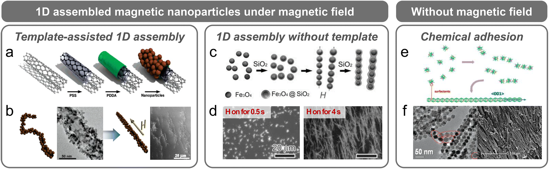 | ||
| Fig. 7 Zero-dimensional MNPs and anisotropic assembly fabricated with various methods. (a) Template-assisted assembly of γ-Fe2O3/Fe3O4 on MWCNTs via layer-by-layer coating and (b) transmission electron microscopy (TEM) images of γ-Fe2O3/Fe3O4 compactly attached to an MWCNT and the alignment of γ-Fe2O3/Fe3O4 under a magnetic field of 0.2 T. The scale bars are 50 nm and 20 μm for the images on the left and right, respectively. Reproduced with permission.81 Copyright 2005, American Chemical Society. (c) Anisotropic assembly of Fe3O4 achieved by applying an external magnetic field followed by fixation with a sol–gel reaction of silica and (d) variation in chain length by controlling the timing and duration of the magnetic field. The scale bar is 20 μm for both images. Reproduced with permission.85 Copyright 2011, John Wiley and Sons. Anisotropic structure induced by chemical adhesion illustrated with (e) a mechanism indicating how the selective adhesion of organic surfactants leads to an α-Fe2O3 nanochain, and (f) TEM images showing the formation of a polyhedron particle by organic surfactants. The scale bars are 50 and 500 nm for the images on the left and right, respectively. Reproduced with permission.90 Copyright 2010, American Chemical Society. | ||
However, in cases where the initially employed template becomes unnecessary in the system, its removal is challenging and requires a complex process. Thus, ongoing research aiming at anisotropic assembly of MNPs without templates is being conducted. An effective strategy for preparing a stable anisotropic assembly of MNPs without templates involves magnetic field-induced alignment, followed by immobilization of the aligned MNPs in a viscous polymer matrix. Sheparovych et al. fabricated magnetite nanowires (NWs) by aligning negatively charged superparamagnetic Fe3O4 NPs under a magnetic field, followed by the slow addition of a positively charged polyelectrolyte to preserve the alignment.84 Hu et al. exploited a sol–gel reaction of silica to fix the anisotropic assembly of peapod-structured MNPs induced by an external magnetic field (Fig. 7c).85 Notably, the morphology of the assembly could be finely tuned by regulating the periodicity and length of the assembly, which was determined by the size of the Fe3O4 NPs and the time for which the magnetic field was applied (Fig. 7d). Xiong et al. adopted polydopamine to lock the anisotropic assembly of Fe3O4 NPs induced by a magnetic field.86 Dopamine can be uniformly deposited on various surfaces and subsequently transform into polydopamine via self-polymerization. This conformal polydopamine coating effectively confined the high-order arrangement of MNPs. As the polydopamine scaffold provided functional groups participating in secondary reactions such as Michael addition or Schiff base reaction,87–89 this polydopamine-coated MNP assembly exhibited reactive functionalities under specific conditions for biosensing applications such as selective antibody capture and prevention of non-specific biofouling.
Under zero-field conditions, several organic compounds help in the fabrication of anisotropic assemblies of MNPs. Meng et al. fabricated a highly uniform α-Fe2O3 nanochain structure via selective adhesion of organic surfactants such as sodium oleate, oleylamine, and oleic acid (Fig. 7e).90 This selective adhesion resulted in metastable polyhedron particles, where the cusp of particles were partially dissolved and bonded to adjacent particles, thereby minimizing the surface energy (Fig. 7f). To enhance the binding stability between particles, a covalent linkage between MNPs can be adopted in an anisotropic assembly. Nakata et al. introduced a binary mixture of immiscible molecules as shells for NPs, and these molecules were self-assembled on the NP shell, leading to a nano-worm structure.91 To further secure the assembled structure, a molecular linker, 11-(10-carboxy-decyldisulfanyl)-undecanoic acid, was used to covalently bind NPs. This strong chemical bonding acted as the primary driving force for the anisotropic assembly of various MNPs, which is different from the magnetic dipole–dipole interaction. However, the chemical bonds that assist the assemblies might also form an undesirable random arrangement or bulk agglomeration of MNPs due to the absence of a guiding field.92 To prevent the random attachment of MNPs, He et al. used maleic anhydride-grafted polypropylene (PP-g-MA), where MA functioned as a surfactant, whereas PP restricted the random aggregation of NPs.93 Considering the co-existing conditions of attraction and repulsion, the weight ratio of PP-g-MA to the metal precursor should be optimized to form chain-like anisotropic assemblies of MNPs rather than a monodisperse or agglomerated MNPs.
3.2. Shape anisotropy of one-dimensional magnetic nanomaterials
Classic nanomaterials, such as nanorods (NRs), nanotubes (NTs), and NWs, characterized by morphological anisotropy demonstrate 1D structures with different length-to-diameter ratios. In cases of magnetic nanomaterials, which are typically crystalline, anisotropy occurs as a natural consequence of the intrinsic variations in interaction strengths among the constituent atomic or molecular building blocks along different directions.94 Anisotropy in 1D magnetic nanomaterials provides a strong directional dependence in their magnetic properties, which is termed shape anisotropy, and notably increases the coercivity and remanence.95–97 In this section, we investigate numerous methods with a specific focus on the preparation of single-crystalline 1D magnetic nanomaterials. Furthermore, we discuss the unique magnetic properties arising from the shape anisotropy inherent to single-crystalline 1D magnetic nanomaterials, which are distinct from those of the 1D assembled MNPs.98Single-crystalline 1D iron oxides have been systematically investigated to exploit their high aspect ratio, leading to distinct magnetic properties because of the high shape anisotropy.96,99 Template-mediated synthesis has yielded well-defined, monodisperse, and size-controlled 1D magnetic nanomaterials using both hard and soft templates, for instance, porous anodic aluminium oxide (AAO) and patterned block copolymer.99,100 Although hard AAO templates have been extensively used to synthesize single-crystalline 1D β-FeOOH, which was further transformed to α-Fe2O3,101 γ-Fe2O3,99 and Fe3O4102 NWs via subsequent heat treatment, the resultant NWs exhibited quasi-1D characteristics and comprised small NPs. As mentioned earlier, classifying these quasi-1D nanostructures as assemblies of zero-dimensional (0D) NPs is more appropriate. The single-crystal growth of 1D nanostructured iron oxides has rarely been described because the corresponding mechanism is based on a “dissolution–reprecipitation” followed by dehydration.103 However, Liu et al. demonstrated epitaxial growth of single-crystal magnetite NTs on a MgO core using pulsed laser deposition (Fig. 8a and b).104 Using “bottlebrush-like block copolymers (BBCPs)” as soft templates, magnetite nanostructures with versatile anisotropic shapes were successfully synthesized.100 Although the template-assisted method is advantageous for synthesizing precisely defined, monodisperse, and finely controlled 1D nanostructures, the post-treatment process required for template removal is challenging.
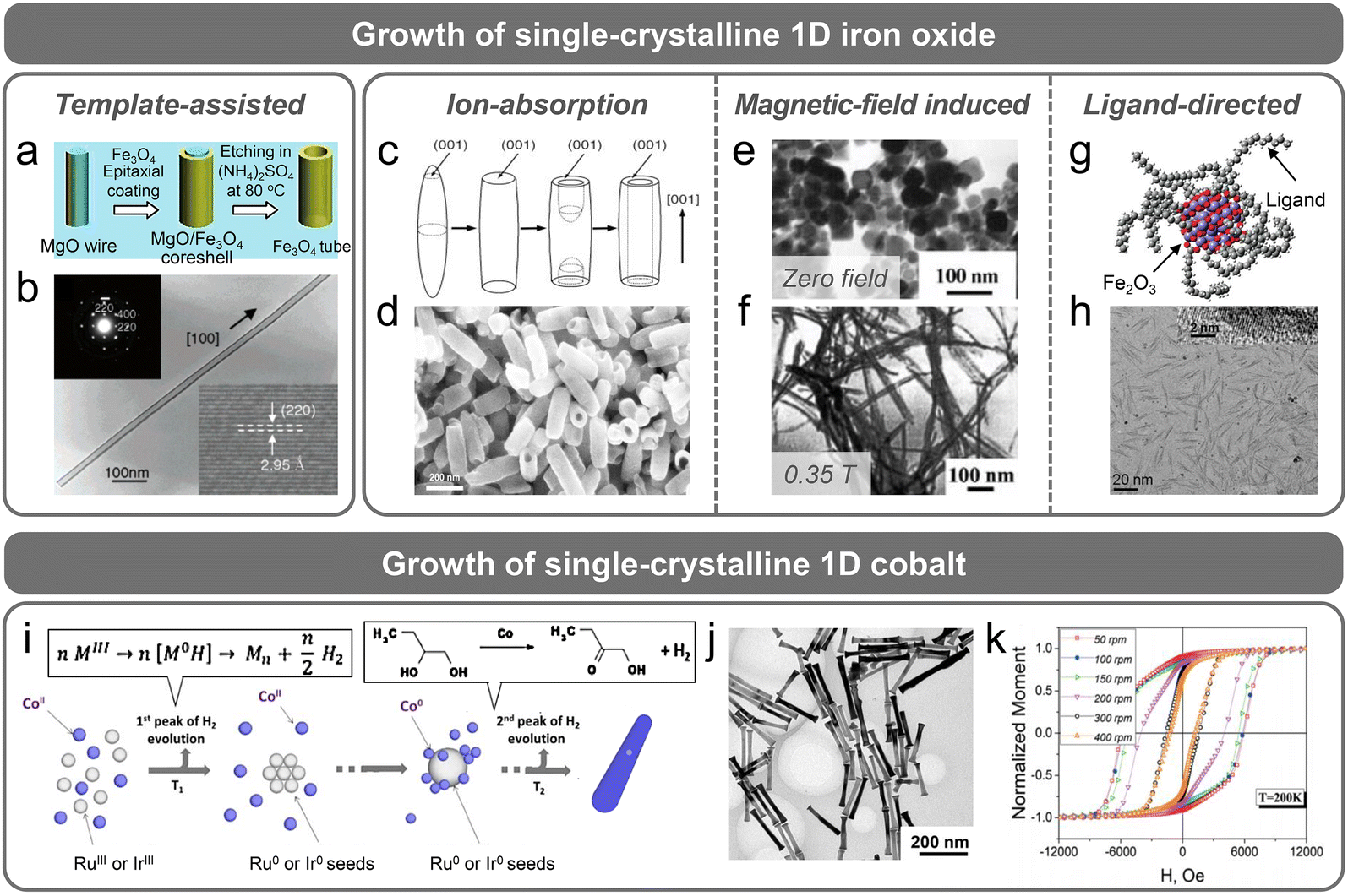 | ||
| Fig. 8 Synthesis of single-crystal 1D magnetic nanomaterials. Template-assisted synthesis of (a) the epitaxial growth of Fe3O4 nanotubes (NTs) on a MgO core via pulsed laser deposition and (b) morphology of an Fe3O4 NT examined using TEM and the corresponding single crystallinity verified by selected area electron diffraction and high-resolution TEM (HR-TEM). The scale bar is 100 nm. Reproduced with permission.104 Copyright 2005, American Chemical Society. Ion absorption during the synthesis of Fe3O4 NTs by (c) selective absorption of phosphate ions on the faces parallel to the c-axis for anisotropic growth of hematite NTs and (d) the anisotropic shape with an AR of 5–6 analyzed by scanning electron microscopy (SEM). The scale bar is 200 nm. Reproduced with permission.105 Copyright 2005, John Wiley and Sons. (e and f) Magnetic-field-assisted growth of single-crystal Fe3O4 NWs during hydrothermal synthesis. (e) Square- or hexagonal-shaped Fe3O4 NPs were formed under zero-field conditions. (f) Fe3O4 NWs with a 20 nm diameter and 0.8 μm length were synthesized under 0.35 T. The scale bar is 100 nm for both (e) and (f). Reproduced with permission.106 Copyright 2004, John Wiley and Sons. (g) Ligand-assisted synthesis of anisotropic Fe2O3 nanowhiskers. The selective decomposition of oleate ligands yielded ultrathin and anisotropic Fe2O3 nanowhiskers verified by (h) TEM and the single crystallinity of these nanowhiskers examined by HR-TEM. The scale bar is 20 nm. Reproduced with permission.109 Copyright 2011, American Chemical Society. Synthesis of single-crystal Co NRs via (i) the polyol method. Reproduced with permission.114 Copyright 2019, American Chemical Society. (j) Synthesized Co NRs showing an AR of 10 in the TEM image. Increasing the stirring rate during the synthesis of Co NRs caused stacking faults resulting in decreasing AR. The scale bar is 200 nm. (k) Magnetic hysteresis curves of Co NRs with different AR obtained at a controlled stirring rate during synthesis. Reproduced with permission.115 Copyright 2017, The Royal Society of Chemistry. | ||
As alternative and straightforward approaches for the growth of 1D magnetic nanomaterials, template-free synthesis has been proposed for 1D anisotropic hematite (α-Fe2O3), maghemite (γ-Fe2O3), and magnetite (Fe3O4). These methods commonly involve anion absorption onto specific crystal planes,105 the application of a magnetic field during the growth of magnetic nanomaterials,106 and the use of complexing agents or ligands.107 Jia et al. represented the synthesis of single-crystalline hematite and maghemite NTs. Hematite NTs were synthesized by a hydrothermal method using NH4H2PO4, which resulted in hollow NT structures.105 Phosphate ions supplied by NH4H2PO4 preferentially adsorbed on the faces parallel to the c-axis of the hematite (Fig. 8c). This preference induced an anisotropic structure by restricting lateral growth (Fig. 8d).108 However, an extended reaction at 220 °C led to dissolution or etching due to the acidic conditions. As a result, the (001) plane, which was less protected by phosphate ions, was more susceptible to dissolution and selectively dissolved, forming a unique NT morphology. Moreover, monodisperse maghemite NTs were synthesized via subsequent reduction and re-oxidation processes, but these methods exhibit limitations in increasing the aspect ratio. Wang et al. synthesized single-crystalline Fe3O4 NWs with a high aspect ratio (≈40) by applying an external magnetic field.106 By varying the strength of the magnetic field using a permanent magnet positioned at the top and bottom of a Teflon-lined hydrothermal reactor, single-crystalline Fe3O4 with various shapes ranging from nanoplate to NW morphologies could be obtained (Fig. 8e and f). The single-crystalline Fe3O4 NWs were readily synthesized at a field strength of 0.35 T and grown along the direction corresponding to one of the magnetic easy axes.
The use of complexing agents or ligands enables the production of single-crystalline iron oxide NWs with a higher aspect ratio.107 Xiong et al. synthesized maghemite NWs with an aspect ratio ≈ 150–300 in the presence of a complexing reagent, 1–10-phenanthroline. 1–10-Phenanthroline formed a stable complex with Fe2+, namely, [Fe(phen)3]2+. Then, spontaneous oxidation of [Fe(phen)3]2+ to an octahedral-structured [Fe(phen)3]3+ caused the oriented growth of maghemite. With the progress of the reaction, [Fe(phen)3]3+ was first degraded to form [Fe(phen)2]3+, which demonstrated a two-dimensional (2D) structure exposing a bare z-direction without a complexing agent. Subsequently, growth primarily occurred along the z-direction, leading to NWs. Similarly, Palchoudhury et al. revealed that the ligand in the iron oleate complex could be selectively decomposed near 150 °C, as determined by density functional theory calculations and thermogravimetric analysis (Fig. 8g).109 This decomposition behavior facilitated the directional growth of γ-Fe2O3 nanowhiskers with an ultrathin morphology (Fig. 8h).
Commercial permanent magnets, characterized by high coercivity and remanence, typically consist of alloys with rare-earth elements, due to their high magnetocrystalline anisotropy. Nevertheless, many researchers have tried to reduce the content of rare-earth elements and replace portions of them with 3d transition metals because of the supply problems of rare-earth metals and their thermal instability. Despite these efforts, magnetic materials fabricated solely from 3d transition metals have limited coercivity and remanence, owing to their limited magnetocrystalline anisotropy. In this context, introducing shape anisotropy into 3d transition metals presents an effective alternative. Among the 3d transition metals, Co has been frequently adopted because of its relatively high intrinsic magnetic anisotropy (i.e., magnetocrystalline anisotropy), which further enhances the coercivity when combined with shape anisotropy. Although there are some technological and economic challenges in utilizing anisotropic Co for permanent magnet applications, there are advantages in soft robotic applications, which will be explained in section 4.2.2.
Dumestre et al. successfully synthesized Co NRs via the thermal decomposition of an organometallic complex, [Co(η3-C8H13)(η4-C8H12)], in anisole with ligands under a H2 atmosphere.110,111 The shapes of the synthesized particles were considerably influenced by the H2 atmosphere and amount of ligands, while the organometallic complex ensured mono-dispersity. However, the preparation and reaction of such organometallic complexes always require a particular gas condition.112 To be free from these strict requirements, Viau's group suggested the polyol method for the synthesis of Co NRs (Fig. 8i).95 In this method, precursors in a reducing agent (1,2-butanediol) were transformed into a solid phase that served as a cation (Co2+) reservoir for the gradual release of Co2+ in an alkaline solution. Controlled liberation of Co2+ from this cation reservoir allowed precise modulation of the growth rate of Co NRs on pre-existing Ru seeds via heterogeneous nucleation. Furthermore, the morphology of Co NRs was diversified by regulating several parameters such as the basicity of the solution, heating/stirring rate, and chain length of the Co precursor.113–115 These Co NRs exhibited high remanence and coercivity in the longitudinal direction, and the further enhancement of magnetic properties could be controlled by modifying the aspect ratio (Fig. 8j and k).116,117 Additionally, Co NRs demonstrated high thermal stability up to 525 K, which was higher than that of the predominantly used permanent magnet, NdFeB. However, owing to the large surface-to-volume ratio in the nanoscale range, Co NRs undergo frequent coalescence and oxidation, which causes a lower Tc than that of pure bulk Co (≈1300 K).118
3.3. Layered magnetic nanomaterials and exchange anisotropy
Numerous magnetic nanomaterials and nanostructures have been proposed to induce exchange anisotropy. Starting with core–shell MNPs,44 a range of alloy and compound systems including Laves phases,119 Mn-based binary alloys,120 and Heusler alloys121 are being investigated.122 In a core–shell NP system, an oxide shell is obtained by the oxidation and chemical surface treatment of NPs.123 For example, when O2 is diffused into a colloidal solution containing Co NPs, NPs with an average diameter of 9 nm are oxidized.123 After undergoing a 4 days’ oxidation process, the average diameters of the core and oxide shell were 3.7 and 3.1 nm, respectively (Fig. 9a). Then, when the magnetization was measured during field cooling from 5 to −5 T, a negative exchange bias of NPs occurred between the FM and AFM layers, where the temperature reduced from 300 to 3 K (Fig. 9b). However, oxidizing the surface of MNPs is challenging because of difficulties in controlling the uniformity of the core size and shell thickness. Therefore, the desired FM ratio is not achieved during the transition from the FM phase to the AFM phases. In this regard, an alternative approach is the in situ oxidation of magnetic nanoclusters during physical vapor deposition (PVD) under controlled O2 gas flow conditions, which offers advantages such as fine-tuning of shell thickness and synthesis of monodisperse core–shell magnetic nanoclusters.124–126 Otherwise, by implanting FM NPs into an AFM matrix or AFM NPs into an FM matrix, interfacial interaction is facilitated for exchange bias through the surface doping of MNPs.127 For example, embedding of FI-NiFe2O4 NPs in an AFM-NiO matrix as a granular system, which is synthesized by high-temperature phase precipitation from an Fe-doped NiO matrix, leads to exchange bias resulting from the pinned ferri-clusters due to frozen spins in the spin-glass-like phase along the cooling-field direction.128 However, interfacial complexities, irregular size, and a broad shape distribution result in limited homogeneity and reproducibility of exchange anisotropy in this type of particle–particle interlayer coupling.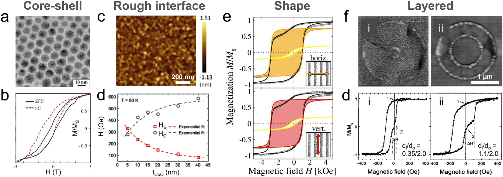 | ||
| Fig. 9 Exchange anisotropy in magnetic interlayers and diverse structures. (a) Interface of Co/CoO core–shell NPs verified using a TEM image and (b) magnetization hysteresis curves exhibiting exchange bias with a red dashed line under field-cooling conditions (5 T with the temperature decreasing from 300 to 3 K). The black solid line represents magnetization hysteresis in the zero-field cooling state. Reproduced with permission.123 Copyright 2023, Elsevier. (c) Atomic force microscopy images of CoO films with 80 nm thickness. (d) Dependence of the exchange bias field (Heb) and coercive field (HC) on the CoO thickness at 80 K. The exchange bias field decreases from −350 to −90 Oe with an increase in CoO film thickness from 5 to 40 nm. Reproduced with permission.144 Copyright 2020, American Physical Society. (e) Exchange bias of the laser-patterned Co/CoO stripes. The upper and lower loops were measured under horizontal and vertical magnetic fields relative to the length direction of the stripes at 10 K, respectively. Reproduced with permission.132 Copyright 2022, IOP Publishing. (f) SEM images of Ta (5 nm)/NiFe (20 nm)/IrMn (7 nm)/Ta (5 nm) rings with inner diameters of (i) 0.35 and (ii) 1.10, and an outer diameter of 2.0 μm. (g) Exchange bias of NiFe/IrMn rings measured at 300 K for each ring (i) and (ii) in (f). Reproduced with permission.131 Copyright 2004, AIP Publishing. | ||
Layered structures of 2D FM and AFM thin films hold a prominent position in the field of spintronic applications based on exchange bias. These layered 2D magnetic thin films provide relatively large-area controllability and easy-tuning of their geometry (dot,129,130 ring,131 stripe,132,133 and wire134), which can be fabricated using various patterning processes including photolithography,53 and electron beam lithography130,131 for micro/nano-fabrication. Moreover, the sequential deposition of FM/AFM layers enables the adjustment of surface roughness,135–137 layer thickness,138–142 and interfacial lattice by regulating the crystallinity of each magnetic thin-film layer.50,143
Exchange anisotropy caused by interlayer coupling is highly dependent on the interfacial properties and surface roughness of the 2D magnetic thin films consisting of layered structures.37,46 A rough and textured surface on 2D magnetic thin films contributes to the reduction of uncompensated spins at the interface within the layered structures, resulting in a decrease in interface magnetization and subsequent contraction of domains to lower the exchange anisotropy energy.46,144 For example, Wu et al. highlighted the effect of surface roughness on the exchange bias field. The root mean square (RMS) roughness (3.7 Å) increased with an increase in the thicknesses (80 nm) of the AFM-CoO films (Fig. 9c).144 As the CoO film thickness increased from 5 to 40 nm, the exchange bias field decreased from −350 to −90 Oe (plotted as red open squares in Fig. 9d) owing to the uncompensated spins at the interface. Dunz et al. investigated the influence of the interaction of a Ta/MnN/CoFeB system with a Ta buffer layer on the exchange bias in the system.145 Thicker Ta layers led to a higher exchange bias field, because this approach improved the crystallinity of MnN and decreased N2 diffusion during annealing. Thus, various approaches have been explored for the roughness control of growing materials, including the thickness control of the buffer layer,135 annealing processes to form oxide layers,136,137 and use of reactive ion-etched substrate.135
Layer thickness not only determines the characteristics of interfacial roughness, but also affects the exchange anisotropy associated with the domains of each layer. The layer thickness can be adjusted by controlling the deposition rate and time during PVD via sputtering or evaporation.139–142 Meinert et al. examined the effect of a variation in the AFM-MnN layer thickness on exchange anisotropy.141 The exchange bias field in the MnN/CoFe bilayer system increased up to a critical MnN layer thickness of approximately 30 nm, while an MnN layer thickness below 6 nm resulted in a zero exchange bias field. This ultrathin MnN–AFM layer (less than 6 nm) was attributed to spin instability and rendered the domain walls either unavailable or extremely small in the confined space.146 Another reason is the temperature-blocking capability of the layered system that depends on the thickness of the AFM layer. With a decrease in the AFM layer thickness, the system loses the capacity to block external temperature variations. Thermal fluctuations destabilize the spins in the AFM layer, which affects the exchange anisotropy. This thickness-dependent change in exchange anisotropy is observed in the FM layer as well.37,147,148 Thus, the most critical factor in interlayer coupling is regulating the thickness of both the AFM and FM layers with the formation of continuous films, rather than isolated island-like grains during deposition.
The crystal orientation in the interlayer region between 2D magnetic thin-film layers is also an important parameter in determining exchange anisotropy. Kohn et al. studied the exchange anisotropy of chemically ordered bcc Fe on L12-IrMn3 and chemically disordered fcc γ-IrMn3 on bcc Fe, grown via molecular-beam epitaxy (MBE).143 The exchange bias field of L12-IrMn3 was considerably greater than that of γ-IrMn3 because of the strong exchange coupling between the Mn atoms and magnetic spins in the ordered crystal lattice. Thanks to the atomic-scale layer-by-layer growth with precise control of the substrate temperature and atom flux during MBE, each layer exhibits a specific plane direction that includes uncompensated spins. This approach was developed to establish crystalline compatibility between the magnetic interlayers. For instance, bilayers such as Fe3O4/NiO149 and CoFe/MnIr50 were fabricated using MBE to ensure a perfect alignment with a (001) crystalline plane at their interface. In addition, exchange anisotropy can be manipulated by tailoring deposition conditions and altering the deposition angle.150,151 Controlling the deposition angle promotes the grain growth in the form of columnar structures, which enables the attainment of uniaxial anisotropy. The controlled aspect ratio of these columns and the resultant crystalline texture collectively contribute to the development of uniaxial anisotropy. Such anisotropy is influenced by both shape and magnetocrystalline anisotropies. For instance, in a study where a NiFe/IrMn bilayer was deposited at an oblique angle between 31° and 45°, the dominant factor contributing to uniaxial anisotropy was the combined effects of shape and magnetocrystalline anisotropy instead of the exchange anisotropy from interlayer coupling.151
As mentioned previously, conventional deposition and patterning processes are highly suitable for manufacturing layered structures in 2D magnetic thin films. This compatibility of fabrication allows for the customizing of shape and size, ranging from the submicron129,130,132 to the nanometer scale,152 in the development of high-performance spintronic devices including spin valve magnetic field sensors,54,153 magnetic storage devices, and non-volatile magnetic random access memories.55,154 Optimization of the geometry and size of patterned magnetic thin-film layers can boost the exchange bias field for spintronics applications.129,155,156 When the dimensions of the patterned layout closely match the domain size of the magnetic layers, the exchange anisotropy energy in the interlayer may be diminished according to the domain state model.157 However, designing patterned magnetic interlayers with small yet optimized sizes can increase the number of uncompensated spins per unit area, thereby strengthening the exchange anisotropy. Perzanowski et al. studied the relationship between patterns of magnetic thin-film layers and exchange bias.132 The hysteresis curve of a striped pattern exhibited a bias of 0.8 kOe away from the center (Fig. 9e), which was higher than that of a flat film. When the dimensions of the patterns were reduced to those of a smaller rectangle, measuring 9.1 μm in length and 4.5 μm in width (SEM images in the inset of Fig. 9e), a larger exchange bias appeared under the vertical field as compared with that in the case of the horizontal field due to the demagnetizing field. This directional difference became more significant with a decrease in the size of the patterns. In contrast, the isotropic square pattern exhibited no difference in exchange bias field with respect to the direction of the applied magnetic field. Thus, high aspect ratio patterns with sizes in the range of several micrometers can exhibit a high exchange bias field. Moreover, the exchange bias field can be varied by modulating the interfacial contact between the magnetic layers. To explore this further, ring-patterned magnetic thin-film layers were designed with different inner and outer diameters (Fig. 9f).131 Ring-patterned magnetic interlayers with a larger inner diameter exhibited a higher exchange bias field as compared with those of the interlayers with a smaller inner diameter, ascribed to the confinement of magnetic domains in the rings.
Layered structures of 2D magnetic thin films are beneficial for fine-tuning the interlayer properties with the goal of adjusting the exchange bias. However, several complexities associated with intermixing, defects, contamination, and lattice mismatches at the interface of bi- or multi-layered structures still exist. Although various comprehensive models have been proposed to explain the mechanisms of exchange anisotropy in different systems and materials, these models are insufficient to offer a complete interpretation of exchange anisotropy. Consequently, the effective management of key parameters, as discussed earlier, is imperative for attaining a reliable exchange anisotropy effect in customized systems and magnetic layers, ensuring the successful application of exchange-biased spintronic devices in practical implementations.
3.4. Synergistic dynamics of magnetic anisotropy in composites
Crystalline magnetic nanomaterials typically maintain a relatively stable magnetic anisotropy, unless severe changes in environmental conditions, such as heating/cooling, pressure, and strong magnetic fields, alter the free energy of the system.158–160 However, ferromagnets, a distinct category of magnetic materials, either undergo dynamic transitions in their magnetic properties in response to mechanical tension or exhibit mechanical strain when subjected to applied magnetic fields.161,162 This section covers three magnetomechanical effects: magnetostriction, magnetoelasticity, and magnetorheology, which are observed in FM materials when these materials are exposed to mechanical forces or when their magnetization states are altered by Hext (Fig. 10a). FM crystals have an easy axis of magnetization, which is determined based on magnetocrystalline anisotropy.163 When a magnetic field is applied along this easy axis, substantial shifts in the domain boundary and the rotation of magnetic domains within the crystals occur.164 Deformation of materials takes place because of magnetic fields and is referred to as the magnetostrictive effect. A similar physical effect is known as the magnetorheological effect, in which the stiffness or modulus of a magnetic soft composite or fluidic system changes when an external magnetic field is applied.165,166 The magnetorheological effect is induced by magnetic forces aligning magnetic nanomaterials within a viscous medium, thereby resisting mechanical deformation. This resistance originates from the dipole–dipole interactions between the magnetic nanomaterials, which are affected by an applied magnetic field.8,167 As mentioned in section 2.4, the phenomenon in which magnetic properties change due to external forces is referred to as the magnetoelastic effect, which is opposite to the magnetostrictive effect. In the case of the magnetoelastic effect, the FM nature considerably affects the stress-induced magnetic anisotropy. Thus, the stress-induced magnetic anisotropy is dependent on other magnetic anisotropies in addition to external stress. The stress-induced response of magnetic properties in conjunction with the other magnetic anisotropies results in a substantial anisotropic response.168 This synergistic anisotropy is also valid for magnetostriction and magnetorheological effects, inducing a remarkable mechanical deformation and strain in response to external fields. The behaviour of a magnetomechanical effect is of great importance for applications in both actuators and sensors.59,169–171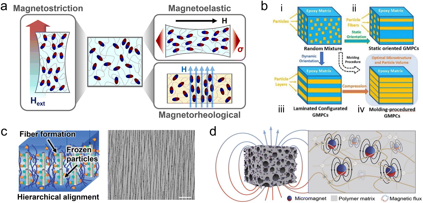 | ||
| Fig. 10 Various magnetomechanical effects based on an anisotropic structure of composites. (a) Stress-induced magnetomechanical effects: magnetostriction, magnetoelastic, and magnetorheological effects. (b) Schematics of isotropic and alignment anisotropic structures: (i) random distribution, (ii) magnetostatic orientation and (iii and iv) routine for the two-step (magnetodynamic orientation and compression) formation of laminated-like structures of Tb–Dy–Fe particles. Reproduced with permission.202 Copyright 2019, Elsevier. (c) Enhanced alignment of an anisotropic structure via a combination of magnetic fields and electrostatic interactions. The scale bar is 50 μm. Reproduced with permission.205 Copyright 2022, John Wiley and Sons. (d) Giant magnetoelastic effect in a soft composite system. Reproduced with permission.213 Copyright 2022, Elsevier. | ||
Since the discovery of the magnetostriction effect in Ni by Joule in 1842 and the giant magnetostriction effect in Fe or rare-earth-metal-based alloys by Clark and Belson in 1972, extensive studies have been conducted to synthesize materials with a high λsi.172,173 Single-crystal Terfenol-D (Tb0.3Dy0.7Fe2), exhibiting an exceptionally high λsi exceeding 1500 ppm owing to its intrinsic magnetocrystalline anisotropy, has found numerous applications such as in sensors, motors, and transducers.174–177 However, a pre-magnetization process is required for domain alignment in polycrystalline Terfenol-D for an optimal magnetostriction effect.178 To address this issue, various efforts aimed at enhancing the magnetic anisotropy in Terfenol-D via several techniques such as free-stand zone melting, modified Bridgman method, sintered powder compact, and mixing polymer matrix with Terfenol-D powder have been made.179–182 Nevertheless, the ordering of polycrystalline Terfenol-D has limitations including the use of expensive elements Tb and Dy, the mechanical brittleness of the resulting materials, and processing challenges.
Oxide-based magnetic materials, such as polycrystalline cobalt ferrite, have emerged as alternatives to Terfenol-D because of their cost effectiveness, superior magnetomechanical coupling factors, and large deformative behaviors under low magnetic fields.183,184 Unlike single-crystal magnetostrictive materials, polycrystalline magnetic materials comprise numerous grains with grain boundaries.185,186 These grain boundaries serve as nucleation sites for stable-to-metastable phase transitions and domain switching triggered by an external field. Accordingly, grains with smaller sizes offer a larger number of nucleation sites, resulting in a more pronounced magnetostriction effect.187 Bhame et al. investigated λsi values across cobalt ferrite NPs with different grain sizes that were prepared by different methods, namely, combustion, reagent addition, coprecipitation, and calcination.188 Cobalt ferrite NPs with grain sizes of 8 μm (combustion), 17 μm (reagent addition), 23 μm (coprecipitation), and larger than 25 μm (calcination) exhibit the maximum λsi values of 197, 184, 159, and 135 parts per million (ppm), respectively. The enriched grain boundaries in the small-grained system induced large domain reversibility and increased the strain response to magnetostriction under low external fields. The λsi value of polycrystalline cobalt ferrite depends on the uniform and smaller grain size. Therefore, the microstructures in polycrystalline cobalt ferrites should be controlled to achieve a high λsi. Despite these efforts, polycrystalline cobalt ferrites still exhibit low λsi values in the range of 130–200 ppm due to the different orientations of magnetic easy axes in the corresponding domains. Magnetic annealing facilitates the production of a higher λsi and strain derivative based on a uniaxial anisotropic structure in the magnetic domains. Wang et al. determined the orientation of polycrystalline cobalt ferrite by applying a magnetic field during calcination.189 A semisolid slurry containing Fe2O3 and Co3O4 powders in a polyvinyl alcohol solution was oriented under a strong magnetic field of 2 T. Thereafter, the mixture was sintered to produce CoFe2O4 with crystal grains of 30 μm oriented in the 〈001〉 direction, a relatively high λsi of 270 ppm, and a strain derivative of 7.7 × 10⁻⁹ m A−1.
Nevertheless, the inherent lack of ductility and mechanical resilience in bulk FM materials hinders their practical use in magnetomechanical applications requiring materials that can withstand repeated deformation.190–192 To overcome this vulnerability, FM materials have been combined with soft polymer matrices aiming for more robust and improved mechanical properties.193–195 To induce a magnetostrictive effect in these soft composite systems, the FM materials are randomly distributed in a polymer matrix via straightforward fabrication (Fig. 10b–i).196 However, randomly-distributed ferromagnets lack magnetic anisotropy, leading to a low magnetostrictive effect.197 In this case, the formation of an anisotropic geometry, composed of aligned FM particles in a fluidic or viscoelastic matrix, enhances the magnetostriction.58 In a soft composite system, when the FM materials are sufficiently close to each other in an appropriate volume ratio, these materials can form an anisotropic structure in the direction of an external magnetic field.198 Consequently, a strong magnetostrictive effect can be generated in the direction of the applied magnetic field. Both isotropic and anisotropic FM materials have been used for anisotropic aligning in a viscous polymer matrix.182,199 A precise alignment of these FM materials commonly requires a fluidic medium that permits the free movement of FM materials while simultaneously guiding these materials under a magnetic field.200,201 For example, Li et al. proposed a method where giant magnetostrictive materials comprising Tb–Dy–Fe were aligned in an epoxy polymer under a magnetic field of 1 T to induce magnetic anisotropy.202 Tb–Dy–Fe particles were manufactured in the form of chain-like structures using a static magnetic field (Fig. 10b-ii), and laminate-like structures via a two-step process based on the combination of a dynamic magnetic field and compression (Fig. 10b-iii). Particularly, an anisotropic composite was produced using a two-step molding process (Fig. 10b-iv), resulting in an optimized microstructure characterized by high density and well-aligned particles. These magnetostrictive composites, which embedded anisotropically aligned FM materials, exhibited superior magnetomechanical properties compared with a random dispersion model. This enhancement in magnetic anisotropy results in a comparable λsi of 1500 ppm to that of a single monolithic Terfenol-D, even with a particle volume fraction of as low as 57%.
The dimensions of FM materials with a relatively low λsi values minimally change with respect to the applied magnetic field.203 However, the anisotropic structure, achieved by the subtle reorientation of magnetized particles, demonstrates the capability to induce a macroscopic magnetostrictive effect, caused by the stretching of the matrix in the direction of the magnetic field.198,204 Guan et al. reported a magnetostrictive effect in a soft composite system containing carbonyl iron particles (CIPs) mixed with silicone rubber.193 CIPs are FM materials with fairly small λsi values (lower than 10 ppm). However, when CIPs are magnetized under a magnetic field, magnetic interactions occur between particles, aligning these particles in the silicone rubber matrix. Consequently, subtle rearrangements of CIPs produce a magnetostrictive effect that allows stretching of the flexible silicone matrix, exhibiting strains of up to 184 ppm. When high-viscosity polymers lack co-solvents for dilution, the anisotropic alignment of magnetic materials in these polymers become challenging, and a strong magnetic field is needed to attain the desired morphology. Furthermore, additional driving forces are required to assist with alignment and freely construct a suitable configuration of magnetic materials. Chen et al. developed a magnetic soft composite in which negatively charged, magnetically responsive Fe3O4 NPs were vertically oriented in a biomimetic hydrogel (Fig. 10c).205 Coupling of the magnetic field and electrostatic repulsion forces MNPs to vertically orient, even when magnetic fields as low as 20 mT are applied. Although Fe3O4 NPs demonstrate a relatively low λsi value of approximately 40 ppm, the densely packed and highly anisotropic configuration indicates their potential to exhibit a magnetostrictive effect.
Soft composite systems where a magnetorheological effect is induced can exhibit high stiffness under a magnetic field, and the magnitude of the reinforcement is influenced by the anisotropic nature.206,207 By controlling the magnetic field during the curing of the matrix, either an isotropic or anisotropic structure is readily introduced, changing the magnetorheological properties.208,209 Jung et al. fabricated two different types of isotropic and anisotropic structure by incorporating CIPs into natural rubber.210 In the case of the anisotropic soft composite, CIPs were aligned in the direction of the applied magnetic field by applying an external magnetic field during the curing process. Although both isotropic and anisotropic composites represented a high modulus after magnetic field application, the anisotropic composite was higher than that of the isotropic composite, by nearly 60%. These results are attributed to the fact that the particle chains are aligned in a similar manner to that of rod-shaped fillers, resulting in a significantly high storage modulus.
The magnetoelastic effect, whose mechanism is opposite to that of magnetostriction, alters the magnetic properties of an FM material in response to external stress and is triggered by a variation in the magnetic domain. Kurita et al. fabricated a soft composite by randomly dispersing Fe–Co–V alloy particles in a polyurethane matrix, and magnetic flux changes were caused by the magnetoelastic effect.211 The fabricated soft composite induced a magnetoelastic effect when subjected to a bending load. An approximately 0.05 mT change in magnetic flux was measured using a Hall sensor positioned at the bottom of the composite. However, variations in the magnetization of FM materials are limited due to the stress distribution in the soft matrix.212,213 To overcome these limitations and enhance the magnetoelastic effect, the concept of the giant magnetoelastic effect was introduced (Fig. 10d). This effect includes a synergistic system of particle movement and rotation, along with changes in the magnetic domain of the FM material in the matrix. Zhou et al. reported a soft composite exhibiting a giant magnetoelastic effect based on a silicone elastomer matrix, which was embedded with both NdFeB hard magnets and Fe3O4 soft magnets.213 In a composite system, magnetic particles with a single dipole configuration could form a unique distribution as “wavy-chains”, even when a polymer and magnetic particles were homogeneously mixed. This was ascribed to the gradual decrease in polymerization degree from the polymer to the particle direction, where the monomeric solution near the FM nanomaterials was not fully polymerized. Therefore, mobile magnetic particles could still survive, and hence change direction and move in response to an applied magnetic field, even after the nanomaterial was solidified from a macroscopic view. Unlike the typical magnetoelastic effect, which is magnetic domain rearrangement for magnetic anisotropy, this magnetic soft composite introduced a different mechanism. In this system, stress led to a relative disordering of the initially well-aligned wavy-chain micromagnets. This disordering changed the magnetic anisotropy and magnetic flux density of the soft composite. Compared with the magnetoelastic behaviors of standalone ferromagnets, the remarkable change in magnetic anisotropy due to elastic deformation highlights the giant magnetoelastic effect. Through this giant magnetoelastic effect, the soft composite achieved high magnetic flux density variations of up to approximately 9 mT. These approaches to enhance magnetoelasticity introduce a new paradigm for the practical applications of magnetostrictive materials in sensing and actuating technologies.
4. Magnetic anisotropy for sensing and actuation in soft robotic applications
Numerous types of magnetic nanomaterial characterized by their magnetic anisotropy have been extensively studied owing to their substantial contributions to the development of both magnetic sensors and actuators. The inherent magnetization direction of these magnetic nanomaterials provides key benefits, including high selectivity, enhanced accuracy, operational efficiency, and customizability, to sensors and actuators in robotic manipulations.In addition, magnetic sensors and actuators are appropriate for tracking motions and subsequently controlling the operations of robots with minimal physical contact in remote sites. Yet, achieving these capabilities with other types of sensor and actuator is quite challenging. The notable features of magnetic sensors and actuators based on magnetic anisotropy demonstrate widespread implications in various fields such as soft manufacturing grippers, rehabilitation robots, advanced prosthetics, human-interactive devices, and the Internet of Things. These contemporary electronic technologies necessitate ultrathin, lightweight, and form-factor-free designs that enable their compact integration and high compatibility with components having irregular shapes and surfaces. Specifically, sensing and actuating devices in the soft robotic systems should possess the capabilities to freely deform and effectively follow the dynamic motions of their pliable bodies to minimize disturbances. In this section, we discuss the numerous advantages of magnetic anisotropy in sensing and actuation. We also explore how magnetic materials can attain the desired softness and flexibility in magnetic sensors and actuators, particularly in the soft robotic applications.
4.1. Flexible and stretchable magnetic sensors
| Types of magnetic sensors | Magnetic materials and device structures | Detection field limit (minimum/maximum) | Linear sensitivity | Flexibility (minimum bending radius/number of bending or stretching cycles/maximum strain) |
|---|---|---|---|---|
| 1 Oe = 0.1 mT. Py = NiFe. Current normalized sensitivity = VH/(Ibias × B) [V/AT]. | ||||
| Hall sensors | Hall cross, bismuth film218 | — | −0.47, −2.29 V/AT | 6 mm/50 cycles with a bending radius of 8 mm/1.25% |
| Hall cross, laser-scribed graphene219 | — | ∼1.12 V/AT | 5 mm/1000 cycles with a bending radius of 5 mm/1.6% | |
| Anomalous Hall sensors | Hall cross, Ta/Co/Pt222 | — | 500 μV kOe−1 | NA/NA/NA |
| AMR sensors | Barber pole, Ta/Py/Ta226 | 150 nT/0.2 mT | 42 T−1 | 5 mm/50 bending cycles with a radius of 5 mm /0.5% |
| Barber pole, Py229 | 50 nT/— | 0.54% mT−1 | 150 μm/2000 cycles with a bending radius of 1 mm/1.33% | |
| Printed AMR sensors | Printed Ta/Py microflake244 | 1 mT/400 mT | 12% T−1 above 5 mT/190% T−1 below 5 mT | 2 mm/NA/NA |
| GMR/GMR spin valve sensors | Stripe, Co/[Co/Cu]50 and Py/[Py/Cu]30241 | — | — | 3 μm/100% stretching for 1000 cycles/∼270% |
| Meander, [Py/Cu]30242 | — | — | 2 mm/NA/NA | |
| Wrinkled, Ta/IrMn/[Py/CoFe]/Cu/[CoFe /Py]318 | 1.2 mT/NA | 0.8% Oe−1 | NA/10% stretching for 500 cycles/∼29% | |
| Wheatstone bridge, Ta/[Py/CoFe]/Cu/[CoFe/Py]/IrMn319 | 2 mT/12 mT | — | 30 μm/NA/4% | |
| Printable GMR sensors | Printed [Py/Cu]30 microflake245 | 1 mT/20 mT | 0.43% mT−1 | 16 μm/NA/100% |
| TMR sensors | Ta/Ru/Ta/NiFe/IrMn/CoFe/Ru/CoFeB/MgO /CoFeB/Ta/Ru251 | — | — | 5 mm/1000 cycles with a bending radius of 15 mm/NA |
| Ta/Ru/Ta/CoFe/Ru/MgO/CoFeB/Ta/Ru320 | — | — | NA/0.2% bending for 20 cycles and 0.4% bending for 20 cycles /∼0.4% | |
| [Ta/CuN]6/Ta/Ru/IrMn/CoFe/Ru/CoFeB/MgO /CoFeB/Ta/NiFe/Ru/IrMn/Ru/Ta/Ru321 | — | 250 μV Oe−1 | 5 mm/NA/NA | |
Hall effect sensors. In fact, the Hall effect may not exhibit a significant correlation with the aforementioned properties of magnetic anisotropy. Nevertheless, it is worth discussing because of its traditional use in most proximity-sensing devices in industry and numerous consumer electronics. In a Hall sensor, a varying external magnetic field orients at right angles to the current flow in a thin-film conductor or semiconductor, generating a Hall voltage. This Hall voltage can be used to detect the presence and magnitude of the magnetic field, as illustrated in Fig. 11a.217 Remarkable progress in the synthesis of 2D nanomaterials and their Hall coefficients has contributed to the development of ultra-thin, mechanically compliant, and field-sensitive Hall sensors (Fig. 11b). Semiconducting 2D bismuth thin films were directly grown and patterned into Hall cross-contact and sensor arrays on plastic substrates including polyimide (PI) and polyetheretherketone, which enabled Hall sensors to be placed on curved surfaces (Fig. 11c–i).218 The flexible bismuth Hall sensor maintained its sensitivity even when subjected to bending stress, with a maximum bending radius of 6 mm (Fig. 11d). By attaching this sensor to the fingertips, magnetic field profiles could be monitored relative to the distance from the sensors on the finger to the magnet (Fig. 11c-ii).
 | ||
| Fig. 11 Hall effect sensor. (a) Schematic of a traditional Hall effect sensor. (b) Components for a 2D thin-film Hall effect sensor, including semiconductors, conductors, and magnetic materials. (c) (i) Flexible printed circuit (FPC) with a bismuth Hall sensor applied to a curved finger. (ii) Real-time monitoring of magnetic field profiles relative to the distance from the Hall sensor on the fingertips to the magnet. (d) Hall sensitivity measured after bending the sensor at various radii, demonstrating consistent sensitivity of the sensor even after bending to a radius of 6 mm. Reproduced with permission.218 Copyright 2014, John Wiley and Sons. (e) Illustration depicting the Hall measurement configuration of a laser-scribed graphene Hall sensor. (f) Sensitivity of the graphene Hall sensor at different bending radii. Reproduced under the Creative Commons CC BY License.219 (g) Images of anomalous Hall sensors mounted on a soft magnetic origami actuator. The magnet approaches the Hall sensors on the soft magnetic origami actuator in the following order: (i) top, (ii) left, (iii) center, and (iv) right. (h) Linear voltage signal response to the external magnetic field and schematic of the four-Hall sensor connection. (i) The voltage output of the anomalous Hall sensors in response to the moving magnet with time. Reproduced under the Creative Commons CC BY License.222 | ||
Two-dimensional nanomaterials, particularly graphene, with its unique electronic properties and mechanical flexibility, are promising for the development of flexible Hall sensors.219,220 A graphene layer grown via chemical vapor deposition and subsequently transferred onto a 50 μm-thick Kapton film demonstrated a maximum voltage and current normalized sensitivities of 0.093 V/VT and 75 V/AT, respectively.220 The sensitivity was stable even after the layer was bent 1000 times with a 5 mm bending radius. Laser-scribed graphene offers a simple and rapid maskless method for the fabrication of Hall crossbars on desired plastic substrates.219 This method involves direct conversion of carbon-rich materials into graphene (Fig. 11e) which is capable of achieving high mechanical stability and a sensitivity of 1.12 V/AT even after being bent with a radius of up to 5 mm (Fig. 11f).
Regarding the enhancement of the Hall coefficient, magnetic nanomaterials offer an anomalous Hall effect (AHE) based on spin-dependent scattering of charge carriers. FM thin films characterized by magnetic anisotropy greatly influence AHEs by altering the path of electrons via interactions of electrons with the localized magnetic moments of the atoms in the FM material. Depending on whether the magnetic field is aligned parallel or perpendicular to the easy axis of magnetization in the FM material, it enables sensitivity adjustments, directional sensing, and performance tunability of AHE sensors for more versatile and effective field-sensing applications.221 Ultra-thin AHE sensors have been proposed for conformal attachment to uneven polymeric surfaces through a series of fabrication processes comprising the deposition of metal-stacked layers with a sub-nanometer thickness onto a 3 μm-thick plastic foil (Fig. 11g).222 Typically, 2D and monolithic Hall sensors measure the Hall voltage that occurs when a magnetic field is perpendicularly applied. However, this planar dimension limits the capability of these sensors to detect changes in magnetic fields along all three spatial coordinate axes. To address this issue, flexible AHE sensor arrays were designed and firmly integrated onto 3D soft deformable composites. The compliant AHE sensor arrays exhibited a high linear sensitivity across a wide range of magnetic field variations, ranging from −400 to 400 Oe (Fig. 11h). The integrated AHE sensor on the three different deformable parts mapped the magnetic field profile with respect to the distance from the center of a fixed reference AHE sensor (Fig. 11g and i). Furthermore, these AHE sensor arrays did not interfere with the motions of magnetic soft actuators. Instead, these AHE sensors supervised the sequential shape-morphing process of actuators by monitoring the sensor output signals resulting from magnetic field changes.
In the case of MR sensors, the dominant influence of magnetic anisotropy leads to variations in the principles of magnetic field detection. These variations depend on the magnetism or magnetization of materials and device structures of the MR sensors. Thus, the extent of resistance changes and magnetic field sensitivity accordingly vary.223 Three representative types of MR sensor are available in terms of device structures and their corresponding operational principles: anisotropic magnetoresistance (AMR), giant magnetoresistance (GMR), and tunnelling magnetoresistance (TMR) sensors.
Anisotropic magnetoresistance (AMR) sensors. First, the AMR effect is significantly dependent on magnetic anisotropy as it varies the electrical resistance of magnetic materials based on the angle between the current and magnetization directions, as illustrated in Fig. 12a. In most materials with positive AMR coefficients, the orientation of the electron orbitals is determined by the direction of the magnetic field originating from spin–orbit coupling. This phenomenon induces greater scattering of a transport electron as the current flows parallel to the applied field.224 As a result, AMR sensors typically consist of thin-film magnetic materials with specific magnetic anisotropy to detect in-plane angular changes of an applied field. However, the straightforward design of AMR sensors exhibits several drawbacks, such as a near-zero sensing capability at low fields. Unlike magnetization, which easily aligns with the magnetic easy axis, guiding the current flow in the uniaxial direction relative to the magnetization direction requires a specific device geometry.225 To ensure linearity in response to changes in the applied field, a barber pole structure is commonly designed to set the angle between the current flow and initial magnetization at 45°. Moreover, a Wheatstone bridge circuit is prevalently used to offset cancellation effects (Fig. 12b). The periodic barber poles on the AMR cells effectively guide the current at either 45 or 135° in alignment with the magnetic easy axis. This configuration exhibited a substantially linear response in terms of variations in output voltage when subjected to a magnetic field ranging from −200 to 200 μT.226 This device structure guarantees that resistance changes are only caused by the magnetic field for improving field sensitivity, reducing thermal noise, and signal calibration and adjustment.152,225–227 The flexible and highly sensitive AMR sensors could detect an extremely low stray field (81 μT) from magnetic strips at a far distance (5 mm, Fig. 12c–f), rendering them suitable for safe and practical applications. Although the barber pole structure enhances the stability and linear sensitivity, challenges occur when dealing with a large area occupied by a complex pattern.53 To address this issue, the AFM layer has been utilized to pin the magnetic moment of the FM layer, within the FM/AFM layered structure, to the predefined magnetization direction of the AFM layer through exchange coupling, thereby achieving self-biasing.53,228 The initial magnetization direction of the FM layer was biased to 45° by optimizing the AFM layer thickness. Consequently, self-biased AMR sensors showed linear sensitivity without the barber pole structure. An ultra-thin AMR sensor consisting of 50 nm-thick FM stripes was developed by combining a Wheatstone bridge circuit with a barber pole structure to assist in thermal noise compensation and to linearize the sensor response.229 This AMR sensor could perceive geomagnetic fields as low as 50 μT and detect in-plane angular variations for an electronic compass, even when placed directly on curved skin.
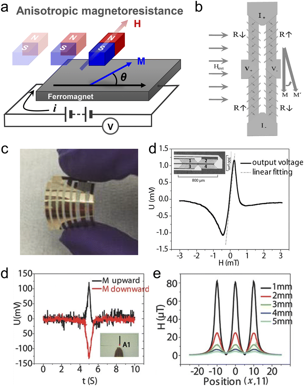 | ||
| Fig. 12 Anisotropic magnetoresistance (AMR) sensor. (a) Schematic of AMR sensor, where θ is the angle between the current flow and the magnetization direction of the ferromagnet. (b) Illustration of the barber pole structures and Wheatstone bridge. Periodic barber poles with AMR cells effectively guided the current at either 45 or 135° in alignment with the magnetic easy axis. (c) Image of a flexible AMR sensor on PET foil. (d) Linear voltage response plotted against magnetic field. (e) Voltage output from the flexible AMR sensor on a finger. The magnetic field approached a single magnetic strip, with voltage changes corresponding to the initial upward or downward magnetization direction of the magnet. (f) Simulation results of the magnetic field profile measured at various distances from the top of the three magnetic strips. Reproduced with permission.226 Copyright 2016, John Wiley and Sons. | ||
Giant magnetoresistance (GMR) sensors. Unlike AMR sensors, GMR sensors with multilayered structures that consist of alternating FM and NM conductive nanomaterials have been proposed, as illustrated in Fig. 13a. The GMR effect significantly changes the electrical resistance compared with the case of the AMR effect due to the spin-dependent scattering mechanism. When the magnetization of these FM layers is parallel, electron scattering is minimized, leading to lower overall resistance (Fig. 13a-i). In contrast, a higher resistance is observed in the GMR multilayer when electron scattering increases owing to the antiparallel alignment of the magnetization in adjacent FM layers (Fig. 13a-ii).230 The performance of GMR sensors is quantified using the GMR ratio, (RAP − RP)/RP, where switching occurs with a shift in the magnetization configuration between the parallel and antiparallel states. RAP and RP represent the resistances for the antiparallel and parallel states, respectively. The field sensitivity of a GMR sensor is calculated as follows:231
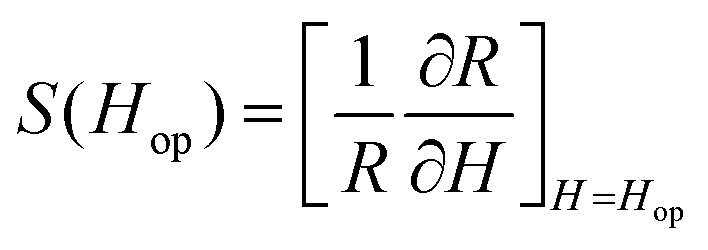 | (20) |
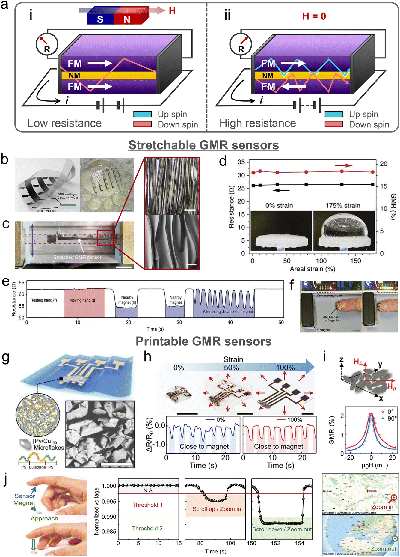 | ||
| Fig. 13 Giant magnetoresistance (GMR) sensor. (a) Schematic of the GMR structure: (i) low-resistance state, where a larger magnetic field than antiFM coupling results in a parallel magnetization configuration, minimizing electron scattering. (ii) High-resistance state, where without an external magnetic field, the magnetization of the FM layers is anti-parallel due to the antiFM coupling, increasing electron scattering. (b–f) Stretchable GMR sensors. (b) Illustration (left) and image (right, scale bar: 10 mm) of the GMR sensor mounted on an ultrathin polyethylene terephthalate (PET) foil. (c) Uniaxial stretching test of the GMR sensor. The GMR sensor was attached to a pre-stretched elastomer substrate and compressed by 50%. Optical image (upper, scale bar: 200 μm) and SEM image (bottom, scale bar: 100 μm) of the wrinkled surface in a compressed state. (d) Resistance measured under various areal strains. The biaxial-wrinkled GMR sensors can be stretched in all lateral directions with an areal strain of up to 175%. (e) Real-time monitoring of resistance from the sensor on the palm by adjusting the distance of the magnet from the sensor. (f) Proximity sensing demonstration of a flexible GMR sensor on the fingertip. The LED light turns on when a permanent magnet is in close proximity. Scale bars: 10 mm. (b–f) are reproduced under the Creative Commons CC BY License.241 (g–j) Printable GMR sensors. (g) Printed GMR sensor composed of Py/Cu multilayer flakes and a poly (styrene–butadiene-styrene) (SBS) matrix on an ultra-thin plastic foil. (h) Stretchability of the GMR sensor. Scale bar: 100 μm. Normalized resistance graph measured under 0 and 100% strain with an approaching magnet. (i) Schematic of the magnetic field direction for resistance measurement of random microflakes and the GMR graph. Random microflakes exhibit magnetic-field sensing capability regardless of the angular change of the sensors. Scale bar: 5 mm. (j) Demonstration of GMR sensor application for augmented reality. When the sensor attached to the finger is close to the magnet, the voltage drops. Below the threshold voltage 1/threshold voltage 2, the map was zoomed in/zoomed out. (g–j) are reproduced under the Creative Commons CC BY License.244 | ||
The GMR ratio varies with the thickness of the FM and NM layers because of the oscillatory exchange coupling effect that can be described by both the Ruderman–Kittel–Kasuya–Yosida theory and quantum confinement effect.236–240 Although stacking multi-layer films with FM and NM layers is necessary for achieving the GMR effect, each layer is still less than a few nanometers in thickness. Therefore, the multilayered yet ultrathin GMR sensors demonstrate not only high field sensitivity as compared with AMR sensors, but also conformal integration onto curved surfaces. Melzer et al. developed multilayered GMR sensors consisting of Py (1.5 nm)/[Py (1.5 nm)/Cu (2.3 nm)]30 on a 1.4 μm-thick polyethylene terephthalate foil (Fig. 13b). These GMR sensors demonstrated remarkable resilience to repeated stretching even at 270% strain.241 Laminating GMR sensors onto biaxially pre-stretched membranes led to the formation of surface wrinkle patterns driven by a large modulus mismatch between the sensors and membranes and imposed strain (Fig. 13c). The biaxial-wrinkled GMR sensors could be stretched in all lateral directions by applying an areal strain of up to 175% (Fig. 13d) without degradation of the GMR performance under multidimensional deformation. The resistance of the GMR sensor on the palm was monitored by adjusting the distance of the magnet from the sensor (Fig. 13e). The real-time monitoring of the magnetic sensor on the skin implied stable resistance with low noise and rapid resistance change over time. The imperceptible and stretchable GMR sensors on a fingertip also demonstrated proximity-sensing capability by monitoring resistance changes, irrespective of the distance of the permanent magnet, thereby activating a light-emitting diode (LED) light (Fig. 13f). Kondo et al. presented a novel active magnetosensory matrix (MSM) system comprising thin-film GMR sensor arrays integrated with a complementary organic thin-film transistor circuit on a 1.5 μm-thick parylene film.242 The flexible MSM system based on an ultrathin plastic substrate with polymer encapsulation demonstrated excellent mechanical stability under severe loading and high surface compliance on uneven skin. The imperceptible and active MSM system performed low-voltage and high-speed operations with multiple integrated components enabling the real-time mapping of the magnetic field. An autonomous battery-powered system and a wireless communication module were incorporated into the MSM system to promote the practical application of the imperceptible MSM system in tracking the 2D magnetic field distribution for position sensing.
Typically, magnetic sensors consisting of rigid-metal thin films on a plastic foil have limited stretchability because of the difference between the Young's modulus of the metal and substrate. The use of ink composed of additives in an elastomeric binder for printing can solve this mechanical property mismatch. The minimal mismatch between the elastomeric substrate and viscoelastic ink results in a printed device that is stretchable and stable when deformed.243 These advantages have led to recent proposals for printable magnetic sensors based on magnetoresistive paste.244,245 A GMR sensor composed of Py/Cu multilayer flakes and a poly(styrene–butadiene–styrene) matrix was printed on an ultrathin plastic foil (Fig. 13g). This printed GMR sensor demonstrated high stability in terms of GMR performance (1.5%) and sensitivity (3.0 T−1) under mechanical impact even with a bending radius of up to 16 μm.245 Due to the surface-wrinkle formation on the pre-stretched substrates, the printed GMR sensors could be repeatedly stretched and released without degrading their magnetic field sensing performance (Fig. 13h). The key function of printed GMR sensors was magnetic field sensing regardless of the angular changes of the sensors (Fig. 13i), which allowed mounting of the sensors on any part of an object. Therefore, these compliant GMR sensors attached to the fingertip demonstrated the potential for augmented reality applications, where the virtual object is controlled by the remote and contactless detection of the magnetic field (Fig. 13j).
Linear sensing capability is important to sensors for predictable, accurate, and simplified data acquisition without complex signal processing. The aforementioned AMR and GMR sensors cannot achieve linear sensitivity near the zero magnetic field range without the barber pole structure. Without the barber pole structure and 45° biasing, the resistance increases as the external magnetic field strengthens when the easy magnetization is aligned along the long or short axis. Thus, the resistance becomes proportional to external magnetic fields, and weak external fields cause low sensitivity according to eqn (20).53 Furthermore, the operation field is limited to the small coercive field of the AMR sensor.223 Therefore, a strategy was proposed to modify the device structure by inserting pinning layers into the GMR multilayers, and the resulting device is known as a GMR spin valve device.246 The basic structure of the GMR spin valve device comprises FM1/NM/FM2/AFM multi-stacked layers, as illustrated in Fig. 14a. In this configuration, the magnetization direction of the FM2 layer is guided by the exchange anisotropy originating from the interaction between the AFM and FM2 layers, as discussed in section 2.3. Notably, only the magnetization direction of the FM1 layer readily undergoes rotation in response to an external magnetic field in the range of the effective anisotropy field. Once the field exceeds the anisotropy threshold, the FM2 layer begins its rotation. As this switching behavior leads to a linear response of the MR sensor within a broad range of magnetic fields, several attempts can be made to adapt the multilayer-stacked GMR spin valve sensors. The GMR spin valve device was composed of a [Py/CoFe]/Cu/[CoFe/Py]/IrMn heterostructure and designed with two Wheatstone bridges and a well-defined magnetic anisotropy axis.229 The inner and outer bridges included in the spin valve sensors that were reverse biased provided a bipolar sine output. The output signals were dependent on the angle between the direction of the applied magnetic field and the magnetic anisotropy of each bridge. Analysis of the arctangent correlation between the two output signals facilitated the real-time reconstruction of the in-plane magnetic field, where the permanent magnet continuously rotated around the spin valve sensor. Furthermore, the spin valve arrays were transferred onto an ultrathin PI foil and encapsulated by polydimethylsiloxane (PDMS). Positioning the GMR multilayers in a neutral plane enhanced the mechanical stability of these multilayers because the GMR multilayers were subjected to bending where both stress and strain were minimized. Thus, the GMR spin valve sensors were conformally attached to the skin and could detect the magnetic field orientation for the touchless manipulation of interactive devices such as a virtual keypad and virtual bulb.
 | ||
| Fig. 14 GMR spin valve sensor. (a) Schematic of the spin valve structure. (b) Overview of the entire stack of the device and synthetic antiferromagnet (SAF) spin valve stack for the self-assembled 3D magnetic field vector angular encoder. (c) Resistance curve during a field sweep in the planar state. The operating range is between 5 and 50 mT, exclusively sensitive to the magnetic field direction. (d) Planar state of a device (upper image) and its transformation into a Swiss-roll structure (lower image). After transformation, the device contains two orthogonal tubes of 250 μm diameter and 8 spin valve sensors. (e) Graph depicting the angular dependence of resistance. ρ is the tile angle of the magnetic field plane with respect to the spin valve. Reproduced with permission.247 Copyright 2019, AAAS. | ||
However, the limitation of MR sensors is that they are only sensitive to the in-plane magnetic field. To address the need for measuring a more complex and 3D magnetic vector field, Becker et al. recently proposed a self-assembled GMR spin valve for 3D magnetic field vector angular encoders.247 A self-assembly method transforms a planar structure into a 3D structure, offering a simpler fabrication process that is cost-effective as compared with the case of direct fabrication of the 3D design. The entire layer stack of the device is shown in Fig. 14b. The synthetic AFM (SAF) spin valve effectively cancels the stray field at the edges of the FM layers (Fig. 14b). After etching the sacrificial layer and inducing swelling of the hydrogel, the thin PI layer was rolled. Thus, four spin valve sensors were integrated into the 3D Swiss-roll structure. In the planar state prior to transformation, the resistance curve was measured using a field sweep (Fig. 14c). The spin valve sensors exhibited a superior GMR ratio (∼8%) and large plateau, indicating a constant resistance range. In this resistance range, the sensors were sensitive to the magnetic field direction rather than to the field strength, making them suitable for angle encoder applications. After transformation, the device contained two orthogonal tubes of 250 μm diameter and eight spin valve sensors (Fig. 14d). The dependence of the resistance on the angle is related to the variation of the tilt angle of the magnetic field plane with respect to the spin valve sensors (Fig. 14e). The results reveal that the sinusoidal angular response remained up to a tilt angle of 45°. After carefully designing the diameter of the Swiss roll, three pairs of spin valve sensors were placed in the two orthogonal tubes, with two sensors in each pair having 90° angular relationships of the pinning directions. This configuration allowed coverage of the 3D orthogonal planes (XY, YX, and XZ). Consequently, the self-assembled 3D magnetic field sensors detected any angular orientation of the magnetic field, magnetic field strength, and distance between the sensors and magnet.
Tunneling magnetoresistance (TMR) sensors. Magnetic field sensors based on the TMR effect are beneficial for detecting weak magnetic fields with high field sensitivity because of their significant MR ratios when compared with those of the AMR and GMR sensors, which are ascribed to the thin-insulating tunneling barrier. TMR devices comprise a magnetic tunnel junction (MTJ) with a FM/tunneling barrier/FM multilayered configuration, as illustrated in Fig. 15a. When two FM layers are parallel (Fig. 15a-i), more electrons tunnel through the layers than when the two FM layers are antiparallel (Fig. 15a-ii), resulting in a lower resistance. Similar to the case of a spin valve device, the magnetization direction of the FM layer is pinned with that of the AFM layer in an MTJ structure based on exchange anisotropy. Conversely, the adjacent FM layer can freely rotate in response to the magnetic field.248 Controlling the magnetization direction of the pinned FM layer with a crossed configuration of the neighboring FM layer results in a linear response of resistance change to the external magnetic field. In an MTJ device structure, the crystalline tunnel barrier and an epitaxial interface between the FM and barrier layers are critical factors. For instance, the largest TMR ratio is 631% in CoFeB/MgO/CoFeB even without an AFM pinning layer at room temperature owing to the optimized sub-nanometer thickness CoFe and Mg, which led to enhanced interface crystallinity.249 Such TMR devices that are capable of weak magnetic field sensing have been widely used in diverse applications including the read heads of hard disk drives, position measurements in industrial robots, and navigation of automotive systems. Willing et al. demonstrated new sensing functionalities of TMR devices with arbitrary intrinsic in-plane anisotropy in each CoFeB layer in the multi-stacked TMR layers formed using oblique-incidence deposition.250 Oblique deposition with an angle of over 70° resulted in highly anisotropic surface roughness and periodic wavy patterns with wave-fronts that were perpendicular to the deposition direction (Fig. 15b). The magnetization direction in thin magnetic layers was parallel to the wave-fronts, which was attributed to in-plane uniaxial magnetic anisotropy originating from shape anisotropy. In the configuration of this TMR device, when the uniaxial anisotropy directions of the two FM layers were perpendicular to each other, the TMR graph took on a triangular shape (Fig. 15b-i). Conversely, when two FM layers shared the same uniaxial anisotropy direction, the TMR graph demonstrated a square shape (Fig. 15b-ii). When the uniaxial anisotropy directions were perpendicular, the TMR response became sinusoidal with respect to the angle of the applied magnetic field (Fig. 15c). Therefore, the ability to manipulate the shape and the exchange anisotropy of the magnetic layers provides a method to customize TMR sensors for diverse applications, offering versatility in sensor design and performance optimization. The remarkable field-sensing capability of the TMR sensor also holds substantial potential for perceiving minute biological magnetic signals. However, the development of TMR devices has primarily focused on improving the MR ratio, and limited efforts have been directed toward the flexibility of these devices.
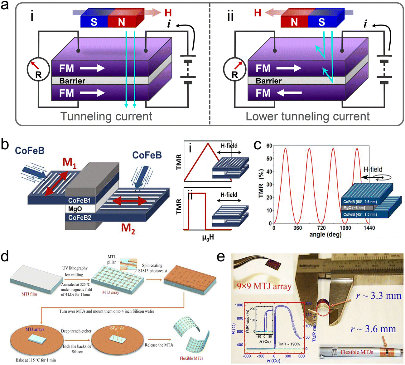 | ||
| Fig. 15 Tunneling magnetoresistance (TMR) sensor. (a) Schematic of the TMR sensor, illustrating the difference between the tunnel current of parallel (i) and antiparallel (ii) states of the FM layer. (b) Schematic of the TMR sensor deposited using oblique incidence deposition with a periodic wave pattern is perpendicular to the deposition direction: (i) triangular-shaped TMR graph and (ii) square-shaped TMR graph. (c) Graph depicting the dependence of resistance on sinusoidal angle. Reproduced under the Creative Commons CC BY License.252 (d) Schematic of flexible MgO barrier magnetic tunnel junction (MTJ) fabrication. (e) Flexible MTJs bent to a radius of 3.3 mm. The TMR graph in the inset shows that the unbent MTJ array demonstrates a TMR ratio of approximately 190%. Reproduced under the Creative Commons CC BY License.251 | ||
Although oxide barriers, such as MgO, are primarily fabricated via PVD, MBE, and atomic layer deposition, the achievement of defect-free, pinhole-free high-quality sub-nanometer oxide barriers still requires high state-of-the-art thin-film technology, which is an expensive and complicated process. Moreover, the fabrication of TMR sensors requires a high-temperature annealing process for the oxidation of the barrier growth, post-annealing for CoFeB crystallization, and pinning of the AFM layer by inducing exchange anisotropy. Commonly used polymer substrates to be applied for thin-film device fabrication have a limited working temperature range and exhibit a relatively higher roughness than those deposited on to Si wafers due to prevention of the growth of high-quality single crystals. Therefore, Chen et al. suggested an alternative for developing flexible TMR sensors by using the transfer method (Fig. 15d).251 It was a sequential fabrication process with the following steps: deposition of magnetic and NM layers on a Si wafer, patterning on the Si wafer, post-annealing for CoFeB crystallization, and magnetic annealing for exchange bias. After finishing the conventional process, the back of the Si wafer was etched, resulting in the only thin MTJ film that could be transferred to a flexible substrate. This flexible MTJ with the MgO barrier demonstrated complete functions with a TMR ratio of approximately 190% before bending. The flexible MTJ could be bent at a radius as small as 3.3 mm without damage (Fig. 15e).
The aforementioned magnetic field sensors exhibit the advantage of maintaining their magnetic performance under bending conditions, making them effective for measuring proximity on curved surfaces. However, MR sensors composed of magnetostrictive materials are affected by stress if they are not engineered in a neutral plane. When subjected to stress, the magnetic domains are aligned by the magnetoelastic effect, and the magnetic anisotropy direction of the FM layer changes. It modifies the angular relationship of external magnetic fields with the easy magnetization direction and induces a change in the resistance output. This resistance variation can be used to determine the strain magnitude or direction with respect to the initial resistance. When the GMR spin valve sensor with positive magnetostrictive Co50Fe50 in the free layer is subjected to tensile stress, the anisotropy field increases in the direction of the strain.252 Due to the anisotropy field, the stress increases the coercive field in the magnetization hysteresis curve, and the increasing trend is saturated at 12% strain. Moreover, Ota et al. performed comprehensive research by measuring the magnitude and direction of strain using a Co/Cu/NiFe GMR strip.253 The bottom NiFe layer was in the single-domain state as the assisting external magnetic field exhibited insensitivity to stress. However, the upper Co layer was strain sensitive. This flexible GMR sensor fabricated on a polyethylene naphthalate sheet showed a resistance change depending on the strain direction and easy magnetization direction angle, confirming the possibility of realizing strain direction sensing using the GMR sensor. FeB-based MTJ as a free layer was designed to integrate spintronics strain gauge sensors with a microelectromechanical systems microphone.254 The magnetization of FeB undergoes rotation in response to both tensile and compressive strains, with a drastic change in the resistance owing to the high λsi of FeB.255 This MTJ also features a high TMR ratio of 190%, resulting in a high strain gauge factor ((ΔR/R)/Δε = 5072). Consequently, a series-connected strain gauge array can promptly respond to diaphragm vibratory motions, even at high frequencies of up to 1 kHz.
Touch sensors based on piezoresistive, triboelectric, capacitive, and piezoelectric effects have been extensively examined in various fields due to their distinctive signal generation mechanisms.261–266 However, their sensitivity to moisture restricts their application as wearable or implantable sensors in specific environments, such as high-humidity conditions and human skin.267,268 Moreover, touch sensors based on triboelectric and piezoelectric effects rely on the alignment of electric dipoles (capacitive conduction) caused by dielectric polarization at the material interface, thus exhibiting low current densities and high impedance values, leading to high signal-to-noise ratios.269,270 In contrast, the magnetic field, which activates a touch sensor using the magnetoelastic effect, remains consistent regardless of moisture and the sensors can operate stably in a wet environment without additional post-processing, including encapsulation. Simultaneously, magnetoelastic-based sensors form an alternating alignment of magnetic dipoles both on and inside the material, which allows them to achieve low impedance.213,271 Magnetoelastic touch sensors exhibit a wide sensing range that is 10 times broader than that of other types of sensor and responds to stimulation ranging from subtle pressure at a low level to intense pressure at a very high level.272
An anisotropic arrangement of magnetic materials in a polymeric matrix is a common feature of magnetoelastic materials. Thus, the touch perception based on the magnetoelastic effect is achieved by detecting the variations in magnetic flux resulting from changes in the net magnetization moment of magnetoelastic materials during mechanical impacts and deformation. For example, in a macroscopic aspect, magnetoelastic composites with an anisotropic arrangement of magnetic particles embedded into the polymers alter the magnetic flux density by disordering the arrangement of magnetic particles under external mechanical stress. At the atomic level, the magnetic domains rotate and move because of the mechanical impact. To reduce the magnetic potential energy, the magnetic moment deviates from the original direction and even jumps to another easy axis created by stress. Thus, the applied pressure varies the magnetic flux density, which can be converted into electrical signals via electromagnetic induction. Zhao et al. proposed stretchable and self-powered biomonitoring sensors by coupling the giant magnetoelastic effect with electromagnetic induction (Fig. 16a).272 As the well-oriented micromagnet wavy chains in the polymer matrix were disrupted because of mechanical deformation, the rearrangement of the magnetic chains significantly affected the difference in magnetic flux, which is termed the giant magnetoelastic effect (Fig. 16a-i). The maximum magnetomechanical coupling factor caused by the giant magnetoelastic effect was 4.16 × 10⁻⁸ TPa−1 (Fig. 16b), which was approximately 3.3 times greater than that of Fe–Co alloys. The variations in magnetic flux induced electric currents within coil-type liquid metal microfibers (Fig. 16a-ii). The fully soft platform combining the giant magnetoelastic composites and electromagnetic coils exhibited high mechanical endurance with a lateral strain of up to 440% and could be worn on all body parts to monitor the different bio-mechanical signals (Fig. 16c). In addition, electromagnetic induction endows the sensors with self-powered operation without a power supply for the prolonged operation of implantable bio-monitoring devices and untethered robots.
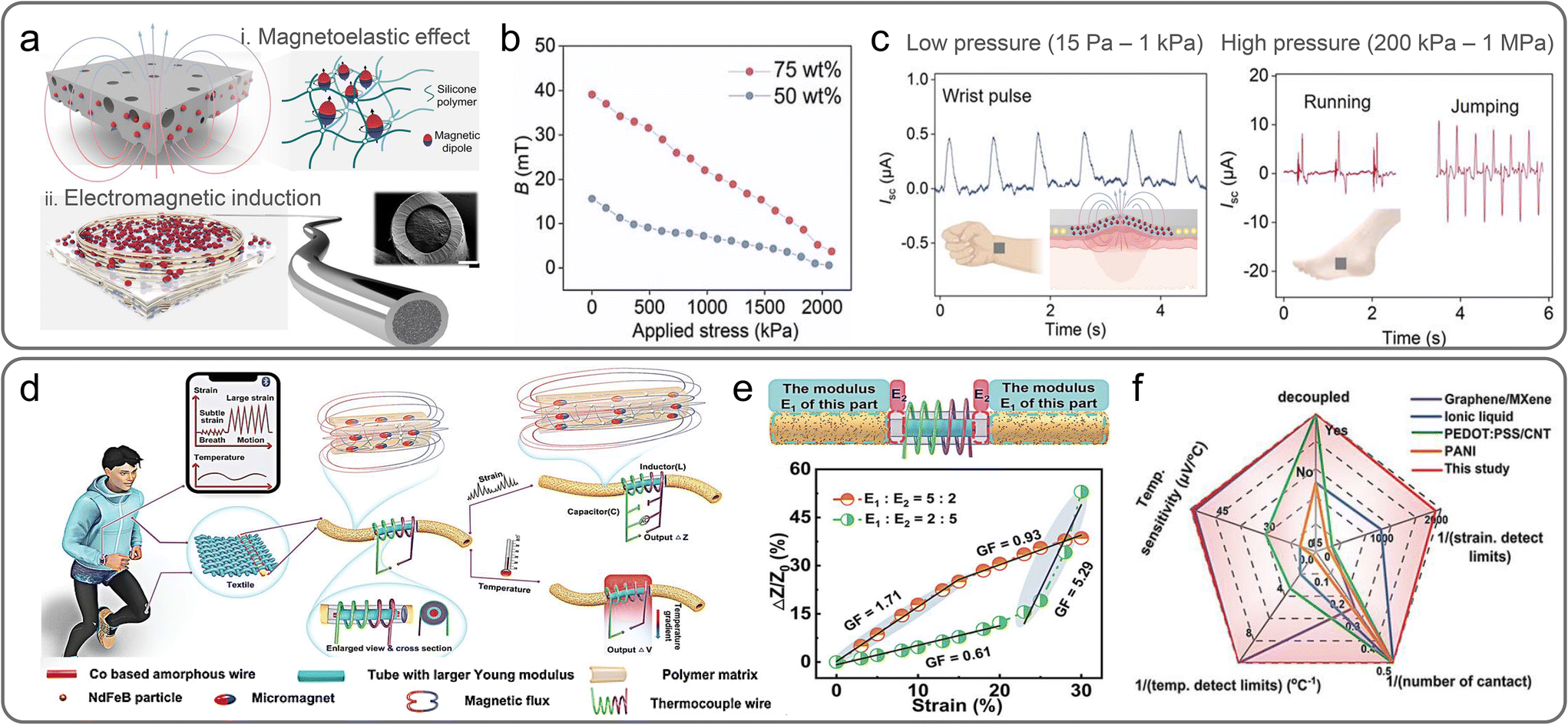 | ||
| Fig. 16 Biomonitoring sensor utilizing the giant magnetoelastic effect. (a) Schematic of the self-powered biomonitoring sensor based on the combination of the (i) giant magnetoelastic effect and (ii) magnetic induction effect, and the internal structure of each component. (b) Magnetic flux density variations of the sensor under different pressures. (c) Output current induced by low-pressure (wrist pulse) and high-pressure (running and jumping) motions with representative diagrams. Reproduced with permission.272 Copyright 2022, American Chemical Society. (d) Schematic of a wearable strain and temperature sensitive dual-mode sensor (STDMS) capable of accurately detecting strain and temperature changes. (e) Schematic of the heterogeneous structure of STDMS and change of relative impedance when strain is applied to the STDMS (E1:E2 = 2:5 (green symbols) and E1:E2 = 5:2 (orange symbols)). E1 and E2 represent the Young's modulus of magnetic composites and non-magnetic cylinders, respectively. The slope of the linear fit is the gauge factor (GF). (f) Comparison of the performance of this sensor with those of other various dual-mode sensors. Reproduced with permission.277 Copyright 2023, John Wiley and Sons. | ||
For intelligent behaviors of robots, the ability to distinguish various stimuli is essential. Sensors in soft robots whose abilities are equivalent to the multi-functional sensing of human skin have been developed based on the concept of multi-modal sensors.273,274 The combination of magnetic sensors with other types of sensing approach has also been suggested to enhance the accuracy, reliability, and functionality of the sensors.275,276 Xiao et al. demonstrated a multimodal sensor combining the giant magnetoelastic and thermoelectric effects (Fig. 16d).277 The multimodal sensor was constructed by wrapping a CuNi-Cu (CNC) thermocouple coil around a magnetoelastic composite in which NdFeB particles were embedded in PDMS. The outputs of the CNC thermocouple coil changed the magnetic flux of the magnetoelastic composite under the applied strain, which was verified by a change in impedance (Fig. 16e). Simultaneously, with a change in temperature, the output voltage varied because of the Seebeck effect caused by the temperature difference between the CuNi and Cu. Therefore, the fabricated textile-type multimodal sensor could detect both strain and temperature gradients, which revealed magnetic field variations of 30 mT under a 30% strain and an output voltage of 2.997 mV in response to a change in temperature from room temperature to 55 °C. Fig. 16f depicts a comparison between the performances of this sensor and various other dual-mode sensors, specifically focusing on the interference-free output for two different stimuli. The strain- and temperature-sensitive dual-mode sensor (STDMS) with a tubular heterogeneous structure detects strain based on the temperature-dependent magnetic field variation and the permeability of Co-based amorphous wires (CoAWs). With an increase in temperature, the remanence of the STDMS originating from strain change was stable, and the permeability of the CoAWs was retained when the temperature was decreased from 100 °C to room temperature. Similarly, the voltage measured by the STDMS thermocouple remained unaffected by the mechanical strain occurring in the wires, and the wires exhibited an identical thermoelectric effect. As a result, the STDMS achieved excellent strain and temperature sensing performances, clearly decoupled the input signals and avoided crosstalk.
For tracking the touch trajectory, magnetoelastic composite-based pressure sensors integrated with Hall sensors were used as multimodal sensing devices.258 Hu et al. demonstrated a wireless magnetic tactile sensor capable of transmitting information about the position and area where external forces were applied (Fig. 17a).278 The integration of Hall sensors with force-sensitive magnetic sensors based on the giant magnetoelastic effect enabled the detection of both the applied force and proximity of the magnetic field. The magnetoelastic composite consisted of permanent NdFeB magnetic particles embedded in a silicone elastomer, which was magnetized in numerous directions to achieve different magnetic flux densities in the x, y, and z directions (Fig. 17b). The difference in the magnetic flux density was monitored in a touchless manner using a Hall sensor located underneath the magnetoelastic composites. Both the tactile and proximity sensing data provided a specific magnetization profile using a clustering k-nearest neighbors algorithm model. The acquired sensing signals were pre-decoded to confirm different contact forces and changes in the magnetic field along the z-direction at 36 to 10000 points, which offered insights into the contact points and corresponding magnitudes for the dexterous and remote manipulation of robots (Fig. 17c). Liu et al. demonstrated a bimodal flexible sensor based on the combination of triboelectricity and the giant magnetoelastic effect, which facilitated touch and touchless sensing according to two distinct modes (Fig. 17d and e).279 In the touchless mode, as a negatively charged external object approaches the magnetoelastic conductive film, potential generation was induced from the charge generation based on contact electrification. Therefore, free electrons flow from the magnetoelastic conductive film to the ground, yielding current. However, when the magnetoelastic conductive film came into contact with an external object, the film underwent deformation because of contact pressure (Fig. 17e). During mechanical deformation, the micromagnet chain structure in the film changed, and thus reduced the surface magnetic flux density (Fig. 17f). Simultaneously, the electromagnetic induction effect caused a liquid metal coil to generate a current that corresponded to changes in the magnetic flux density. A sensor mounted on a soft robotic hand obtained signals regarding the surface roughness of the object, by sliding the robotic hand over the surface of the object and scanning the shape in a non-contact mode (Fig. 17g). The system also included a convolutional neural network (CNN) model that was pre-trained using the distinct electrical signals generated by the triboelectric effect, which depended on the shape of the object. Furthermore, when the robotic hand slid on the surface of objects with different roughnesses, the magnetic flux change caused by the magnetoelastic effect was converted into an electrical signal via electromagnetic induction. The CNN was also pre-trained using the different electrical signals generated according to the surface roughness. Consequently, the object was recognized by combining the electrical signals acquired as the robotic hand approached a specific material and slid on its surface. The combination of these two sensing modes (Fig. 17h-i and ii), coupled with a CNN model, resulted in a sensor with superior sensitivity and high accuracy (Fig. 17h-iii). These findings highlight the potential of soft electronic systems in response to multiple stimuli and are used for the development of intelligent magnetoelastic devices, which are essential for object recognition and discrimination.
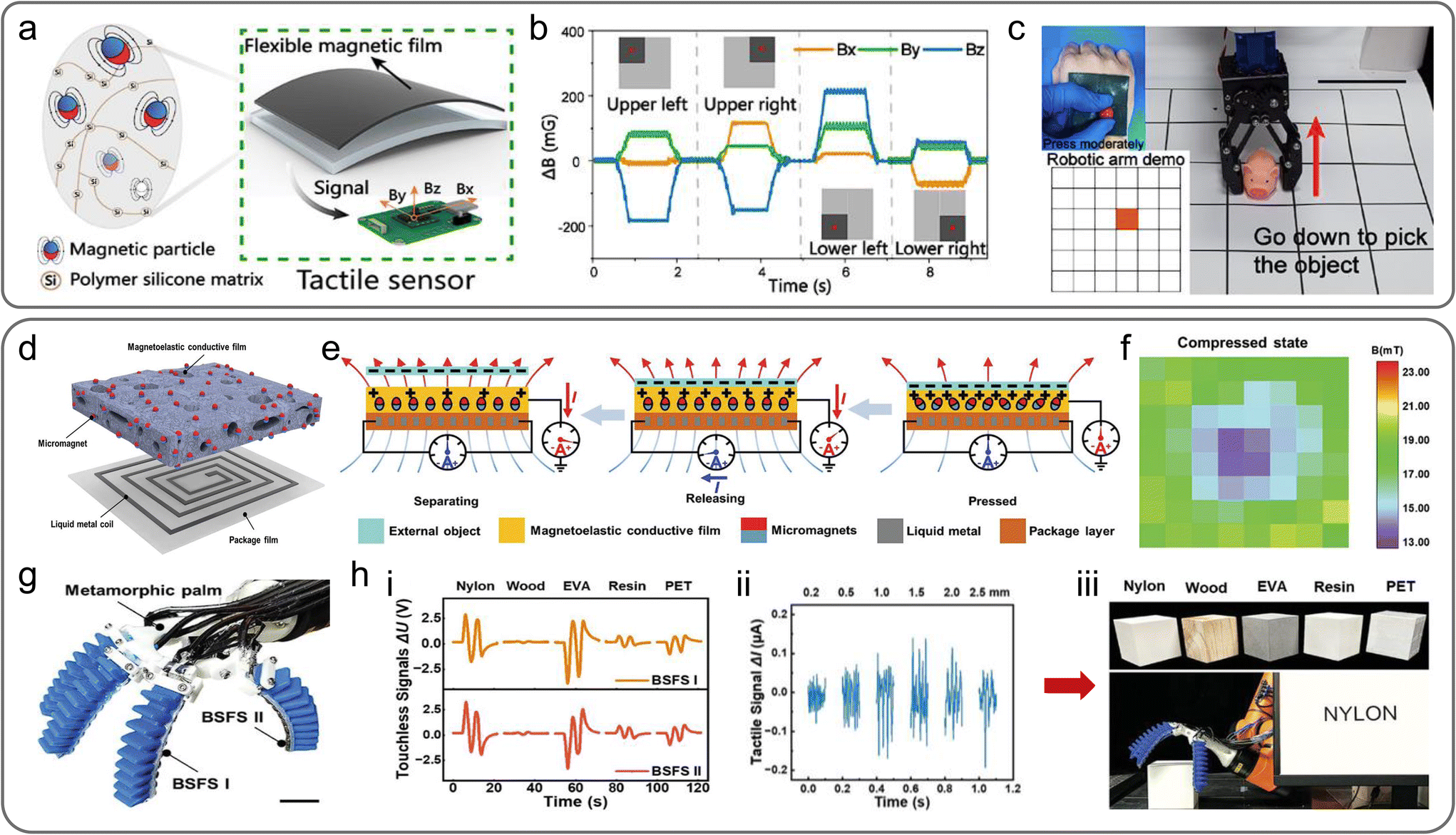 | ||
| Fig. 17 Multifunctional sensor based on the giant magnetoelastic effect. (a) Configuration of a wireless flexible magnetic tactile sensor (FMTS) with its internal structure, and the directions of the magnetic flux density signals (Bx, By, and Bz). The left inset image exhibits the distribution of magnetic particles (red and blue spheres) in the polymer matrix (orange lines). (b) Various magnetic flux density signals generated in the x, y, and z directions upon touching different regions of a “symmetrical up and down” magnetized FMTS. (c) Demonstration of a real-time remotely operated robotic arm control system based on the FMTS. Reproduced with permission.278 Copyright 2022, American Chemical Society. (d) Components of a bimodal self-powered flexible sensor (BSFS): a packaged liquid metal coil and a magnetoelastic conductive film. (e) Operating mechanism based on the triboelectric and giant magnetoelastic effects of the BSFS. (f) Variation of magnetic flux density mappings under pressure in a soft magnetoelastic film. (g) Schematic of a flexible robotic hand equipped with a BSFS. The scale bar is 3 cm. (h) Demonstration of an intelligent robotic system capable of sensing and describing various objects. (i) Triboelectric output signals, (ii) giant magnetoelastic output signals, and (iii) an image of a robotic hand recognizing various objects. A total of five materials are included: nylon, wood, ethylene vinyl acetate (EVA), photosensitive resin, and polyethylene terephthalate (PET). Reproduced with permission.279 Copyright 2023, John Wiley and Sons. | ||
4.2. Magnetic soft actuators
Soft actuators have great importance for qualifying key parameters of locomotion, manipulation, and safety in soft robotics, which mimics the dexterity and adaptability of natural organisms.4,280,281 Unlike traditional electrical, pneumatic, or hydraulic motor-mounted robots, soft actuators facilitate dynamic motion and enable robots to navigate through intricate and unpredictable environments. Owing to their high compliancy, soft actuators can conform to irregular shapes and handle fragile items for delicate manipulation. Moreover, compatible soft actuators can accommodate variations in the environments with good adaptability and minimize the risk of damage when the robots come into contact and interact with humans or soft objects. Considering their significant importance, a lot of strategies have been proposed for soft actuators using stimuli-responsive soft materials that are activated by heat,282 humidity,283 pH,284 electric field,285,286 and magnetic field.287,288 Among these stimuli-responsive materials, magnetic field-responsive soft materials offer many advantages such as remote manipulation of actuators, fast response time, and versatility for operation across a wide range of environments, even in water.289,290 Forces that trigger the mechanical responses of magnetic soft composites can be classified into translational force, magnetostriction effect, and torque. However, the translational force and magnetostriction exhibit some limitations in application for magnetic actuators. Translational force is referred to as a mechanical source that causes spatial movements of magnetic materials under a gradient magnetic field. Although the strength of the force is relatively high, it rarely induces deformation of a matrix, thus resulting in limited freedom of magnetic actuators. Magnetostriction in magnetic soft composites changes in the dimensions of the material under a uniform magnetic field due to the magnetostrictive properties of the filler or its tendency to be aligned along the magnetic field. However, magnetostriction cannot generate a large deformation as the unit of strain is typically measured in ppm, as presented in section 3.4. Meanwhile, the magnetic torque that induces the rotation of magnetic materials under a uniform magnetic field causes a large deformation. The deformation can be finely tuned by programming magnetic anisotropy resulting in a complex shape-morphing capability. In this section, we review the recent advances in magnetic soft actuators achieved by manipulating magnetic anisotropy to control the deformation of magnetic soft composites based on the torque.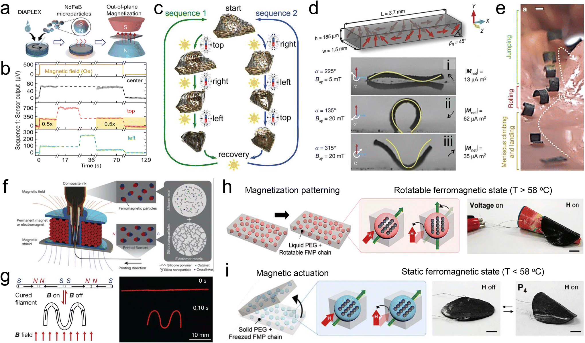 | ||
| Fig. 18 Magnetic soft actuators with rare-earth magnet-based soft composite. Reconfigurable soft origami actuator with responsiveness to light and magnetic field fabricated by (a) mixing a shape memory polymer with NdFeB particles followed by out-of-plane magnetization, (b) position sensing capability by integrating a flexible anomalous Hall effect sensor into the soft composite, and (c) localized actuation without hinges by light and magnetic field. Reproduced with permission.222 Copyright, the Creative Commons CC BY License. Small-scale magnetic soft actuator with (d) the harmonic magnetization profile enabling (i) sine-shape, (ii) C-curve, (iii) V-shape deformations by modulating the strength and direction of the magnetic field and (e) locomotion in an artificial stomach. Reproduced with permission.294 Copyright 2018, Springer Nature. (f) 3D printing method to encode a complex magnetization profile with pre-magnetized NdFeB particles and (g) a simple filament case showing how the generated magnetization profile makes a deformation under an external magnetic field. Reproduced with permission.288 Copyright 2018, Springer Nature. (h) Reprogrammable magnetic moment above the Tm owing to the mobility in medium (left) and encoding magnetization profile simply by wrapping the soft composite around a certain object during magnetization (right). (i) Fixing oriented magnetic moments by cooling under Tm (left) and reproduction of the shape of the wrapped object under an external magnetic field resulting from the programmed magnetic profile (right). Reproduced with permission.296 Copyright 2020, American Chemical Society. | ||
To enhance DoFs and controllable actuation, strategies to assign directionality have been studied by varying the magnetization states of soft magnetic materials.293,294 Hu et al. proposed small, untethered, and soft bodies composed of NdFeB microparticles embedded in a silicone elastomer and programmed a harmonic magnetization profile by simply wrapping these soft composites around a cylinder (Fig. 18d).294 The programmed soft robot was controlled using a time-varying magnetic field to realize different locomotion modes. According to the magnetization direction of the soft bodies with respect to the strength and orientation of the applied field, these magnetic soft composites could be deformed into various structures and complex transformation like a sinewave shape, C-curve or V-shape (Fig. 18d-i, d-ii, and d-iii). This complex 3D shape morphing can propel magnetic soft bodies for forward movement, which can be applied to a magnetic swimmer in underwater conditions. The soft robot demonstrates its potential in navigating across unstructured and moist environments by executing a series of movements to explore a surgical stomach phantom (Fig. 18e).
In the aforementioned studies, magnetic nanomaterials are typically dispersed randomly in solidified polymer matrices, thereby lacking the alignment of their magnetic easy axes. Therefore, the net magnetic moment in the direction of magnetization tends to be relatively modest when the external magnetic field is removed, attributed to the diminished remanence. To enhance the net magnetic moment, the magnetic easy axis should be oriented along the magnetization direction. Thus, the re-orientation of pre-magnetized magnetic fillers has been proposed before the magnets are fixed within a polymer matrix. The pre-magnetization of magnets along their magnetic easy axes is easily conducted to maximize the net magnetic moment because magnets are suspended in a viscous polymer solution with high mobility during the curing process.295 Kim et al. demonstrated a novel 3D printing method combined with a magnetic field to reorient pre-magnetized NdFeB microparticles along the field using fumed silica as a rheological modifier (Fig. 18f).288 Upon switching the direction of the magnetic field wrapped around the nozzle, the magnetic composite encoded a complex magnetic moment profile (Fig. 18g). Furthermore, the fumed silica additives in the composite ink enabled the as-printed resin to maintain its reoriented magnetic moment due to the presence of yield stress originating from the rheological properties of the fumed silica (Fig. 18f). Encoding for a high-resolution magnetic moment profile was proposed by using ultraviolet (UV) lithography.291 Pre-magnetized particles were initially aligned in a UV-curable resin, and then UV light was illuminated on the confined region to achieve a localized magnetic moment profile as small as 250 μm. In this method, a highly responsive magnetic actuator with a digitalized magnetic moment profile can be fabricated enabling complex shape morphing.
Since soft robots are required to be versatile and have an adaptive configuration for task-specific morphing and adaptation to environmental changes, reconfigurable and reprogrammable actuations are necessary. Reversible locking–unlocking processes have been suggested to reprogram actuations by encapsulating magnetic particles with phase-transition materials. Song et al. designed oligomeric polyethylene glycol (PEG)-encapsulated NdFeB microparticles, and embedded the encapsulated particles in a silicone matrix to develop reprogrammable magnetic soft actuators (Fig. 18h and i).296 Because the PEG shell with a low melting temperature (Tm) of 58 °C facilitated the solid-to-liquid phase transition over the Tm for the mobility of NdFeB microparticles, this phase transition resulted in the easy reorientation of NdFeB microparticles along the direction of the external magnetic field. During this process, a particular magnetization profile was simply encoded by wrapping the soft composite around a specific object (Fig. 18h). A cooling process triggered a liquid-to-solid phase transition of the PEG, which then locked the orientation of the magnetic moments, endowing the magnetic composites with the ability to respond to a magnetic field. Interestingly, the magnetic soft composite could be deformed into the shape of the object that was wrapped during encoding, highlighting its potential for use as a surface-copying device (Fig. 18i). Thermally induced phase transition and a subsequent increase in the mobility of magnets enabled the decoding of the magnetization profile and reprograming of a new profile. For a similar yet more localized transition of the magnetization profile, Deng et al. adopted a photothermal process with laser scanning, because a high spatial resolution of the laser beam can heat a specific region, inducing the photothermal effects of NdFeB microparticles.295 The NdFeB microparticles were encapsulated with polycaprolactone (PCL) that underwent a solid-to-liquid phase transition above its Tm (∼60 °C) via localized laser irradiation. After the phase transition in the selected space, the external magnetic field could align the magnetic moment of NdFeB microparticles in a certain direction and the microparticles were fixed immediately after removing the laser source. The programmed patterns showed a high resolution of ∼300 μm due to the fast and reversible phase transition of the PCL shell and highly localized laser spot for phase transition.
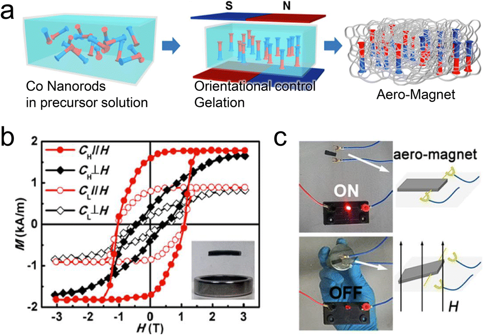 | ||
| Fig. 19 Ultralight magnetic soft composite with an anisotropic magnetic nanomaterial. (a) Fabrication of a silica-aerogel-based ultralight magnetic composite. Mixing Co NRs with an AR of 9 in a precursor solution (left), aligning Co NRs during gelation by applying a magnetic field (middle), and the aero-magnet achieved by subsequent solvent exchange and critical point drying (right). (b) Magnetic hysteresis curves of the aerogel composite with a high doping ratio of 30 wt% (CH) and low doping ratio of 15 wt% (CL) under parallel and vertical magnetic fields. Levitating behavior above the hollow ring magnet (∼11 mT) resulting from the light weight of the aerogel matrix (inset image). (c) Possible application of the ultralight magnet in a switching device. Reproduced with permission.298 Copyright 2019, American Chemical Society. | ||
Uniaxial arrangements of 0D MNPs can be adopted for manipulating magnetic soft actuators by inducing magnetic anisotropy. For example, superparamagnetic iron oxide NPs (SPIONs) are extensively used magnetic nanomaterials as this MNP exhibits various advantages such as high saturation magnetization,299 cost-effective production,300 and biocompatibility when compared with the other magnetic nanomaterials like Co and Ni.301 However, individual SPIONs do not exhibit coercivity and remanence, which can only show a translational force under an external magnetic field but not torque. The absence of a magnetization orientation limits the programmability of magnetic composites and decreases the complexity of actuation modes.8 However, if SPIONs can be aligned by a magnetic field to form a chain configuration, the ensemble of SPIONs can undergo a torque following the applied field.287 Initially, SPIONs mixed in photocurable resins formed a chain configuration in the direction of the external magnetic field and were subsequently fixed in the resin via photopolymerization (Fig. 20a). As a result, the magnetic easy axis appeared along the longitudinal direction of the SPION assembly, similar to the magnetic anisotropy. When applying an external magnetic field, a torque could be generated as the longitudinal direction of SPION assemblies has a tendency to rotate, aligning their easy axes along the magnetic field (Fig. 20b). Finally, by connecting two elements where SPIONs were aligned along the in-plane direction and another two elements with vertically aligned SPIONs, a micro-actuator mimicking a “looper caterpillar” was fabricated showing potential for application in micro-actuators (Fig. 20c).
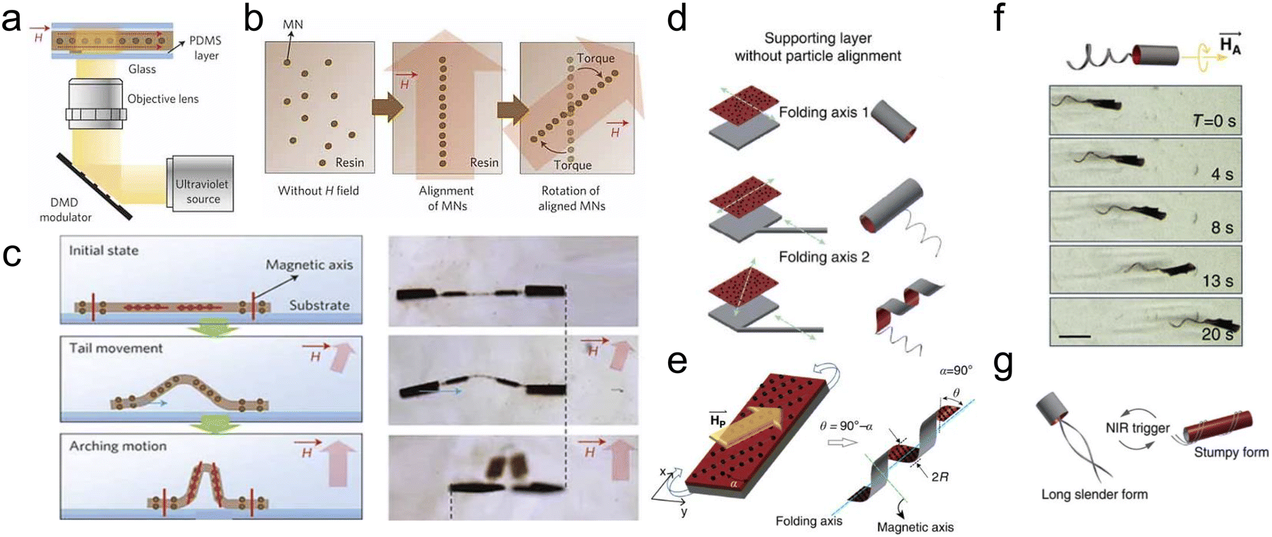 | ||
| Fig. 20 Magnetic soft actuators with an anisotropic assembly of SPIONs. (a) Polymeric magnetic micro-actuators with an anisotropic assembly of SPIONs prepared by photolithography. (b) SPIONs during fabrication and actuation: randomly dispersed SPIONs under a zero-magnetic field (left), alignment of SPIONs under a magnetic field followed by fixation during curing (middle), and torque generation of the fixed assembly by an external magnetic field (right). (c) Application for a micro-actuator mimicking “looper caterpillar”. Configuration of the micro-actuator composed of two elements with in-plane aligned SPIONs as the body and another two elements with vertically aligned SPIONs as the head and tail (left). Real images of the actuation of the micro-actuator (right). Reproduced with permission.287 Copyright 2011, Springer Nature. Microorganism-inspired actuator with versatile motility appropriate for certain environments: (d) structure of the actuator and its self-folding behavior which is determined by the location of the tail and shape of the body. (e) Controlling the self-folding behavior by programming the alignment of the MNPs. (f) Propulsion of the micro-actuator under a rotating magnetic field and (g) versatile designs of the micro-actuator upon heat treatment enabling various shape morphing modes. Reproduced with Creative Commons CC BY License.302 | ||
The assembled SPIONs can act as stiffness reinforcing agents.302 Inspired by microorganisms, a micro-robot consisting of body and tails was designed with motility under a magnetic field by incorporating SPIONs into a hydrogel matrix (Fig. 20d). Microorganisms adjust their shape in response to an environmental change appropriate for the surroundings. The robot body mimicking these functions was made of a bilayer structure comprising a thermo-responsive hydrogel and passive hydrogel matrix, which could be self-folded into a 3D structure. However, the folding sequence was mainly affected by the geometric constraints imposed by the body and tail configuration, which, then, restricted the flexibility in designing the actuation mode. To achieve a high DoF in shape morphing, MNPs were aligned in a specific direction, and the self-folding behavior was manually controlled, irrespective of the body–tail configuration (Fig. 20e). Furthermore, the anisotropic alignments of the MNPs produced a torque upon exposure to an external magnetic field, enabling the propulsion of the micro-actuator in a fluidic environment (Fig. 20f). The thermo-responsive matrix allowed a reversible transition in the structure, facilitating precise control of the actuation mode in response to external conditions (Fig. 20g). Magnetic soft actuators with various magnetic materials and magnetization profiles are summarized in Table 3.
| Materials | Polymer matrix | Magnetization or alignment step | Magnetization profile/encoding method | Content of magnetic particles/dimension of actuators |
|---|---|---|---|---|
| NdFeB | Shape memory polymer222 | Magnetization after solidifying | — | 40 wt%/50 × 50 × 0.06 mm |
| Ecoflex294 | Magnetization after solidifying | 3D profile/wrapping cylinder under a magnetic field | 50 wt%/1.5 × 3.7 × 0.185 mm | |
| UV resin291 | Reorientation of pre-magnetized particles | 3D profile/UV lithographic method | 50 wt%/millimeter-scale | |
| PCL for encapsulation | Reorientation of pre-magnetized particles | 3D profile/encapsulating PCL and laser reprogramming | 50 wt% for NdFeB@PCL | |
| Ecoflex matrix295 | 50 wt% of NdFeB@PCL in Ecoflex/millimeter scale | |||
| Ecoflex287 | Reorientation of pre-magnetized particles | 3D profile/magnetic field-assisted 3D printing | 20 vol%/millimeter-scale | |
| Ni nanowires | Spiropyran297 | Magnetization during solidifying | 3D profile/deformation of matrix | 5 mg ml−1/millimeter-scale |
| Co nanorods | Silica aerogel298 | Magnetization during solidifying | Two domains/two opposite magnetic field | 30 wt%/centimeter-scale |
| Co nanowires | Ecoflex322 | Magnetization during solidifying | — | 10 wt%/4.5 × 15 mm |
| Fe3O4 | PEGDA287 | Magnetization during solidifying | 3D profile/UV lithographic method | —/micrometer-scale |
| Thermal responsive matrix: PNIPAAm-Aam-PEGDA | Magnetization during solidifying | 3D profile/deformation of matrix | 4.7 wt% for thermally responsive matrix | |
| Passive matrix: PEGDA302 | 1.2 wt% for passive matrix /micrometer-scale | |||
| Commercial iron oxide ferrofluid | Gelatin methacryloyl hydrogel323 | Magnetization during solidifying | 3D profile/alignment under a gradient field | 2.9 vol%/millimeter-scale |
The stiffness of the anisotropic assembly of MNPs could be further strengthened by subjecting this assembly to an external magnetic field because of the magnetic dipole–dipole interaction, a phenomenon known as the magnetorheological effect, introduced in section 3.4. In the field of robotics, the ability to adjust stiffness is important for gripper technologies.303–305 While soft grippers offer benefits, such as safer handling and adaptability to different object shapes due to their flexibility, they often lack sufficient grasping force.306 To address this issue, recent research has focused on soft grippers whose stiffness can be varied to enhance their grasping power.307 Thus, magnetic soft composites demonstrate potential for application in soft grippers owing to stiffness tunability arising from magnetorheological effects. To maximize the change of stiffness, magnetic soft composites embedded with magnetorheological fluid have been used.308 Without a magnetic field, the composite shows a very low modulus, enabling conformal attachment to various shapes. Subsequently, by switching on the magnetic field, the magnetic particles in the fluid align into chains along the field lines, significantly increasing the stiffness of the composites. This transformation results in a higher grip strength, thus improving the ability of the gripper to securely hold objects. When the magnetic field is switched off, the stiffness of the composites reduces to its original state, facilitating the release of the gripped object. Therefore, the magnetomechanical effects induced by magnetic anisotropy not only enhance the functionality of magnetic soft actuators but also expand their potential applications.
As magnetic anisotropy in soft actuating materials can be achieved by numerous strategies, precise and digitalized programming of the magnetization profile can be realized. Not only hard magnets but also soft magnets can enhance the actuation performance with high selectivity and controllability based on the magnetic anisotropy. Furthermore, the magnetic anisotropy in magnetic soft composites contributes to the decrease of magnetic filler contents required in the composites and the strength of magnetic field for triggering actuation. The magnetomechanical effect driven by magnetic anisotropy provides additional functionality to the actuators which can be favorable for certain applications including soft grippers. Therefore, actuators with magnetic anisotropy behavior can be applied to wearable rehabilitation robots, ultralight inspection drones, untethered explorers, and intelligent humanoids.
5. Conclusions and future perspective
In summary, this review highlighted the critical role of the magnetic anisotropy of various materials in flexible sensors and soft actuators for granting advanced features and functions to soft robots. Initially, we discussed the fundamental principles of magnetic anisotropy, driven by intrinsic and extrinsic factors including the crystallographic nature of magnetic materials, dependency on shape and dimension, interlayer coupling for exchange bias, and external stress-induced magnetic anisotropy. Magnetic anisotropy does not arise from a single, isolated factor among the aforementioned influencing factors; rather, it emerges as a result of a complex interplay of these factors, with certain dominant elements governing the overall energy of the system. Moreover, we presented and categorized strategies to enhance magnetic anisotropy via alignments, shapes, layered structures, and external energy sources. We examined the magnetic anisotropy effects caused by magnetic domain alignment or the arrangement of magnetic nanomaterials across different dimensions and the related novel fabrication methodologies. Such recent breakthroughs in the implications of magnetic anisotropy have significantly improved the performance of magnetic sensors and actuators for their applications in soft robotics. Thanks to magnetic anisotropy, which boosts the magnetic properties of materials toward a specific orientation, magnetic sensors demonstrate outstanding sensitivity, precision, and selectivity. This sensing capability is particularly evident in the detection of changes in both the direction and intensity of magnetic fields. Moreover, magnetic anisotropy facilitates the programming of magnetization in actuating soft bodies. The anisotropy nature of soft actuators enables complex shape transformations without pre-designed hinges and effective operation even in weak magnetic fields. These advancements are highly advantageous for magnetic soft robots, which can enhance their response speeds, facilitate both proximity and touch sensing, offer remote control and adjustable actuation, and lead to overall better performance. Therefore, magnetic soft robots have potential applications in fields like biomedical micro-robots, micro-swimmers, and soft grippers. Among them, biomedical micro-robots have been thoroughly explored and are considered the most promising application in the field of implantable and therapeutic robots. With a biocompatible soft matrix, magnetic soft robots can aid in targeted drug delivery and minimally invasive surgery. Additionally, there is a growing need to remove microplastics from the ocean environment. In this context, micro-swimmers capable of navigating through the water environment and collecting microplastics would be another practical application. Also, soft grippers, with the ability to manipulate various materials delicately, offer a feasible application of magnetic actuators by taking advantage of remote controllability, stiffness tunability from the magnetorheological effect, and the softness of the matrix. These applications are anticipated to contribute to improving medical devices, resolving environmental pollution, and enhancing efficiency in industrial lines.However, several challenges should be addressed for the practical applications of magnetic soft robots in real-world scenarios. Firstly, the high compatibility of these integrated sensors and actuators with surrounding electronic devices, which often face electromagnetic interference (EMI), should be considered.309–311 The implementation of suitable EMI shielding materials is necessary to ensure reliable signal transmission between sensors and actuators with the precise control of magnetic soft robots, irrespective of the environmental conditions. Novel design and fabrication methods, such as inducing magnetic anisotropy in managing the geometry of materials or device structures, are essential to minimize external EMI and achieve the selective manipulation of magnetic components in soft robots.
Secondly, safety and biocompatibility are significant concerns, particularly because magnetic soft robots are designed for human interaction in cooperative tasks or for biomedical applications like micro-robotic drug delivery and surgical assistance. While the assistance from highly flexible and functionalized materials combined with the feedback control systems makes soft robots highly relevant in our daily lives, the biocompatibility of magnetic elements remains a debated topic. SPIONs, preferred for their relatively lower toxicity,312,313 do not offer substantial benefits in terms of biocompatibility versus performance for robotic applications.314 Although encapsulating magnetic components in biocompatible materials is a potential solution, ensuring the inherent biocompatibility of magnetic materials is crucial for long-term and harsh environment applications.315 Developing low-toxicity magnetic NPs with adjustable spin states can provide safer and more biocompatible options for magnetic soft robots.316,317
Finally, the development of wearable and untethered soft robots aims to assist human movement or replace manpower in risky environments. To establish these features, the required strength of the magnetic field source should be minimized for efficient and high DoF operation. Traditional magnetic actuators, which operate in response to the strong and uniform magnetic fields generated by bulky electromagnetic coils, have limitations in terms of their versatility and being bound to ground. To overcome this obstacle, compact electromagnetic coil systems need to be integrated into robot bodies serving as actuation sources. Combining these systems with sensors and actuators is crucial for efficient signal processing and feedback control of soft robots. Moreover, additional components such as wireless communication modules, signal processors, and permanent power sources, all governed by sophisticated closed-loop signal-processing algorithms, ultimately find a way to develop an autonomous, untethered, and intelligent soft robot.
Conflicts of interest
There are no conflicts to declare.Acknowledgements
This work was supported by the National Research Foundation of Korea (NRF) grant funded by the Korea government (MIST) (No. NRF-2022R1C1C1004845, RS-2023-00207836).References
- A. Billard and D. Kragic, Science, 2019, 364, eaat8414 CrossRef CAS PubMed.
- X. Dong, X. Luo, H. Zhao, C. Qiao, J. Li, J. Yi, L. Yang, F. J. Oropeza, T. S. Hu, Q. Xu and H. Zeng, Soft Matter, 2022, 18, 7699–7734 RSC.
- W. Dou, G. Zhong, J. Cao, Z. Shi, B. Peng and L. Jiang, Adv. Mater. Technol., 2021, 6, 2100018 CrossRef.
- N. El-Atab, R. B. Mishra, F. Al-Modaf, L. Joharji, A. A. Alsharif, H. Alamoudi, M. Diaz, N. Qaiser and M. M. Hussain, Adv. Intell. Syst. Comput., 2020, 2, 2000128 CrossRef.
- Z. Shen, F. Chen, X. Zhu, K.-T. Yong and G. Gu, J. Mater. Chem. B, 2020, 8, 8972–8991 RSC.
- B. Chen and Z. L. Wang, Small, 2022, 18, 2107034 CrossRef CAS PubMed.
- I. Apsite, S. Salehi and L. Ionov, Chem. Rev., 2022, 122, 1349–1415 CrossRef CAS PubMed.
- Y. Kim and X. Zhao, Chem. Rev., 2022, 122, 5317–5364 CrossRef CAS PubMed.
- H. Kim, S.-k. Ahn, D. M. Mackie, J. Kwon, S. H. Kim, C. Choi, Y. H. Moon, H. B. Lee and S. H. Ko, Mater. Today, 2020, 41, 243–269 CrossRef CAS.
- P. Won, K. K. Kim, H. Kim, J. J. Park, I. Ha, J. Shin, J. Jung, H. Cho, J. Kwon, H. Lee and S. H. Ko, Adv. Mater., 2021, 33, 2002397 CrossRef CAS PubMed.
- H. Lee, H. Kim, I. Ha, J. Jung, P. Won, H. Cho, J. Yeo, S. Hong, S. Han, J. Kwon, K. J. Cho and S. H. Ko, Soft Robot., 2019, 6(6), 760–767 CrossRef PubMed.
- H. Kim, H. Lee, I. Ha, J. Jung, P. Won, H. Cho, J. Yeo, S. Hong, S. Han, J. Kwon, K.-J. Cho and S. H. Ko, Adv. Funct. Mater., 2018, 28, 1801847 CrossRef.
- C. Hegde, J. Su, J. M. R. Tan, K. He, X. Chen and S. Magdassi, ACS Nano, 2023, 17, 15277–15307 CrossRef CAS PubMed.
- C. Han, Y. Jeong, J. Ahn, T. Kim, J. Choi, J.-H. Ha, H. Kim, S. H. Hwang, S. Jeon, J. Ahn, J. T. Hong, J. J. Kim, J.-H. Jeong and I. Park, Adv. Sci., 2023, 10, 2302775 CrossRef PubMed.
- H. Wang, M. Totaro and L. Beccai, Adv. Sci., 2018, 5, 1800541 CrossRef PubMed.
- A. Tsay, T. J. Allen, U. Proske and M. J. Giummarra, Neurosci. Biobehav. Rev., 2015, 52, 221–232 CrossRef CAS PubMed.
- Z. Lin, Z. Wang, W. Zhao, Y. Xu, X. Wang, T. Zhang, Z. Sun, L. Lin and Z. Peng, Adv. Intell. Syst. Comput., 2023, 5, 2200329 CrossRef.
- D. Alatorre, D. Axinte and A. Rabani, IEEE Trans. Robot., 2022, 38, 526–535 Search PubMed.
- P. Gambardella, S. Rusponi, M. Veronese, S. S. Dhesi, C. Grazioli, A. Dallmeyer, I. Cabria, R. Zeller, P. H. Dederichs, K. Kern, C. Carbone and H. Brune, Science, 2003, 300, 1130–1133 CrossRef CAS PubMed.
- P. Bruno, Phys. Rev. B: Condens. Matter Mater. Phys., 1989, 39, 865–868 CrossRef PubMed.
- B. D. Cullity and C. D. Graham, in Introduction to Magnetic Materials, ed. L. Hanzo, Wiley, New Jersey, 2nd edn, 2009, ch. 7, pp. 197–239 Search PubMed.
- Q. Fan, Z. Li, C. Wu and Y. Yin, Precis. Chem., 2023, 1, 272–298 CrossRef CAS PubMed.
- R. C. O'handley, Modern magnetic materials: principles and applications, Wiley, 2000 Search PubMed.
- J. M. D. Coey, in Magnetism and Magnetic Materials, Cambridge University Press, Cambridge, 2010, ch. 5, pp. 128–194 Search PubMed.
- L. Sagnotti, in Encyclopedia of Solid Earth Geophysics, ed. H. K. Gupta, Springer, Netherlands, Dordrecht, 2011, pp. 717–729.
- Z. Yang, Y. Chen, W. Liu, Y. Wang, Y. Li, D. Zhang, Q. Lu, Q. Wu, H. Zhang and M. Yue, Nanomaterials, 2022, 12, 1261 CrossRef CAS PubMed.
- A. P. Guimarães, in Principles of Nanomagnetism, ed. A. P. Guimarães, Springer Berlin Heidelberg, Berlin, Heidelberg, 2009, pp. 57–104. DOI:10.1007/978-3-642-01482-6_3.
- J. Mohapatra, M. Xing and J. P. Liu, Materials, 2019, 12, 3208 CrossRef CAS PubMed.
- J. Mohapatra and J. P. Liu, in Handbook of Magnetic Materials, ed. E. Brück, Elsevier, 2018, vol. 27, pp. 1–57 Search PubMed.
- E. C. Stoner and E. P. Wohlfarth, Nature, 1947, 160, 650–651 CrossRef.
- A.-H. Lu, E. L. Salabas and F. Schüth, Angew. Chem., Int. Ed., 2007, 46, 1222–1244 CrossRef CAS PubMed.
- Z. Ma, J. Mohapatra, K. Wei, J. P. Liu and S. Sun, Chem. Rev., 2023, 123, 3904–3943 CrossRef CAS PubMed.
- M. D. Nguyen, H.-V. Tran, S. Xu and T. R. Lee, Appl. Sci., 2021, 11, 11301 CrossRef CAS PubMed.
- J. S. Lee, J. M. Cha, H. Y. Yoon, J. K. Lee and Y. K. Kim, Sci. Rep., 2015, 5, 12135 CrossRef PubMed.
- M. Unni, A. M. Uhl, S. Savliwala, B. H. Savitzky, R. Dhavalikar, N. Garraud, D. P. Arnold, L. F. Kourkoutis, J. S. Andrew and C. Rinaldi, ACS Nano, 2017, 11, 2284–2303 CrossRef CAS PubMed.
- F. Chen, N. Ilyas, X. Liu, Z. Li, S. Yan and H. Fu, Front. Energy Res., 2021, 9, 780008 CrossRef.
- J. Nogués and I. K. Schuller, J. Magn. Magn. Mater., 1999, 192, 203–232 CrossRef.
- W. H. Meiklejohn, J. Appl. Phys., 2004, 33, 1328–1335 CrossRef.
- M. Tsunoda, M. Naka, D. Y. Kim and M. Takahashi, J. Magn. Magn. Mater., 2006, 304, e88–e90 CrossRef CAS.
- Z. Swiatkowska-Warkocka, K. Kawaguchi, H. Wang, Y. Katou and N. Koshizaki, Nanoscale Res. Lett., 2011, 6, 226 CrossRef PubMed.
- M. Heigl, C. Vogler, A.-O. Mandru, X. Zhao, H. J. Hug, D. Suess and M. Albrecht, ACS Appl. Nano Mater., 2020, 3, 9218–9225 CrossRef CAS PubMed.
- H. Singh, R. Gupta, T. Chakraborty, A. Gupta and C. Mitra, IEEE Trans. Magn., 2014, 50, 1–4 Search PubMed.
- W. B. Rui, Y. Hu, A. Du, B. You, M. W. Xiao, W. Zhang, S. M. Zhou and J. Du, Sci. Rep., 2015, 5, 13640 CrossRef CAS PubMed.
- W. H. Meiklejohn and C. P. Bean, Phys. Rev., 1957, 105, 904–913 CrossRef CAS.
- R. Jungblut, R. Coehoorn, M. T. Johnson, J. aan de Stegge and A. Reinders, J. Appl. Phys., 1994, 75, 6659–6664 CrossRef CAS.
- A. P. Malozemoff, Phys. Rev. B: Condens. Matter Mater. Phys., 1987, 35, 3679–3682 CrossRef PubMed.
- D. Mauri, H. C. Siegmann, P. S. Bagus and E. Kay, J. Appl. Phys., 1987, 62, 3047–3049 CrossRef.
- T. Ambrose and C. L. Chien, J. Appl. Phys., 1998, 83, 6822–6824 CrossRef CAS.
- U. Nowak, K. D. Usadel, J. Keller, P. Miltényi, B. Beschoten and G. Güntherodt, Phys. Rev. B: Condens. Matter Mater. Phys., 2002, 66, 014430 CrossRef.
- T. Kobayashi, H. Kato, T. Kato, S. Tsunashima and S. Iwata, J. Phys.: Conf. Ser., 2010, 200, 072052 Search PubMed.
- W. Zhang and K. M. Krishnan, Mater. Sci. Eng., R, 2016, 105, 1–20 CrossRef.
- J. Nogués, T. J. Moran, D. Lederman, I. K. Schuller and K. V. Rao, Phys. Rev. B: Condens. Matter Mater. Phys., 1999, 59, 6984–6993 CrossRef.
- W. Su, Z. Wang, T. Wen, Z. Hu, J. Wu, Z. Zhou and M. Liu, IEEE Electron Device Lett., 2019, 40, 969–972 CAS.
- M. F. Hansen and G. Rizzi, IEEE Trans. Magn., 2017, 53, 1–11 Search PubMed.
- J. C. S. Kools, IEEE Trans. Magn., 1996, 32, 3165–3184 CrossRef CAS.
- S. S. P. Parkin, K. P. Roche, M. G. Samant, P. M. Rice, R. B. Beyers, R. E. Scheuerlein, E. J. O'Sullivan, S. L. Brown, J. Bucchigano, D. W. Abraham, Y. Lu, M. Rooks, P. L. Trouilloud, R. A. Wanner and W. J. Gallagher, J. Appl. Phys., 1999, 85, 5828–5833 CrossRef CAS.
- E. W. Lee, Rep. Prog. Phys., 1955, 18, 184 CrossRef.
- B. D. Cullity and C. D. Graham, in Introduction to Magnetic Materials, ed. L. Hanzo, Wiley, New Jersey, 2nd edn, 2009, ch. 8, pp. 241–273 Search PubMed.
- A. G. Olabi and A. Grunwald, Mater. Des., 2008, 29, 469–483 CrossRef CAS.
- N. B. Ekreem, A. G. Olabi, T. Prescott, A. Rafferty and M. S. J. Hashmi, J. Mater. Process. Technol., 2007, 191, 96–101 CrossRef CAS.
- M. S. Amiri, M. Thielen, M. Rabung, M. Marx, K. Szielasko and C. Boller, J. Magn. Magn. Mater., 2014, 372, 16–22 CrossRef.
- Z. Deng and M. J. Dapino, Smart Mater. Struct., 2018, 27, 113001 CrossRef.
- A. Bieńkowski and J. Kulikowski, J. Magn. Magn. Mater., 1980, 19, 120–122 CrossRef.
- R. Q. Wu, L. J. Chen, A. Shick and A. J. Freeman, J. Magn. Magn. Mater., 1998, 177–181, 1216–1219 CrossRef CAS.
- F. Wang, X. Dai, Y. Li, H. Jia, X. Wang and G. Liu, Presented in part at the 2015IEEE International Magnetics Conference, Beijing, May, 2015.
- A. Speliotis and D. Niarchos, Sens. Actuators, A, 2003, 106, 298–301 CrossRef CAS.
- A. Pateras, R. Harder, S. Manna, B. Kiefer, R. L. Sandberg, S. Trugman, J. W. Kim, J. de la Venta, E. E. Fullerton, O. G. Shpyrko and E. Fohtung, NPG Asia Mater., 2019, 11, 59 CrossRef CAS.
- G. O. Fulop, M. B. S. Dias, H. R. Z. Sandim and C. Bormio-Nunes, J. Magn. Magn. Mater., 2021, 527, 167702 CrossRef CAS.
- K. Y. Ho, X. Y. Xiong, J. Zhi and L. Z. Cheng, J. Appl. Phys., 1993, 74, 6788–6790 CrossRef CAS.
- D. Vanoost, S. Steentjes, J. Peuteman, G. Gielen, H. De Gersem, D. Pissoort and K. Hameyer, J. Magn. Magn. Mater., 2016, 414, 168–179 CrossRef CAS.
- P. Ruuskanen and P. Kettunen, J. Magn. Magn. Mater., 1991, 98, 349–358 CrossRef CAS.
- A. E. Clark and K. B. Hathaway, MRS Bull., 1993, 18, 34–41 CrossRef.
- S. Kotapati, A. Javed, N. Reeves-McLaren, M. R. J. Gibbs and N. A. Morley, J. Magn. Magn. Mater., 2013, 331, 67–71 CrossRef CAS.
- H. Wang, Y. N. Zhang, R. Q. Wu, L. Z. Sun, D. S. Xu and Z. D. Zhang, Sci. Rep., 2013, 3, 3521 CrossRef PubMed.
- M. Klokkenburg, C. Vonk, E. M. Claesson, J. D. Meeldijk, B. H. Erné and A. P. Philipse, J. Am. Chem. Soc., 2004, 126, 16706–16707 CrossRef CAS PubMed.
- L. N. Donselaar, P. M. Frederik, P. Bomans, P. A. Buining, B. M. Humbel and A. P. Philipse, J. Magn. Magn. Mater., 1999, 201, 58–61 CrossRef CAS.
- R. Massart, IEEE Trans. Magn., 1981, 17, 1247–1248 CrossRef.
- J. Park, K. An, Y. Hwang, J.-G. Park, H.-J. Noh, J.-Y. Kim, J.-H. Park, N.-M. Hwang and T. Hyeon, Nat. Mater., 2004, 3, 891–895 CrossRef CAS PubMed.
- S. Sun, H. Zeng, D. B. Robinson, S. Raoux, P. M. Rice, S. X. Wang and G. Li, J. Am. Chem. Soc., 2004, 126, 273–279 CrossRef CAS PubMed.
- H. Deng, X. Li, Q. Peng, X. Wang, J. Chen and Y. Li, Angew. Chem., Int. Ed., 2005, 44, 2782–2785 CrossRef CAS PubMed.
- M. A. Correa-Duarte, M. Grzelczak, V. Salgueiriño-Maceira, M. Giersig, L. M. Liz-Marzán, M. Farle, K. Sierazdki and R. Diaz, J. Phys. Chem. B, 2005, 109, 19060–19063 CrossRef CAS PubMed.
- X. Fan, F. Tan, G. Zhang and F. Zhang, Mater. Sci. Eng., A, 2007, 454–455, 37–42 CrossRef.
- K. Liu, L. Han, P. Tang, K. Yang, D. Gan, X. Wang, K. Wang, F. Ren, L. Fang, Y. Xu, Z. Lu and X. Lu, Nano Lett., 2019, 19, 8343–8356 CrossRef CAS PubMed.
- R. Sheparovych, Y. Sahoo, M. Motornov, S. Wang, H. Luo, P. N. Prasad, I. Sokolov and S. Minko, Chem. Mater., 2006, 18, 591–593 CrossRef CAS.
- Y. Hu, L. He and Y. Yin, Angew. Chem., Int. Ed., 2011, 50, 3747–3750 CrossRef CAS PubMed.
- Q. Xiong, C. Y. Lim, J. Ren, J. Zhou, K. Pu, M. B. Chan-Park, H. Mao, Y. C. Lam and H. Duan, Nat. Commun., 2018, 9, 1743 CrossRef PubMed.
- J. Saiz-Poseu, J. Mancebo-Aracil, F. Nador, F. Busqué and D. Ruiz-Molina, Angew. Chem., Int. Ed., 2019, 58, 696–714 CrossRef CAS PubMed.
- H. Lee, S. M. Dellatore, W. M. Miller and P. B. Messersmith, Science, 2007, 318, 426–430 CrossRef CAS PubMed.
- B. K. Ahn, J. Am. Chem. Soc., 2017, 139, 10166–10171 CrossRef CAS PubMed.
- L. r. Meng, W. Chen, C. Chen, H. Zhou, Q. Peng and Y. Li, Cryst. Growth Des., 2010, 10, 479–482 CrossRef CAS.
- K. Nakata, Y. Hu, O. Uzun, O. Bakr and F. Stellacci, Adv. Mater., 2008, 20, 4294–4299 CrossRef CAS.
- G. A. DeVries, M. Brunnbauer, Y. Hu, A. M. Jackson, B. Long, B. T. Neltner, O. Uzun, B. H. Wunsch and F. Stellacci, Science, 2007, 315, 358–361 CrossRef CAS PubMed.
- Q. He, T. Yuan, S. Wei, N. Haldolaarachchige, Z. Luo, D. P. Young, A. Khasanov and Z. Guo, Angew. Chem., Int. Ed., 2012, 51, 8842–8845 CrossRef CAS PubMed.
- Y. Xia, Q. Chen and U. Banin, Chem. Rev., 2023, 123, 3325–3328 CrossRef CAS PubMed.
- Y. Soumare, C. Garcia, T. Maurer, G. Chaboussant, F. Ott, F. Fiévet, J.-Y. Piquemal and G. Viau, Adv. Funct. Mater., 2009, 19, 1971–1977 CrossRef CAS.
- Y. C. Zhang, J. Y. Tang and X. Y. Hu, J. Alloys Compd., 2008, 462, 24–28 CrossRef CAS.
- V. F. Puntes, K. M. Krishnan and A. P. Alivisatos, Science, 2001, 291, 2115–2117 CrossRef CAS PubMed.
- L. Hu, R. Zhang and Q. Chen, Nanoscale, 2014, 6, 14064–14105 RSC.
- L. Y. Zhang, D. S. Xue, X. F. Xu, A. B. Gui and C. X. Gao, J. Phys.: Condens. Matter, 2004, 16, 4541 CrossRef CAS.
- X. Pang, Y. He, J. Jung and Z. Lin, Science, 2016, 353, 1268–1272 CrossRef CAS PubMed.
- D. S. Xue, C. X. Gao, Q. F. Liu and L. Y. Zhang, J. Phys.: Condens. Matter, 2003, 15, 1455 CrossRef CAS.
- D.-S. Xue, L.-Y. Zhang, C.-X. Gao, X.-F. Xu and A.-B. Gui, Chin. Phys. Lett., 2004, 21, 733 CrossRef CAS.
- J. Cha, J. S. Lee, S. J. Yoon, Y. K. Kim and J.-K. Lee, RSC Adv., 2013, 3, 3631–3637 RSC.
- Z. Liu, D. Zhang, S. Han, C. Li, B. Lei, W. Lu, J. Fang and C. Zhou, J. Am. Chem. Soc., 2005, 127, 6–7 CrossRef CAS PubMed.
- C.-J. Jia, L.-D. Sun, Z.-G. Yan, L.-P. You, F. Luo, X.-D. Han, Y.-C. Pang, Z. Zhang and C.-H. Yan, Angew. Chem., Int. Ed., 2005, 44, 4328–4333 CrossRef CAS PubMed.
- J. Wang, Q. Chen, C. Zeng and B. Hou, Adv. Mater., 2004, 16, 137–140 CrossRef CAS.
- Y. Xiong, Y. Xie, Z. Li, R. Zhang, J. Yang and C. Wu, New J. Chem., 2003, 27, 588–590 RSC.
- W. Wu, X. Xiao, S. Zhang, J. Zhou, L. Fan, F. Ren and C. Jiang, J. Phys. Chem. C, 2010, 114, 16092–16103 CrossRef CAS.
- S. Palchoudhury, W. An, Y. Xu, Y. Qin, Z. Zhang, N. Chopra, R. A. Holler, C. H. Turner and Y. Bao, Nano Lett., 2011, 11, 1141–1146 CrossRef CAS PubMed.
- F. Dumestre, B. Chaudret, C. Amiens, M.-C. Fromen, M.-J. Casanove, P. Renaud and P. Zurcher, Angew. Chem., Int. Ed., 2002, 41, 4286–4289 CrossRef CAS PubMed.
- F. Dumestre, B. Chaudret, C. Amiens, M. Respaud, P. Fejes, P. Renaud and P. Zurcher, Angew. Chem., Int. Ed., 2003, 42, 5213–5216 CrossRef CAS PubMed.
- F. Fiévet, S. Ammar-Merah, R. Brayner, F. Chau, M. Giraud, F. Mammeri, J. Peron, J. Y. Piquemal, L. Sicard and G. Viau, Chem. Soc. Rev., 2018, 47, 5187–5233 RSC.
- K. A. Atmane, C. Michel, J.-Y. Piquemal, P. Sautet, P. Beaunier, M. Giraud, M. Sicard, S. Nowak, R. Losno and G. Viau, Nanoscale, 2014, 6, 2682–2692 RSC.
- R. K. Ramamoorthy, A. Viola, B. Grindi, J. Peron, C. Gatel, M. Hytch, R. Arenal, L. Sicard, M. Giraud, J.-Y. Piquemal and G. Viau, Nano Lett., 2019, 19, 9160–9169 CrossRef CAS PubMed.
- K. Mrad, F. Schoenstein, H. T. T. Nong, E. Anagnostopoulou, A. Viola, L. Mouton, S. Mercone, C. Ricolleau, N. Jouini, M. Abderraba, L. M. Lacroix, G. Viau and J. Y. Piquemal, CrystEngComm, 2017, 19, 3476–3484 RSC.
- H.-H. Xu, Q. Wu, M. Yue, C.-L. Li and H.-J. Li, Rare Met., 2023, 42, 1994–1999 CrossRef CAS.
- S. Ener, E. Anagnostopoulou, I. Dirba, L.-M. Lacroix, F. Ott, T. Blon, J.-Y. Piquemal, K. P. Skokov, O. Gutfleisch and G. Viau, Acta Mater., 2018, 145, 290–297 CrossRef CAS.
- K. A. Atmane, F. Zighem, Y. Soumare, M. Ibrahim, R. Boubekri, T. Maurer, J. Margueritat, J. Y. Piquemal, F. Ott, G. Chaboussant, F. Schoenstein, N. Jouini and G. Viau, J. Solid State Chem., 2013, 197, 297–303 CrossRef.
- P. D. Kulkarni, S. K. Dhar, A. Provino, P. Manfrinetti and A. K. Grover, Phys. Rev. B: Condens. Matter Mater. Phys., 2010, 82, 144411 CrossRef.
- J. S. Kouvel and C. D. Graham, J. Phys. Chem. Solids, 1959, 11, 220–225 CrossRef.
- Y. Ma, Y. Yang, Y. Gao and Y. Hu, Phys. Chem. Chem. Phys., 2021, 23, 17365–17373 RSC.
- S. Giri, M. Patra and S. Majumdar, J. Phys.: Condens. Matter, 2011, 23, 073201 CrossRef CAS PubMed.
- M. Ávila-Gutiérrez, A. Moisset, A.-T. Ngo, S. Costanzo, G. Simon, P. Colomban, M. Petit, C. Petit and I. Lisiecki, Colloids Surf., A, 2023, 676, 132281 CrossRef.
- J. A. González, J. P. Andrés, J. A. De Toro, P. Muñiz, T. Muñoz, O. Crisan, C. Binns and J. M. Riveiro, J. Nanopart. Res., 2009, 11, 2105–2111 CrossRef.
- O. Crisan, K. von Haeften, A. M. Ellis and C. Binns, J. Nanopart. Res., 2008, 10, 193–199 CrossRef CAS.
- S. A. Koch, G. Palasantzas, T. Vystavel, J. T. M. De Hosson, C. Binns and S. Louch, Phys. Rev. B: Condens. Matter Mater. Phys., 2005, 71, 085410 CrossRef.
- V. Skumryev, S. Stoyanov, Y. Zhang, G. Hadjipanayis, D. Givord and J. Nogués, Nature, 2003, 423, 850–853 CrossRef CAS PubMed.
- Z. M. Tian, S. L. Yuan, S. Y. Yin, L. Liu, J. H. He, H. N. Duan, P. Li and C. H. Wang, Appl. Phys. Lett., 2008, 93, 222505 CrossRef.
- Y. Shen, Y. Wu, H. Xie, K. Li, J. Qiu and Z. Guo, J. Appl. Phys., 2002, 91, 8001–8003 CrossRef CAS.
- K. Temst, E. Popova, H. Loosvelt, M. J. Van Bael, S. Brems, Y. Bruynseraede, C. Van Haesendonck, H. Fritzsche, M. Gierlings, L. H. A. Leunissen and R. Jonckheere, J. Magn. Magn. Mater., 2006, 304, 14–18 CrossRef CAS.
- Z. B. Guo, Y. K. Zheng, K. B. Li, Z. Y. Liu, P. Luo and Y. H. Wu, J. Appl. Phys., 2004, 95, 4918–4921 CrossRef CAS.
- M. Perzanowski, O. Polit, J. Chojenka, W. Sas, A. Zarzycki and M. Marszalek, Nanotechnology, 2022, 33, 495707 CrossRef PubMed.
- A. Hoffmann, M. Grimsditch, J. E. Pearson, J. Nogués, W. A. A. Macedo and I. K. Schuller, Phys. Rev. B: Condens. Matter Mater. Phys., 2003, 67, 220406 CrossRef.
- W. Zhang, D. N. Weiss and K. M. Krishnan, J. Appl. Phys., 2010, 107, 09D724 CrossRef.
- C. Liu, C. Yu, H. Jiang, L. Shen, C. Alexander and G. J. Mankey, J. Appl. Phys., 2000, 87, 6644–6646 CrossRef CAS.
- D. Kumar, S. Singh and A. Gupta, J. Appl. Phys., 2016, 120, 085307 CrossRef.
- B.-Y. Wang, C.-J. Chen, K. Lin, C.-Y. Hsu, J.-Y. Ning, M.-S. Tsai, T.-H. Chuang, D.-H. Wei and S.-C. Weng, Appl. Surf. Sci., 2020, 533, 147501 CrossRef CAS.
- K.-i. Imakita, M. Tsunoda and M. Takahashi, J. Appl. Phys., 2005, 97, 10K106 CrossRef.
- M. S. Lund, W. A. A. Macedo, K. Liu, J. Nogués, I. K. Schuller and C. Leighton, Phys. Rev. B: Condens. Matter Mater. Phys., 2002, 66, 054422 CrossRef.
- S. K. Mishra, J. Magn. Magn. Mater., 2019, 488, 165374 CrossRef CAS.
- M. Meinert, B. Büker, D. Graulich and M. Dunz, Phys. Rev. B: Condens. Matter Mater. Phys., 2015, 92, 144408 CrossRef.
- N. T. Thanh, M. G. Chun, N. D. Ha, K. Y. Kim, C. O. Kim and C. G. Kim, J. Magn. Magn. Mater., 2006, 305, 432–435 CrossRef CAS.
- A. Kohn, A. Kovács, R. Fan, G. J. McIntyre, R. C. C. Ward and J. P. Goff, Sci. Rep., 2013, 3, 2412 CrossRef CAS PubMed.
- R. Wu, M. Xue, T. Maity, Y. Peng, S. K. Giri, G. Tian, J. L. MacManus-Driscoll and J. Yang, Phys. Rev. B, 2020, 101, 014425 CrossRef CAS.
- M. Dunz and M. Meinert, J. Appl. Phys., 2020, 128, 153902 CrossRef CAS.
- H. Lu, J. F. Bi, K. L. Teo, T. Liew and T. C. Chong, J. Appl. Phys., 2010, 107, 09D717 CrossRef.
- T. Blachowicz and A. Ehrmann, Coatings, 2021, 11, 122 CrossRef CAS.
- M. Öztürk, E. Demirci, R. Topkaya, S. Kazan, N. Akdoğan, M. Obaida and K. Westerholt, J. Supercond. Novel Magn., 2012, 25, 2597–2603 CrossRef.
- H.-C. Wu, R. Ramos, R. G. S. Sofin, Z.-M. Liao, M. Abid and I. V. Shvets, Appl. Phys. Lett., 2012, 101, 052402 CrossRef.
- N. N. Phuoc, G. Chai and C. K. Ong, J. Appl. Phys., 2012, 112, 083925 CrossRef.
- N. N. Phuoc and C. K. Ong, J. Appl. Phys., 2013, 114, 043911 CrossRef.
- F. Liu and C. A. Ross, J. Appl. Phys., 2014, 116, 194307 CrossRef.
- Y. Guo, Y. Ouyang, N. Sato, C. C. Ooi and S. X. Wang, IEEE Sens. J., 2017, 17, 3309–3315 CAS.
- S. Bhatti, R. Sbiaa, A. Hirohata, H. Ohno, S. Fukami and S. N. Piramanayagam, Mater. Today, 2017, 20, 530–548 CrossRef.
- J. Nogués, J. Sort, V. Langlais, V. Skumryev, S. Suriñach, J. S. Muñoz and M. D. Baró, Phys. Rep., 2005, 422, 65–117 CrossRef.
- A. Nemoto, Y. Otani, S. G. Kim, K. Fukamichi, O. Kitakami and Y. Shimada, Appl. Phys. Lett., 1999, 74, 4026–4028 CrossRef CAS.
- V. Baltz, J. Sort, S. Landis, B. Rodmacq and B. Dieny, Phys. Rev. Lett., 2005, 94, 117201 CrossRef CAS PubMed.
- E. Elahi, G. Dastgeer, G. Nazir, S. Nisar, M. Bashir, H. A. Qureshi, D.-k. Kim, J. Aziz, M. Aslam, K. Hussain, M. A. Assiri and M. Imran, Comput. Mater. Sci., 2022, 213, 111670 CrossRef CAS.
- C. Favieres, J. Vergara, C. Magén, M. R. Ibarra and V. Madurga, J. Alloys Compd., 2016, 664, 695–706 CrossRef CAS.
- M. Li, H. Yang, Y. Xie, K. Huang, L. Pan, W. Tang, X. Bao, Y. Yang, J. Sun, X. Wang, S. Che and R.-W. Li, Nano Lett., 2023, 23, 8073–8080 CrossRef CAS PubMed.
- J. Ryu, S. Priya, K. Uchino and H.-E. Kim, J. Electroceram., 2002, 8, 107–119 CrossRef CAS.
- T. A. Baudendistel and M. L. Turner, IEEE Sens. J., 2007, 7, 245–250 Search PubMed.
- A. Chernyshov, M. Overby, X. Liu, J. K. Furdyna, Y. Lyanda-Geller and L. P. Rokhinson, Nat. Phys., 2009, 5, 656–659 Search PubMed.
- Z.-Y. Jia, H.-F. Liu, F.-J. Wang, W. Liu and C.-Y. Ge, Measurement, 2011, 44, 88–95 CrossRef.
- P. P. Phulé, Smart Mater. Bull., 2001, 2001, 7–10 CrossRef.
- L. Chen and S. Jerrams, J. Appl. Phys., 2011, 110, 013513 CrossRef.
- Y. Han, W. Hong and L. E. Faidley, Int. J. Solids Struct., 2013, 50, 2281–2288 CrossRef CAS.
- J. O'Donnell, M. S. Rzchowski, J. N. Eckstein and I. Bozovic, Appl. Phys. Lett., 1998, 72, 1775–1777 CrossRef.
- F. T. Calkins, A. B. Flatau and M. J. Dapino, J. Intell. Mater. Syst. Struct., 2007, 18, 1057–1066 CrossRef.
- H.-C. Chang, S.-C. Liao, H.-S. Hsieh, J.-H. Wen, C.-H. Lai and W. Fang, Sens. Actuators, A, 2016, 238, 25–36 CrossRef CAS.
- A. Grunwald and A. G. Olabi, Sens. Actuators, A, 2008, 144, 161–175 CrossRef CAS.
- J. P. Joule, Ann. Electr. Magn. Chem., 1842, 8, 219–224 Search PubMed.
- A. E. Clark and H. S. Belson, Phys. Rev. B: Solid State, 1972, 5, 3642–3644 CrossRef.
- J. D. Lopez, A. Dante, A. O. Cremonezi, R. M. Bacurau, C. C. Carvalho, R. C. d. S. B. Allil, E. C. Ferreira and M. M. Werneck, IEEE Sens. J., 2020, 20, 3572–3578 CAS.
- M. A. Anjanappa and J. Bi, Smart Struct. Intel. Sys., 1993, 1917, 908–918 Search PubMed.
- J. M. Vranish, D. P. Naik, J. B. Restorff and J. P. Teter, IEEE Trans. Magn., 1991, 27, 5355–5357 Search PubMed.
- D. Satpathi, J. A. Moore and M. G. Ennis, IEEE Sens. J., 2005, 5, 1057–1065 Search PubMed.
- C. Y. Lo, S. W. Or and H. L. W. Chan, IEEE Trans. Magn., 2006, 42, 3111–3113 CAS.
- J. D. Snodgrass and O. D. McMasters, J. Alloys Compd., 1997, 258, 24–29 CrossRef CAS.
- A. E. Clark, J. P. Teter, M. Wun-Fogle, M. Moffett and J. Lindberg, J. Appl. Phys., 1990, 67, 5007–5009 CrossRef CAS.
- S. Zhao, H. Liu, X. Han, X. Meng, J. Qu, Y. Li and S. Li, J. Appl. Phys., 2006, 99, 08M708 CrossRef.
- C. Rodríguez, A. Barrio, I. Orue, J. L. Vilas, L. M. León, J. M. Barandiarán and M. L. F. Ruiz, Sens. Actuators, A, 2008, 142, 538–541 CrossRef.
- K. K. Mohaideen and P. A. Joy, ACS Appl. Mater. Interfaces, 2012, 4, 6421–6425 CrossRef CAS PubMed.
- S. E. Shirsath, D. Wang, S. S. Jadhav, M. L. Mane and S. Li, in Handbook of Sol-Gel Science and Technology: Processing, Characterization and Applications, ed. L. Klein, M. Aparicio and A. Jitianu, Springer International Publishing, Cham, 2018, pp. 695–735, DOI:10.1007/978-3-319-32101-1_125.
- H. Lim, M. G. Lee, J. H. Kim, B. L. Adams and R. H. Wagoner, Int. J. Plast., 2011, 27, 1328–1354 CrossRef CAS.
- H. Van Swygenhoven, D. Farkas and A. Caro, Phys. Rev. B: Condens. Matter Mater. Phys., 2000, 62, 831–838 CrossRef CAS.
- C.-C. Hu, Z. Zhang, T.-N. Yang, Y.-G. Shi, X.-X. Cheng, J.-J. Ni, J.-G. Hao, W.-F. Rao and L.-Q. Chen, Appl. Phys. Lett., 2019, 115, 162402 CrossRef.
- S. D. Bhame and P. A. Joy, Sens. Actuators, A, 2007, 137, 256–261 CrossRef CAS.
- J. Wang, X. Gao, C. Yuan, J. Li and X. Bao, J. Magn. Magn. Mater., 2016, 401, 662–666 CrossRef CAS.
- S. Guruswamy, M. R. Loveless, N. Srisukhumbowornchai, M. K. McCarter and J. P. Teter, IEEE Trans. Magn., 2000, 36, 3219–3222 CrossRef CAS.
- D. Davino, A. Giustiniani and C. Visone, Phys. B, 2012, 407, 1427–1432 CrossRef CAS.
- Q. B. Hu, Y. Hu, S. Zhang, W. Tang, X. J. He, Z. Li, Q. Q. Cao, D. H. Wang and Y. W. Du, Appl. Phys. Lett., 2018, 112, 052404 CrossRef.
- X. Guan, X. Dong and J. Ou, J. Magn. Magn. Mater., 2008, 320, 158–163 CrossRef CAS.
- S. H. Lim, S. R. Kim, S. Y. Kang, J. K. Park, J. T. Nam and D. Son, J. Magn. Magn. Mater., 1999, 191, 113–121 CrossRef CAS.
- R. Elhajjar, C.-T. Law and A. Pegoretti, Prog. Mater. Sci., 2018, 97, 204–229 CrossRef CAS.
- X. Guan, X. Dong and J. Ou, J. Magn. Magn. Mater., 2009, 321, 2742–2748 CrossRef CAS.
- Z. Yang, Z. Wang, K. Nakajima, D. Neyama and F. Narita, Compos. Sci. Technol., 2021, 210, 108840 CrossRef CAS.
- K. Danas, S. V. Kankanala and N. Triantafyllidis, J. Mech. Phys. Solids, 2012, 60, 120–138 CrossRef CAS.
- A. D. M. Charles, A. N. Rider, S. A. Brown and C.-H. Wang, J. Mater. Chem. C, 2022, 10, 16865–16877 RSC.
- X. Dong, M. Qi, X. Guan and J. Ou, Polym. Test., 2010, 29, 369–374 CrossRef CAS.
- T. A. Duenas and G. P. Carman, J. Appl. Phys., 2000, 87, 4696–4701 CrossRef CAS.
- B. Li, T. Zhang, Y. Wu and C. Jiang, J. Alloys Compd., 2019, 805, 1266–1270 CrossRef CAS.
- K. K. Mohaideen and P. A. Joy, Appl. Phys. Lett., 2012, 101, 072405 CrossRef.
- S. Bednarek, Appl. Phys. A, 1999, 68, 63–67 CrossRef CAS.
- W. Chen, Z. Zhang and P. H. J. Kouwer, Small, 2022, 18, 2203033 CrossRef CAS PubMed.
- A. K. Bastola, M. Paudel, L. Li and W. Li, Smart Mater. Struct., 2020, 29, 123002 CrossRef CAS.
- L. C. Davis, J. Appl. Phys., 1999, 85, 3348–3351 CrossRef CAS.
- Y. Li, J. Li, W. Li and H. Du, Smart Mater. Struct., 2014, 23, 123001 CrossRef.
- T. H. Nam, I. Petríková and B. Marvalová, Polym. Test., 2020, 81, 106272 CrossRef CAS.
- H. S. Jung, S. H. Kwon, H. J. Choi, J. H. Jung and Y. G. Kim, Compos. Struct., 2016, 136, 106–112 CrossRef.
- H. Kurita, T. Keino, T. Senzaki and F. Narita, Sens. Actuators, A, 2022, 337, 113427 CrossRef CAS.
- S. Datta, J. Atulasimha, C. Mudivarthi and A. B. Flatau, J. Magn. Magn. Mater., 2010, 322, 2135–2144 CrossRef CAS.
- Y. Zhou, X. Zhao, J. Xu, Y. Fang, G. Chen, Y. Song, S. Li and J. Chen, Nat. Mater., 2021, 20, 1670–1676 CrossRef CAS PubMed.
- M. A. Khan, J. Sun, B. Li, A. Przybysz and J. Kosel, Eng. Res. Express, 2021, 3, 022005 CrossRef.
- S. Mostufa, P. Yari, B. Rezaei, K. Xu and K. Wu, ACS Appl. Nano Mater., 2023, 6, 13732–13765 CrossRef CAS.
- M. Melzer, D. Makarov and O. G. Schmidt, J. Phys. D: Appl. Phys., 2020, 53, 083002 CrossRef CAS.
- J. E. Lenz, Proc. IEEE, 1990, 78, 973–989 CrossRef.
- M. Melzer, J. I. Mönch, D. Makarov, Y. Zabila, G. S. C. Bermúdez, D. Karnaushenko, S. Baunack, F. Bahr, C. Yan, M. Kaltenbrunner and O. G. Schmidt, Adv. Mater., 2015, 27, 1274–1280 CrossRef CAS PubMed.
- B. A. Kaidarova, W. Liu, L. Swanepoel, A. Almansouri, N. R. Geraldi, C. M. Duarte and J. Kosel, npj Flexible Electron., 2021, 5, 2 CrossRef CAS.
- Z. Wang, M. Shaygan, M. Otto, D. Schall and D. Neumaier, Nanoscale, 2016, 8, 7683–7687 RSC.
- E. Roman, Y. Mokrousov and I. Souza, Phys. Rev. Lett., 2009, 103, 097203 CrossRef PubMed.
- M. Ha, G. S. C. Bermúdez, J. A.-C. Liu, E. S. Oliveros Mata, B. A. Evans, J. B. Tracy and D. Makarov, Adv. Mater., 2021, 33, 2008751 CrossRef CAS PubMed.
- B. Lim, M. Mahfoud, P. T. Das, T. Jeon, C. Jeon, M. Kim, T.-K. Nguyen, Q.-H. Tran, F. Terki and C. Kim, APL Mater., 2022, 10, 051108 CrossRef CAS.
- T. McGuire and R. Potter, IEEE Trans. Magn., 1975, 11, 1018–1038 CrossRef.
- W. Kwiatkowski and S. Tumanski, J. Phys. E: Sci. Instrum., 1986, 19, 502 CrossRef.
- Z. Wang, X. Wang, M. Li, Y. Gao, Z. Hu, T. Nan, X. Liang, H. Chen, J. Yang, S. Cash and N.-X. Sun, Adv. Mater., 2016, 28, 9370–9377 CrossRef CAS PubMed.
- S. Tumański, Przegl. Elektrotech., 2013, 1–12 Search PubMed.
- J. Yu, X. Tang, H. Su and Z. Zhong, J. Magn. Magn. Mater., 2020, 493, 165695 CrossRef CAS.
- G. S. C. Bermúdez, H. Fuchs, L. Bischoff, J. Fassbender and D. Makarov, Nat. Electron., 2018, 1, 589–595 CrossRef.
- R. Coehoorn, J. C. S. Kools, T. G. S. M. Rijks and K. M. H. Lenssen, Philips J. Res., 1998, 51, 93–124 CrossRef CAS.
- T. G. S. M. Rijks, R. F. O. Reneerkens, R. Coehoorn, J. C. S. Kools, M. F. Gillies, J. N. Chapman and W. J. M. de Jonge, J. Appl. Phys., 1997, 82, 3442–3451 CrossRef CAS.
- Y. Gong, Z. Cevher, M. Ebrahim, J. Lou, C. Pettiford, N. X. Sun and Y. H. Ren, J. Appl. Phys., 2009, 106, 063916 CrossRef.
- A. Siritaratiwat, E. W. Hill, I. Stutt, J. M. Fallon and P. J. Grundy, Sens. Actuators, A, 2000, 81, 40–43 CrossRef CAS.
- C. Rizal, B. Moa, J. Wingert and O. G. Shpyrko, IEEE Trans. Magn., 2015, 51, 1–6 CAS.
- C. S. Rizal and Y. Ueda, IEEE Trans. Magn., 2009, 45, 2399–2402 CAS.
- I. Bakonyi, E. Simon, B. G. Tóth, L. Péter and L. F. Kiss, Phys. Rev. B: Condens. Matter Mater. Phys., 2009, 79, 174421 CrossRef.
- K. Takanashi, in Spintronics for Next Generation Innovative Devices, 2015, pp. 1–20. DOI:10.1002/9781118751886.ch1.
- S. N. Okuno and K. Inomata, Phys. Rev. Lett., 1994, 72, 1553–1556 CrossRef CAS PubMed.
- A. Tekgül, M. Alper and H. Kockar, J. Magn. Magn. Mater., 2017, 421, 472–476 CrossRef.
- K. Inomata and Y. Saito, J. Magn. Magn. Mater., 1993, 126, 425–429 CrossRef CAS.
- M. Melzer, M. Kaltenbrunner, D. Makarov, D. Karnaushenko, D. Karnaushenko, T. Sekitani, T. Someya and O. G. Schmidt, Nat. Commun., 2015, 6, 6080 CrossRef CAS PubMed.
- M. Kondo, M. Melzer, D. Karnaushenko, T. Uemura, S. Yoshimoto, M. Akiyama, Y. Noda, T. Araki, O. G. Schmidt and T. Sekitani, Sci. Adv., 2020, 6, eaay6094 CrossRef CAS PubMed.
- J. Lv, G. Thangavel and P. S. Lee, Nanoscale, 2023, 15, 434–449 RSC.
- E. S. O. Mata, G. S. C. Bermúdez, M. Ha, T. Kosub, Y. Zabila, J. Fassbender and D. Makarov, Appl. Phys. A, 2021, 127, 280 CrossRef.
- M. Ha, G. S. C. Bermúdez, T. Kosub, I. Mönch, Y. Zabila, E. S. O. Mata, R. Illing, Y. Wang, J. Fassbender and D. Makarov, Adv. Mater., 2021, 33, 2005521 CrossRef CAS PubMed.
- D. E. Heim, R. E. Fontana, C. Tsang, V. S. Speriosu, B. A. Gurney and M. L. Williams, IEEE Trans. Magn., 1994, 30, 316–321 CrossRef CAS.
- C. Becker, D. Karnaushenko, T. Kang, D. D. Karnaushenko, M. Faghih, A. Mirhajivarzaneh and O. G. Schmidt, Sci. Adv., 2019, 5, eaay7459 CrossRef CAS PubMed.
- J.-G. Zhu and C. Park, Mater. Today, 2006, 9, 36–45 CrossRef CAS.
- T. Scheike, Z. Wen, H. Sukegawa and S. Mitani, Appl. Phys. Lett., 2023, 122, 112404 CrossRef CAS.
- S. Willing, K. Schlage, L. Bocklage, M. M. R. Moayed, T. Gurieva, G. Meier and R. Röhlsberger, ACS Appl. Mater. Interfaces, 2021, 13, 32343–32351 CrossRef CAS PubMed.
- J.-Y. Chen, Y.-C. Lau, J. M. D. Coey, M. Li and J.-P. Wang, Sci. Rep., 2017, 7, 42001 CrossRef CAS PubMed.
- T. Uhrmann, L. Bär, T. Dimopoulos, N. Wiese, M. Rührig and A. Lechner, J. Magn. Magn. Mater., 2006, 307, 209–211 CrossRef CAS.
- S. Ota, A. Ando and D. Chiba, Nat. Electron., 2018, 1, 124–129 CrossRef.
- Y. Fuji, Y. Higashi, S. Kaji, K. Masunishi, T. Nagata, A. Yuzawa, K. Okamoto, S. Baba, T. Ono and M. Hara, Jpn. J. Appl. Phys., 2019, 58, SD0802 CrossRef CAS.
- J. M. Barandiarán, J. Gutiérrez and A. García-Arribas, Phys. Status Solidi A, 2011, 208, 2258–2264 CrossRef.
- Y.-C. Lai, J. Deng, R. Liu, Y.-C. Hsiao, S. L. Zhang, W. Peng, H.-M. Wu, X. Wang and Z. L. Wang, Adv. Mater., 2018, 30, 1801114 CrossRef PubMed.
- M. Fattori, S. Cardarelli, J. Fijn, P. Harpe, M. Charbonneau, D. Locatelli, S. Lombard, C. Laugier, L. Tournon, S. Jacob, K. Romanjek, R. Coppard, H. Gold, M. Adler, M. Zirkl, J. Groten, A. Tschepp, B. Lamprecht, M. Postl, B. Stadlober, J. Socratous and E. Cantatore, Nat. Electron., 2022, 5, 289–299 CrossRef CAS.
- S. Pyo, J. Lee, K. Bae, S. Sim and J. Kim, Adv. Mater., 2021, 33, 2005902 CrossRef CAS PubMed.
- D. Sengupta, J. Romano and A. G. P. Kottapalli, npj Flexible Electron., 2021, 5, 29 CrossRef CAS.
- G. Gu, N. Zhang, H. Xu, S. Lin, Y. Yu, G. Chai, L. Ge, H. Yang, Q. Shao, X. Sheng, X. Zhu and X. Zhao, Nat. Biomed. Eng., 2023, 7, 589–598 CrossRef PubMed.
- Q. Wei, G. Chen, H. Pan, Z. Ye, C. Au, C. Chen, X. Zhao, Y. Zhou, X. Xiao, H. Tai, Y. Jiang, G. Xie, Y. Su and J. Chen, Small Methods, 2022, 6, 2101051 CrossRef CAS PubMed.
- X. Cao, J. Zhang, S. Chen, R. J. Varley and K. Pan, Adv. Funct. Mater., 2020, 30, 2003618 CrossRef CAS.
- R. Guo, Y. Fang, Z. Wang, A. Libanori, X. Xiao, D. Wan, X. Cui, S. Sang, W. Zhang, H. Zhang and J. Chen, Adv. Funct. Mater., 2022, 32, 2204803 CrossRef CAS.
- X. He, Z. Liu, G. Shen, X. He, J. Liang, Y. Zhong, T. Liang, J. He, Y. Xin, C. Zhang, D. Ye and G. Cai, npj Flexible Electron., 2021, 5, 17 CrossRef.
- G. Tian, W. Deng, Y. Gao, D. Xiong, C. Yan, X. He, T. Yang, L. Jin, X. Chu, H. Zhang, W. Yan and W. Yang, Nano Energy, 2019, 59, 574–581 CrossRef CAS.
- T. Liu, W. Mo, X. Zou, B. Luo, S. Zhang, Y. Liu, C. Cai, M. Chi, J. Wang, S. Wang, D. Lu and S. Nie, Adv. Funct. Mater., 2023, 33, 2304321 CrossRef CAS.
- J. Wang, J. He, L. Ma, Y. Yao, X. Zhu, L. Peng, X. Liu, K. Li and M. Qu, Chem. Eng. J., 2021, 423, 130200 CrossRef CAS.
- L. Yang, J. Ma, W. Zhong, Q. Liu, M. Li, W. Wang, Y. Wu, Y. Wang, X. Liu and D. Wang, J. Mater. Chem. C, 2021, 9, 5217–5226 RSC.
- R. D. I. G. Dharmasena, J. H. B. Deane and S. R. P. Silva, Adv. Energy Mater., 2018, 8, 1802190 CrossRef.
- M. S. Woo, J. H. Ahn, J. H. Eom, W. S. Hwang, J. H. Kim, C. H. Yang, G. J. Song, S. D. Hong, J. P. Jhun and T. H. Sung, Sens. Actuators, A, 2018, 269, 524–534 CrossRef CAS.
- G. Chen, X. Zhao, S. Andalib, J. Xu, Y. Zhou, T. Tat, K. Lin and J. Chen, Matter, 2021, 4, 3725–3740 CrossRef CAS PubMed.
- X. Zhao, G. Chen, Y. Zhou, A. Nashalian, J. Xu, T. Tat, Y. Song, A. Libanori, S. Xu, S. Li and J. Chen, ACS Nano, 2022, 16, 6013–6022 CrossRef CAS PubMed.
- Y. Yan, Z. Hu, Z. Yang, W. Yuan, C. Song, J. Pan and Y. Shen, Sci. Robot, 2021, 6, eabc8801 CrossRef PubMed.
- R. Yang, W. Zhang, N. Tiwari, H. Yan, T. Li and H. Cheng, Adv. Sci., 2022, 9, 2202470 CrossRef PubMed.
- Q. Zhou, B. Ji, F. Hu, J. Luo and B. Zhou, Nano-Micro Lett., 2021, 13, 197 CrossRef CAS PubMed.
- W. Liu, Y. Duo, J. Liu, F. Yuan, L. Li, L. Li, G. Wang, B. Chen, S. Wang, H. Yang, Y. Liu, Y. Mo, Y. Wang, B. Fang, F. Sun, X. Ding, C. Zhang and L. Wen, Nat. Commun., 2022, 13, 5030 CrossRef CAS PubMed.
- H. Xiao, S. Li, Z. He, Y. Wu, Z. Gao, C. Hu, S. Hu, S. Wang, C. Liu, J. Shang, M. Liao, D. Makarov, Y. Liu and R.-W. Li, Adv. Funct. Mater., 2023, 33, 2214907 CrossRef CAS.
- H. Hu, C. Zhang, C. Pan, H. Dai, H. Sun, Y. Pan, X. Lai, C. Lyu, D. Tang, J. Fu and P. Zhao, ACS Nano, 2022, 16, 19271–19280 CrossRef CAS PubMed.
- W. Liu, Y. Duo, X. Chen, B. Chen, T. Bu, L. Li, J. Duan, Z. Zuo, Y. Wang, B. Fang, F. Sun, K. Xu, X. Ding, C. Zhang and L. Wen, Adv. Funct. Mater., 2023, 33, 2306368 CrossRef CAS.
- P. Rothemund, Y. Kim, R. H. Heisser, X. Zhao, R. F. Shepherd and C. Keplinger, Nat. Mater., 2021, 20, 1582–1587 CrossRef CAS PubMed.
- M. Cianchetti, C. Laschi, A. Menciassi and P. Dario, Nat. Rev. Mater., 2018, 3, 143–153 CrossRef.
- Z. Ding, C. Yuan, X. Peng, T. Wang, H. J. Qi and M. L. Dunn, Sci. Adv., 2017, 3, e1602890 CrossRef PubMed.
- T. Jia, Y. Wang, Y. Dou, Y. Li, M. Jung de Andrade, R. Wang, S. Fang, J. Li, Z. Yu, R. Qiao, Z. Liu, Y. Cheng, Y. Su, M. Minary-Jolandan, R. H. Baughman, D. Qian and Z. Liu, Adv. Funct. Mater., 2019, 29, 1808241 CrossRef.
- W. Wang, X. Xu, C. Zhang, H. Huang, L. Zhu, K. Yue, M. Zhu and S. Yang, Adv. Sci., 2022, 9, 2105764 CrossRef CAS PubMed.
- X. Duan, J. Yu, Y. Zhu, Z. Zheng, Q. Liao, Y. Xiao, Y. Li, Z. He, Y. Zhao, H. Wang and L. Qu, ACS Nano, 2020, 14, 14929–14938 CrossRef CAS PubMed.
- R. E. Pelrine, R. D. Kornbluh and J. P. Joseph, Sens. Actuators, A, 1998, 64, 77–85 CrossRef CAS.
- J. Kim, S. E. Chung, S.-E. Choi, H. Lee, J. Kim and S. Kwon, Nat. Mater., 2011, 10, 747–752 CrossRef CAS PubMed.
- Y. Kim, H. Yuk, R. Zhao, S. A. Chester and X. Zhao, Nature, 2018, 558, 274–279 CrossRef CAS PubMed.
- H.-J. Chung, A. M. Parsons and L. Zheng, Adv. Intell. Syst. Comput., 2021, 3, 2000186 CrossRef.
- L. Hines, K. Petersen, G. Z. Lum and M. Sitti, Adv. Mater., 2017, 29, 1603483 CrossRef PubMed.
- T. Xu, J. Zhang, M. Salehizadeh, O. Onaizah and E. Diller, Sci. Robot, 2019, 4, eaav4494 CrossRef PubMed.
- J. Thévenot, H. Oliveira, O. Sandre and S. Lecommandoux, Chem. Soc. Rev., 2013, 42, 7099–7116 RSC.
- S. Yi, L. Wang, Z. Chen, J. Wang, X. Song, P. Liu, Y. Zhang, Q. Luo, L. Peng, Z. Wu, C. F. Guo and L. Jiang, Nat. Commun., 2022, 13, 4177 CrossRef CAS PubMed.
- W. Hu, G. Z. Lum, M. Mastrangeli and M. Sitti, Nature, 2018, 554, 81–85 CrossRef CAS PubMed.
- H. Deng, K. Sattari, Y. Xie, P. Liao, Z. Yan and J. Lin, Nat. Commun., 2020, 11, 6325 CrossRef CAS PubMed.
- H. Song, H. Lee, J. Lee, J. K. Choe, S. Lee, J. Y. Yi, S. Park, J.-W. Yoo, M. S. Kwon and J. Kim, Nano Lett., 2020, 20, 5185–5192 CrossRef CAS PubMed.
- C. Li, G. C. Lau, H. Yuan, A. Aggarwal, V. L. Dominguez, S. Liu, H. Sai, L. C. Palmer, N. A. Sather, T. J. Pearson, D. E. Freedman, P. K. Amiri, M. O. de la Cruz and S. I. Stupp, Sci. Robot, 2020, 5, eabb9822 CrossRef PubMed.
- Y. Li, Q. Liu, A. J. Hess, S. Mi, X. Liu, Z. Chen, Y. Xie and I. I. Smalyukh, ACS Nano, 2019, 13, 13875–13883 CrossRef CAS PubMed.
- L. S. Ganapathe, M. A. Mohamed, R. M. Yunus and D. D. Berhanuddin, Magnetochemistry, 2020, 6, 68 CrossRef CAS.
- T. Mi, Y. Cai, Q. Wang, N. Habibul, X. Ma, Z. Su and W. Wu, RSC Adv., 2020, 10, 10309–10314 RSC.
- B. Wang, D. Liu, Y. Liao, Y. Huang, M. Ni, M. Wang, Z. Ma, Z. Wu and Y. Lu, ACS Nano, 2022, 16, 20985–21001 CrossRef CAS PubMed.
- H.-W. Huang, M. S. Sakar, A. J. Petruska, S. Pané and B. J. Nelson, Nat. Commun., 2016, 7, 12263 CrossRef CAS PubMed.
- J. Shintake, V. Cacucciolo, D. Floreano and H. Shea, Adv. Mater., 2018, 30, 1707035 CrossRef PubMed.
- K. Ham, J. Han and Y.-J. Park, Int. J. Precis., 2018, 19, 487–494 CrossRef.
- B. Fang, F. Sun, L. Wu, F. Liu, X. Wang, H. Huang, W. Huang, H. Liu and L. Wen, Soft Rob., 2022, 9, 233–249 CrossRef PubMed.
- J. Cramer, M. Cramer, E. Demeester and K. Kellens, Procedia CIRP, 2018, 76, 127–132 CrossRef.
- Y. Haibin, K. Cheng, L. Junfeng and Y. Guilin, Mech. Mach. Theory, 2018, 128, 254–274 CrossRef.
- D. Wang, H. Hu, S. Li, H. Tian, W. Fan, X. Li, X. Chen, A. C. Taylor and J. Shao, Sci. Adv., 2023, 9, eadf4051 CrossRef CAS PubMed.
- M. Tan, D. Chen, Y. Cheng, H. Sun, G. Chen, S. Dong, G. Zhao, B. Sun, S. Wu, W. Zhang, J. Han, W. Han and X. Zhang, Adv. Funct. Mater., 2022, 32, 2202057 CrossRef CAS.
- A. Iqbal, P. Sambyal and C. M. Koo, Adv. Funct. Mater., 2020, 30, 2000883 CrossRef CAS.
- L. Omana, A. Chandran, R. E. John, R. Wilson, K. C. George, N. V. Unnikrishnan, S. S. Varghese, G. George, S. M. Simon and I. Paul, ACS Omega, 2022, 7, 25921–25947 CrossRef CAS PubMed.
- L. Leyssens, B. Vinck, C. Van Der Straeten, F. Wuyts and L. Maes, Toxicology, 2017, 387, 43–56 CrossRef CAS PubMed.
- G. Genchi, A. Carocci, G. Lauria, M. S. Sinicropi and A. Catalano, Int. J. Environ. Res. Public Health, 2020, 17, 679 CrossRef CAS PubMed.
- B. Ankamwar, T. C. Lai, J. H. Huang, R. S. Liu, M. Hsiao, C. H. Chen and Y. K. Hwu, Nanotechnology, 2010, 21, 075102 CrossRef CAS PubMed.
- R. Qiao, C. Yang and M. Gao, J. Mater. Chem., 2009, 19, 6274–6293 RSC.
- G. L. Nealon, B. Donnio, R. Greget, J.-P. Kappler, E. Terazzi and J.-L. Gallani, Nanoscale, 2012, 4, 5244–5258 RSC.
- K. C. Kwon, E. Jo, Y.-W. Kwon, B. Lee, J. H. Ryu, E. J. Lee, K. Kim and J. Lee, Adv. Mater., 2017, 29, 1701146 CrossRef PubMed.
- M. Melzer, G. Lin, D. Makarov and O. G. Schmidt, Adv. Mater., 2012, 24, 6468–6472 CrossRef CAS PubMed.
- G. S. C. Bermúdez, D. D. Karnaushenko, D. Karnaushenko, A. Lebanov, L. Bischoff, M. Kaltenbrunner, J. Fassbender, O. G. Schmidt and D. Makarov, Sci. Adv., 2018, 4, eaao2623 CrossRef PubMed.
- L. M. Loong, W. Lee, X. Qiu, P. Yang, H. Kawai, M. Saeys, J.-H. Ahn and H. Yang, Adv. Mater., 2016, 28, 4983–4990 CrossRef CAS PubMed.
- J. Gaspar, H. Fonseca, E. Paz, M. Martins, J. Valadeiro, S. Cardoso, R. Ferreira and P. P. Freitas, IEEE Trans. Magn., 2017, 53, 1–4 Search PubMed.
- P. I. Baburova, D. V. Kladko, A. Lokteva, A. Pozhitkova, V. Rumyantceva, V. Rumyantceva, I. V. Pankov, S. Taskaev and V. V. Vinogradov, ACS Nano, 2023, 17, 20925–20938 CrossRef PubMed.
- S. R. Goudu, I. C. Yasa, X. Hu, H. Ceylan, W. Hu and M. Sitti, Adv. Funct. Mater., 2020, 30, 2004975 CrossRef CAS.
| This journal is © The Royal Society of Chemistry 2024 |