
High-throughput growth of organic scintillating single crystal fibers for neutron/gamma pulse shape discrimination†
Jinke
Jiang
a,
Leilei
Zhang
b,
Jiashuai
Chen
a,
Cuicui
Li
a,
Xin
Ye
c,
Xiaoxin
Zheng
 d,
Huimin
Li
a,
Hanlin
Zhao
a,
Shuwen
Wang
a,
Xutang
Tao
*a and
Yang
Liu
*a
d,
Huimin
Li
a,
Hanlin
Zhao
a,
Shuwen
Wang
a,
Xutang
Tao
*a and
Yang
Liu
*a
aState Key Laboratory of Crystal Materials, Institute of Crystal Materials, Shandong University, Jinan 250100, P. R. China. E-mail: txt@sdu.edu.cn; liuyangicm@sdu.edu.cn
bState Key Laboratory of NBC Protection for Civilian, Beijing 102205, P. R. China
cKey Laboratory of Organic Integrated Circuits, Ministry of Education, Tianjin Key Laboratory of Molecular Optoelectronic Sciences, Department of Chemistry, School of Science, Tianjin University, Tianjin 300072, P. R. China
dKey Laboratory of In-fiber Integrated Optics of Ministry of Education, College of Physics and Optoelectronic Engineering, Harbin Engineering University, Harbin 150001, P. R. China
First published on 6th May 2025
Abstract
The organic scintillation single crystals grown by conventional methods typically exhibit intrinsic crystal habits and lack machinability to be processed into suitable forms to match different application scenarios. Here, a mold-embedded growth (MEG) method is reported for the batch growth of organic scintillating single crystal fibers (SCFs). The SCFs, each embedded in a capillary glass shell, manifest homogeneous orientation, high crystallinity and satisfactory optical properties. Moreover, they can be directly integrated into pixelated scintillator arrays with various shapes. As exemplified by the organic scintillation crystal of 9,10-diphenylanthrance (DPA)-doped p-terphenyl, the pixelated fiber array shows higher performance in neutron detection compared to the monolithic organic single crystal, with the figure of merit (FOM) value of 3.684 versus 2.824 for neutron/gamma pulse shape discrimination (PSD), which could be attributed to the light-guiding effect and lower lateral optical crosstalk in the array composed of SCFs.
1. Introduction
In both daily life and applications, materials such as wood, glass, and steel are often processed into specific shapes to adapt to practical application scenarios. Single crystals, being the thermodynamically most stable form of matter, exhibit the highest performance under defined conditions, making them the preferred candidate for integration into various functional devices.1–5 However, single crystals obtained by conventional growth techniques typically exhibit intrinsic crystal habits, and their inherent brittleness poses significant challenges for subsequent machining and shaping processes.6–9 To minimize these post-processing difficulties and improve the overall fabrication efficiency, it is imperative to develop a growth strategy capable of yielding single crystals with geometries that are precisely tailored to meet the specific requirements of intended application scenarios.Scintillation crystals, which are utilized to convert high-energy rays into visible light, are the key component in radiation detectors for X-rays, gamma rays, and neutrons.10–15 Traditionally, a large-sized monolithic crystal is selected as the scintillator. However, due to significant crosstalk within the bulk crystals, the optical coupling from the scintillation crystal to the detector is poor, which would inevitably degrade the detection efficiency and imaging resolution.16–18 To optimize the detection performance of scintillators, pixelated scintillators through structural engineering have been investigated further. Therefore, needle-like CsI:Tl crystals prepared by the sublimation method become a preferred choice.19,20 The individual micro-/nanocrystals effectively minimize lateral light transport, while simultaneously enhancing the optical waveguide effect, thereby improving the X-ray imaging resolution. Additionally, creating a photonic crystal geometry by two-dimensional periodic etching of the scintillator surface can effectively enhance the light outcoupling.21 In this respect, anodic aluminum oxide (AAO) has become an ideal template for fabricating pixelated scintillators due to its adjustable nanochannel size, low refractive index, high melting point and cost-effectiveness.22–25 Zhao et al. reported the fabrication of a pixelated Cs3Cu2I5–AAO film via the hot-pressing method.26 The incorporation of the AAO template not only promotes oriented crystal growth but also enhances the light-guiding effect by leveraging the lower refractive index of AAO relative to the Cs3Cu2I5 at the emission wavelength of 440 nm. Similarly, Hui et al. employed an AAO template in combination with a solution synthesis method to achieve in situ growth of Cs3Cu2I5 nanowires, wherein an X-ray imaging resolution of ≥20 lp mm−1 was realized.27 Additionally, silicon micropore array templates and capillary glass array templates with aluminum reflective coatings have also been explored for the fabrication of pixelated scintillators.28,29 However, due to the limitations of flat-panel porous templates and fabrication methods such as hot-pressing or solution evaporation, achieving uniform filling of scintillation materials in the porous matrices remains challenging, resulting in the inhomogeneity of radioluminescence within the scintillation films.30,31 Consequently, there is a critical need to develop a crystal growth technique that ensures uniform crystal distribution and supports straightforward incorporation into array structures.
In this study, we report a high-throughput method for growing organic scintillating single crystal fibers, which can be directly used for the fabrication of pixelated scintillator arrays. This method, coined mold-embedded growth (MEG), utilizes hundreds of glass capillaries as molds combined with the melt growth method.32–34 This approach offers the following advantages: (i) by embedding the mold into the melt, the desired shape of the scintillator can be customized according to the shape of the mold; (ii) all the single crystal fibers exhibit homogeneous orientation and high crystallinity, as they originate from the same seed crystal; (iii) it enables the growth of hundreds of single crystal fibers in one batch, facilitating large-scale production; (iv) the reserved glass capillary molds after growth protect the internal organic crystals, keeping their integrity during post-processing, and producing a light-guiding effect by leveraging their lower refractive index than that of the interior organic crystals; (v) the single crystal fibers could be assembled into arrays with various shapes. Exemplified by the growth of organic scintillator 9,10-diphenylanthrance-doped p-terphenyl, a large number of high-quality single crystal fibers (SCFs) were prepared and SCF arrays were assembled. In neutron detection, the pixelated fiber array shows higher performance compared to the monolithic bulk organic single crystal, with the figure of merit (FOM) value of 3.684 versus 2.824 for neutron/gamma pulse shape discrimination (PSD), which should be attributed to the light-guiding effect and lower lateral optical crosstalk.
2. Results and discussion
2.1. MEG protocols for scintillating single crystal fibers
Fig. 1a and b illustrate schematics of the optical propagation paths in the monolithic bulk scintillator and SCF array. High-energy radiation interacts with the scintillator through the photoelectric effect and Compton scattering. This interaction leads to the emission of scintillation light. In the monolithic bulk crystal, the scintillation light propagates in all directions, potentially causing optical crosstalk. Given that bulk scintillators are typically coupled directly with a photomultiplier tube (PMT) via a single polished surface, this configuration results in significant losses of scintillation light from the uncoupled surfaces, thereby reducing overall detection efficiency. On the contrary, the pixelated scintillating SCFs suppress lateral light propagation effectively. Furthermore, the refractive index of the cladding is lower than that of the internal scintillator, enhancing the optical waveguide effect within the scintillating single crystal fiber, thereby significantly improving its radiation detection efficiency.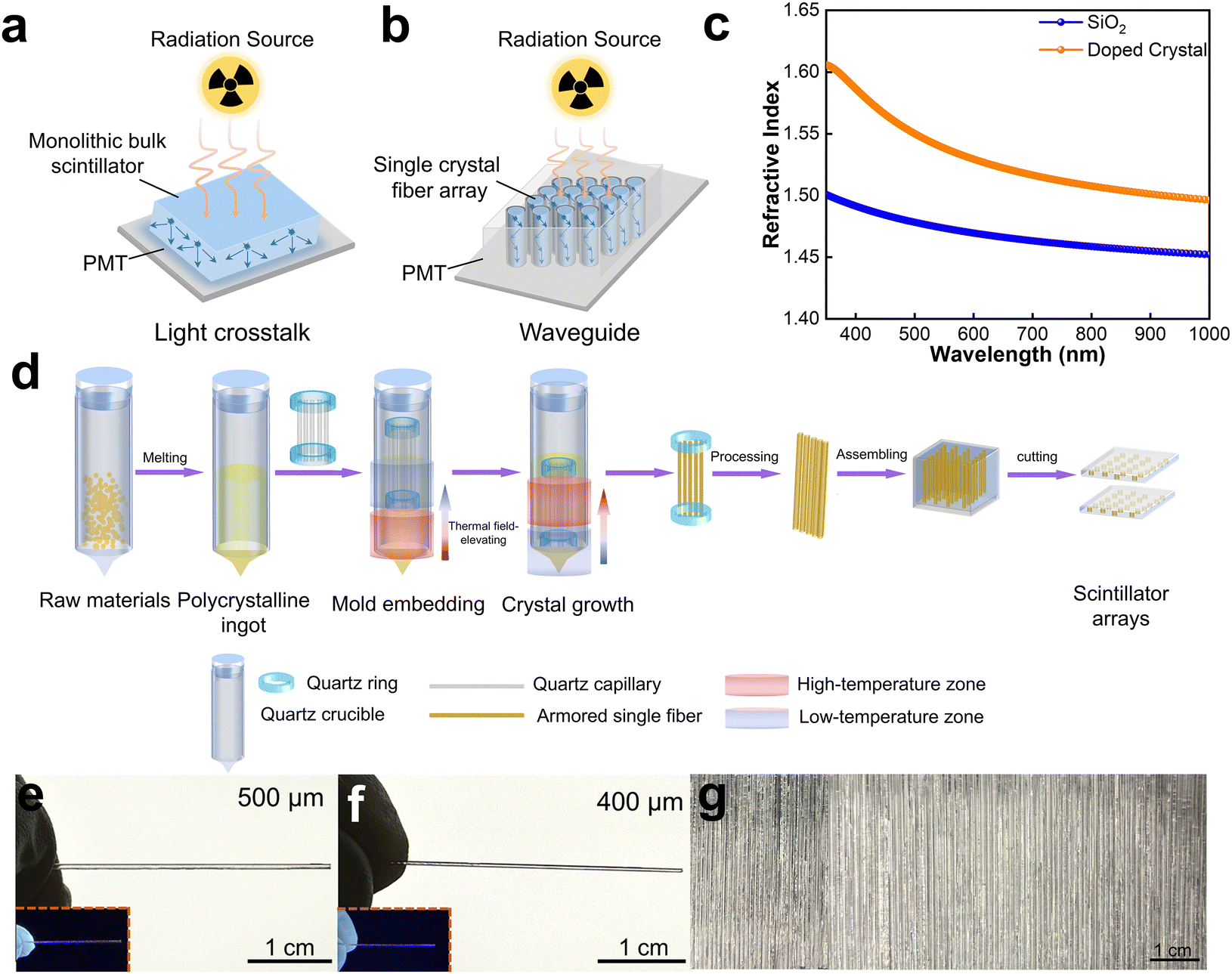 | ||
| Fig. 1 MEG protocols for organic single crystal fibers. Light propagation mechanisms in the monolithic bulk scintillation crystal (a) and SCF array (b). (c) Refractive indexes of doped crystals and SiO2. (d) Schematic of MEG, assembly of SCFs and fabrication of SCF arrays. (e) and (f) Photograph of SCFs with inner diameters of 500 and 400 μm, respectively. The insets are the corresponding fluorescence images under 365-nm excitation. (g) Photographs of 140 pieces of 0.01 wt% DPA-doped p-terphenyl SCFs as-grown by MEG in one batch. | ||
This study employed representative organic molecules, p-terphenyl as the host and 9,10-diphenylanthrance as the dopant, to grow doped organic scintillating single crystal fibers for the application of neutron detection. We selected a quartz capillary with a refractive index lower than that of the doped crystal as the cladding to suppress lateral light propagation and enhance the optical waveguide effect, as illustrated in Fig. 1c. The growth of the organic scintillating single crystal fiber was carried out using the MEG method. This method integrates melt growth with the confinement effect of the capillary, and is carried out using the thermal field-elevating technique previously reported by our group. As shown in Fig. 1d, firstly, p-terphenyl and DPA with a defined doping ratio were loaded into a quartz crucible and heated at 230 °C for two hours. After complete melting, the temperature was gradually reduced to room temperature to obtain a polycrystalline ingot. Subsequently, several hundred O2-plasma treated quartz capillaries (Fig. S1, ESI†), fixed with quartz rings at both ends, were carefully placed atop the polycrystalline ingot. Then, mold embedding was implemented by gradually lowering down the high-temperature zone while keeping the crucible still. The temperature of the high-temperature zone was set to 245 °C, while that of the low-temperature zone was maintained at 185 °C, with the growth rate of 0.5 mm h−1. As the polycrystalline ingot continued to melt, the molten substance was gradually drawn into the capillaries via capillary action. Once the capillaries were fully immersed in the melt, the growth of the scintillating single crystal fiber began. The crystallization began at the tip of the quartz crucible, with controlled nucleation to ensure uniform crystallographic orientation within all the fibers. Upon completion of the growth process, scintillating fibers armored by capillary molds were obtained. Finally, the scintillating fibers were assembled and encapsulated, resulting in scintillating fiber arrays. It is important to emphasize that the mold used is not separated from the scintillating single crystal fiber during processing, which provides two advantages: (1) organic crystals usually exhibit relatively poor mechanical properties and are difficult to process. The outer quartz capillary molds protect the inner crystal during subsequent processing. (2) By exploiting the refractive index difference between the mold and the organic crystal, the optical waveguiding effect can effectively suppress the lateral light propagation. While the mold is not removed, this does not significantly increase the overall cost. On the contrary, retaining the capillary simplifies the fabrication process, effectively eliminates the need for post-growth processing steps such as demolding and structural reinforcement, and provides mechanical support and environmental protection for the crystal. Fig. 1e and f present the DPA-doped p-terphenyl single crystal fibers with a doping ratio of 0.01 wt% grown using quartz capillaries with inner diameters of 0.5 mm and 0.4 mm. The fibers exhibit smooth surfaces and uniform transparency, reflecting their high crystallization quality. The insets are fluorescence images of the 0.01 wt% DPA-doped p-terphenyl scintillating single crystal fibers under 365-nm excitation. The organic scintillating single crystal fibers (SCFs) were grown using quartz capillaries with an inner diameter of 500 μm at different growth rates (0.5 mm h−1 and 1.0 mm h−1), as shown in Fig. S2 (ESI†). It is evident that a slower growth rate is found to produce SCFs with a smoother surface, free of cracks, exhibiting more pronounced extinction patterns under a polarizing microscope—an indication of enhanced crystalline quality. Fig. 1g shows 140 pieces of 0.01 wt% DPA-doped p-terphenyl scintillating single crystal fibers in one batch. The number of fibers is governed by the ratio of the outer diameter of the quartz capillary to the inner diameter of the growth crucible, and more higher throughput growth of scintillating single crystal fibers in one batch can be achieved easily by adjusting this diameter ratio.
2.2. Uniformity characterization of the scintillating single crystal fibers
To further investigate the crystallinity and uniformity of the scintillating single crystal fibers grown by MEG, a series of physical characterizations were performed. As shown in Fig. 2a, the optical microscopy images reveal the crack-free and smooth surface of a SCF at different positions. Under the polarizing microscope, consistent extinction patterns are observed upon rotation of the polarizer, indicating a uniform crystallographic orientation across the single crystal fiber. The Laue back-reflection measurements were conducted at three positions to assess the crystal quality. As shown in Fig. 2b, the diffraction patterns obtained from all three positions are bright, clear and uniform, indicating its nice crystallinity. The cross-sectional surface of a SCF of inner diameter 500 μm was examined using both optical microscopy (OM) and scanning electron microscopy (SEM). The images reveal a well-defined core-cladding configuration with a crack-free single crystal/quartz shell boundary (Fig. 2c and d). The elemental mapping of the cross-sectional surface illustrates that the C element is distributed in the inner crystal, while the Si and O elements are distributed in the quartz cladding, forming a clear boundary, as shown in Fig. 2d. To verify the crystalline orientation of the grown SCFs, X-ray diffraction (XRD) measurements were conducted along the cross-sectional surface of the fiber. As shown in Fig. 2e, compared to the standard XRD pattern, the SCFs exhibit prominent diffraction peaks corresponding to the (110), (111), (201), and (211) planes, with the (110) plane displaying the highest diffraction intensity. Among all the diffraction peaks, the (110) peak has the largest texture coefficient of 2.87 (Fig. 2f), indicating a preferred orientation of the (110) plane along the cross-sectional surface.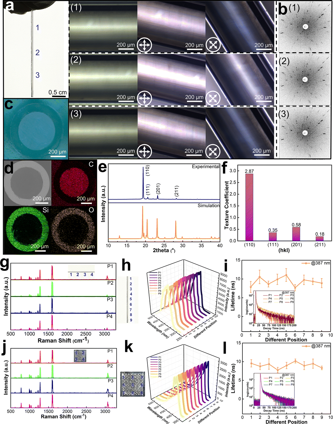 | ||
| Fig. 2 Characterization of SCFs and the SCF array. (a) The optical microscopy (left) and cross-polarized optical microscopy (right) images of different positions of a SCF with an inner diameter of 500 μm. (b) Laue patterns at different locations of a SCF. (c) Optical microscopy image of the cross-sectional surface of a SCF. (d) Scanning electron microscopy image and elemental mapping of the cross-sectional surface of a SCF. (e) The X-ray diffraction of the cross-section of a SCF. (f) The texture coefficients of all labeled diffraction peaks of a SCF. (g) Raman spectra of different locations of a SCF. (h) The photoluminescence spectra of different locations of a SCF. (i) The PL lifetimes at an emission wavelength of 387 nm for different locations of a SCF. The inset is the corresponding time-resolved PL spectra. (j) The Raman spectra at different locations of an SCF array. (k) The PL spectra at different locations of an array. (l) The PL lifetimes at an emission wavelength of 387 nm for different locations of an array. The inset is the corresponding time-resolved PL spectra. | ||
A SCF with an inner diameter of 500 μm was employed to perform Raman scattering measurements at different positions. As shown in Fig. 2g, the Raman peaks from four test positions are identical and consistent with previous reports, confirming the homogeneity of the chemical composition and crystallinity of the single crystal fiber. The photoluminescence (PL) spectra and PL lifetimes were also measured at various positions along the fiber. The PL emission intensity and peak position remain consistent across different positions under the consistent testing conditions (Fig. 2h). Additionally, the PL lifetimes are consistent across different positions at the same emission wavelength (Fig. 2i and Fig. S4, S5, ESI†). The inset shows the lifetime spectra at 387 nm at different positions along a SCF. These results further confirm the uniform crystallinity as well as the uniform distribution of the doped organic molecule DPA within the host matrix of p-terphenyl. Furthermore, to verify the uniformity between different SCFs, a certain number of fibers were assembled and encapsulated to form an array. Raman spectroscopy measurements were conducted at various positions on the array. The Raman peaks remain consistent (Fig. 2j). PL spectra and lifetime tests were also performed. The emission intensities and the fluorescence lifetimes are nearly identical across different positions (Fig. 2k, l and Fig. S6, S7, ESI†). The inset shows the fluorescence lifetime spectra at different positions of the fiber array at 387 nm. In conclusion, because all of the scintillating SCFs are from the same seed crystal, the uniformity across different fibers is effectively maintained. Such uniformity provides a foundation for the application of these SCFs in array-based devices.
2.3. Optical properties of a scintillating single crystal fiber
The optical properties of 0.01 wt% DPA-doped p-terphenyl SCFs were characterized further. DPA was selected as the guest molecule in p-terphenyl matrix to extend the emission range to align it with the sensitive range of PMT. As shown in Fig. 3a, the DPA solution with a concentration of 10−5 mol L−1 exhibits a broad absorption band ranging from 300 nm to 420 nm, with three main peaks at 355 nm, 375 nm, and 395 nm, which significantly overlaps with the PL emission spectrum of p-terphenyl. This spectral overlap favors efficient energy transfer from p-terphenyl to DPA. In addition, the lowest unoccupied molecular orbital (LUMO) of p-terphenyl is higher than that of DPA, while the highest occupied molecular orbital (HOMO) of p-terphenyl is lower than that of DPA, indicating feasible energy transfer from p-terphenyl to DPA (Fig. S8, ESI†). The PL spectra were measured for the doped scintillating single crystal fiber, p-terphenyl crystal, DPA solution and crystal, respectively. As shown in Fig. 3b, comparative analysis of the emission spectra from these samples demonstrates that the emission peaks of the doped crystal closely align with those of both the p-terphenyl crystal and DPA solution. This proves that DPA molecules have been successfully doped into the p-terphenyl matrix, where they predominantly exist in a monomeric form rather than aggregates. This is further validated through UV-Vis absorption spectra of the doped crystal and p-terphenyl crystal. The doped crystal exhibits two distinct absorption peaks of DPA at 372 nm and 394 nm in comparison to the absorption spectrum of p-terphenyl, as shown in Fig. 3c. | ||
| Fig. 3 Optical properties of SCFs. (a) The absorption and PL spectra of DPA and p-terphenyl (10−5 mol L−1 in CH2Cl2). (b) Comparison of the PL spectra of the doped crystal, p-terphenyl, DPA solution and DPA crystal. (c) Comparison of the absorption spectra of p-terphenyl and the doped crystal. (d) PL spectra of doped crystals with different concentrations of 0.01 wt%, 0.05 wt%, 0.1 wt% and 1 wt%. (e) PL quantum yields of crystals with different doping concentrations of 0 wt%, 0.01 wt%, 0.05 wt%, 0.1 wt% and 1 wt%. (f) The lifetime of a 0.01 wt%-doped SCF at 387 nm. | ||
For pure p-terphenyl, the overlap between its PL and absorption can lead to quenching of the scintillation light. Doping of DPA into the p-terphenyl host can shift the emission spectrum by leveraging Förster resonance energy transfer (FRET) from p-terphenyl to DPA to mitigate the quenching effect of the scintillation light. By carefully optimizing the DPA concentration, the extent of reabsorption can be alleviated while still maintaining efficient energy transfer. This helps preserve high radiative output without significant quenching. Moreover, by utilization of the waveguiding structure of the fiber geometry and cladding layer, the guided photon emission would minimize the reabsorption loss.
Furthermore, SCFs with different doping concentrations were grown, and their PL emission spectra, lifetimes, and photoluminescence quantum yields (PLQYs) were characterized. As shown in Fig. 3d, as the doping concentration increases, the energy transfer from p-terphenyl to DPA becomes more pronounced, resulting in a gradual reduction in the PL intensity of the p-terphenyl matrix while the emission intensity of the DPA guest increased. However, as the doping concentration increases to higher than 0.05 wt%, the overall PL intensity of the doped crystals decreases, which is likely due to fluorescence quenching caused by aggregation of the DPA molecules within the p-terphenyl matrix. The PLQYs of the SCFs with different doping concentrations were measured, as shown in Fig. 3e. The PLQY of the 0.01 wt% doped crystal was measured to be 80%, approximately 1.3 times that of p-terphenyl, and is the highest among the four tested samples with different doping concentrations. As shown in Fig. 3f and Fig. S9, S10 (ESI†), the fluorescence lifetime measurements for 0.01 wt% DPA-doped p-terphenyl were also performed at 387, 411, and 428 nm, with corresponding lifetimes of 8, 5, and 3 ns, respectively. Compared to those of commercial inorganic scintillators such as CsI:Tl, Bi4Ge3O12, and YAG:Ce, the exceptionally short lifetimes of organic scintillators significantly mitigate the influence of background noise on the detector, thereby improving the energy resolution.35
2.4. Neutron detection properties
In the domain of high-energy radiation detection, neutron detection relies on the (n, p) nuclear reaction between neutrons and light elements, which provides a unique advantage to organic scintillators with abundant carbon (C) and hydrogen (H) content.36 Therefore, an array of organic SCFs was fabricated and the neutron detection performances were investigated by the charge integration method using 252Cf as a radioactive source, which emits both neutrons and gamma rays. The figure of merit (FOM) was quantitatively determined to assess the neutron/gamma discrimination performance. The neutron/gamma pulse shape discrimination (PSD) and FOM are defined from the following eqn (1) and (2):| PSD = (Qlong − Qshort)/Qshort | (1) |
| FOM = (S/(δneutron + δgamma)) | (2) |
As shown in Fig. 4a, the neutron detection configuration consists of a 252Cf radioactive source, scintillator, PMT, multi-channel analyzer, and signal processing computer. The radioluminescence (RL) spectrum of the 0.01 wt% DPA-doped p-terphenyl SCF aligns well with the sensitive range of the PMT CR173-01 (Fig. 4b). As shown in Fig. 4c and e, the SCF array achieved better PSD with a gate time of 80 ns, where the separation between the neutron and gamma peaks was clearly pronounced. The calculated FOM value was 3.684 within the energy selection range of 300–700 channels (Table S1 and Fig. S11, ESI†). The inset shows the PSD plot in the energy selection range of 300–700 channels. As a contrast, the PSD of the monolithic bulk crystal with the same doping concentration was tested under identical conditions, and the results are shown in Fig. 4d and f. The FOM value was determined to be 2.824 within the energy selection range of 300–700 channels, which is lower than that of the SCF array. These results indicate that the scintillating SCFs effectively mitigate light crosstalk and enhance the optical waveguide effect, leading to improved neutron/gamma pulse shape discrimination performance in comparison to the monolithic bulk crystal.37 In addition to their utility in neutron/gamma pulse shape discrimination, the organic scintillating single crystal fiber array holds promise for soft X-ray imaging.
 | ||
| Fig. 4 Neutron detection properties comparison of the doped organic SCF array with the monolithic bulk crystal. (a) The setup of neutron detection. (b) The response plot of PMT and radioluminescence spectrum of the doped crystal. (c) Neutron/gamma PSD of the SCF array at a gate time of 80 ns. (d) Neutron/gamma PSD pattern of the monolithic bulk crystal at a gate time of 80 ns. (e) Calculation of the FOM value of the SCF array based on the PSD profile with an energy selection range of 300–700 channels. The inset is the corresponding neutron/gamma PSD pattern. (f) Calculation of the FOM value of the monolithic bulk crystal based on PSD profile with an energy selection range of 300–700 channels. The inset is the corresponding neutron/gamma PSD pattern. | ||
3. Conclusion
In conclusion, we report a high-throughput growth method for growing organic scintillating single crystal fibers, which can be directly used for fabricating pixelated scintillator arrays. The single crystal fibers grown by MEG exhibit homogeneous orientation, high crystallinity and satisfactory optical properties. Additionally, the single crystal fibers can be assembled into arrays. Exemplified by the growth of organic scintillator 9,10-diphenylanthrance-doped p-terphenyl, a large number of high-quality single crystal fibers were prepared and fiber arrays were assembled. Compared to the monolithic bulk scintillation single crystals, the fiber array shows superior neutron/gamma pulse shape discrimination, with the FOM value of 3.684, due to its lower lateral optical crosstalk and enhanced light-guiding effect. Although the current demonstrations are not yet abundant, their scalability to higher-throughput and more advanced products is undoubtedly anticipated.Author contributions
Xutang Tao and Yang Liu performed conceptualization; Xutang Tao, Yang Liu and Jinke Jiang performed methodology; Jinke Jiang, Leilei Zhang, Jiashuai Chen, Cuicui Li, Xin Ye, and Xiaoxin Zheng performed investigation; Yang Liu and Jinke Jiang wrote the original draft, reviewed and edited; Xutang Tao and Yang Liu performed funding acquisition; Huimin Li, Hanlin Zhao and Shuwen Wang provided resources. All authors discussed the results and commented on the manuscript.Data availability
The data supporting this article have been included as part of the ESI.†Conflicts of interest
There are no conflicts to declare.Acknowledgements
The authors gratefully acknowledge the financial support from the National Natural Science Foundation of China (Grant no. 52273185, 51973106, 52102006 and 51932004), the National Key Research and Development Program of China (Grant no. 2018YFB0406502), and the 111 Project 2.0 in China (Grant no. PB2018013). Yang Liu is thankful for the support from the Distinguished Young Scholars of Shandong Province (Grant no. ZR2019JQ03), and Qilu (Zhongying) Young Scholars. Xin Ye is thankful for the support from the Natural Science Foundation of Shandong Province (Grant no. ZR2021QE091) and the China Postdoctoral Science Foundation (Grant no. 2021M701973). Xiaoxin Zheng is thankful for the support from the State Key Laboratory of Particle Detection and Electronics (SKLPDE-KF-202414).References
- H. Wei and J. Huang, Nat. Commun., 2019, 10, 1066 CrossRef PubMed.
- L. Liu, M. Xu, X. Xu, X. Tao and Z. Gao, Adv. Mater., 2024, 36, 2406443 CrossRef CAS.
- J. Zhang, W. Li, Y. Yang, Z. Tang, H. Wei, J. Zhang and B. Yang, Adv. Funct. Mater., 2025, 2422003 CrossRef.
- J. Pang, H. Wu, H. Li, T. Jin, J. Tang and G. Niu, Nat. Commun., 2024, 15, 1769 CrossRef PubMed.
- Y. Hua, G. Zhang, X. Sun, P. Zhang, Y. Hao, Y. Xu, Y. Yang, Q. Lin, X. Li, Z. Zhai, F. Cui, H. Liu, J. Liu and X. Tao, Nat. Photonics, 2024, 18, 870–877 CrossRef CAS.
- X. Zhang, D. Chu, B. Jia, Z. Zhao, J. Pi, Z. Yang, Y. Li, J. Hao, R. Shi, X. Dong, Y. Liang, J. Feng, A. Najar, Y. Liu and S. (Frank) Liu, Adv. Mater., 2024, 36, 2305513 CrossRef CAS PubMed.
- D. Fu, Z. Chen, Y. Chang and X.-M. Zhang, Chem. Mater., 2023, 35, 9806–9816 CrossRef CAS.
- X. Li, X. Du, P. Zhang, Y. Hua, L. Liu, G. Niu, G. Zhang, J. Tang and X. Tao, Sci. China Mater., 2021, 64, 1427–1436 CrossRef CAS.
- W. Li, H. Li, J. Song, C. Guo, H. Zhang, H. Wei and B. Yang, Sci. Bull., 2021, 66, 2199–2206 CrossRef CAS PubMed.
- J. Wang, C. Wang, X. Sun, Z. Wang, Y. Li, H. Li, G. Ren and Y. Wu, J. Synth. Crys., 2023, 52, 1582–1588 CAS.
- Q. Wang, C. Wang, Z. Wang, X. Sun, M. Nikl, X. Ouyang and Y. Wu, J. Phys. Chem. Lett., 2022, 13, 9066–9071 CrossRef CAS PubMed.
- M. Xia, G. Niu, L. Liu, R. Gao, T. Jin, P. Wan, W. Pan, X. Zhang, Z. Xie, S. Teale, Z. Cai, J. Luo, S. Zhao, H. Wu, S. Chen, Z. Zheng, Q. Xie, X. Ouyang, E. H. Sargent and J. Tang, InfoMat, 2022, 4, e12325 CrossRef CAS.
- X. Hu, D. Rigamonti, I. Villa, L. Pollice, M. Mauri, A. D. Molin, M. Tardocchi, F. Meinardi, C. Weder and A. Monguzzi, Adv. Mater., 2024, 36, 2400443 CrossRef CAS.
- H. Ling, L. Xu, S. Chen, Y. Tang, H. Sun, X. Guo, Y. Feng and Q. Hu, J. Synth. Crys., 2024, 53, 1121–1126 CAS.
- Y. Li, L. Chen, B. Liu, P. Jin, R. Gao, L. Zhou, P. Wan, Q. Xu and X. Ouyang, J. Mater. Chem. C, 2021, 9, 17124–17128 RSC.
- S. Cheng, M. Nikl, A. Beitlerova, R. Kucerkova, X. Du, G. Niu, Y. Jia, J. Tang, G. Ren and Y. Wu, Adv. Opt. Mater., 2021, 9, 2100460 CrossRef CAS.
- B. Jia, D. Chu, N. Li, Y. Zhang, Z. Yang, Y. Hu, Z. Zhao, J. Feng, X. Ren, H. Zhang, G. Zhao, H. Sun, N. Yuan, J. Ding, Y. Liu and S. F. Liu, ACS Energy Lett., 2023, 8, 590–599 CrossRef CAS.
- B. Yang, X. Ouyang, X. Zhao, J. Su, Y. Li, S. Zhang and X. Ouyang, InfoMat, 2024, e12648 Search PubMed.
- Z. Sun, M. Gu, X. Liu, B. Liu, J. Zhang, S. Huang and C. Ni, Sci. Rep., 2022, 12, 8748 CrossRef CAS PubMed.
- H. Chen, M. Gu, Z. Sun, X. Liu, B. Liu, J. Zhang, S. Huang and C. Ni, Opt. Express, 2019, 27, 14871 CrossRef CAS.
- C. Roques-Carmes, N. Rivera, A. Ghorashi, S. E. Kooi, Y. Yang, Z. Lin, J. Beroz, A. Massuda, J. Sloan, N. Romeo, Y. Yu, J. D. Joannopoulos, I. Kaminer, S. G. Johnson and M. Soljačić, Science, 2022, 375, eabm9293 CrossRef CAS.
- B. Wang, X. Ouyang, X. He, Z. Deng, Y. Zhou and P. Li, Adv. Opt. Mater., 2023, 11, 2300388 CrossRef CAS.
- H. Dierks, Z. Zhang, N. Lamers and J. Wallentin, Nano Res., 2023, 16, 1084–1089 CrossRef CAS.
- Z. Zhang, H. Dierks, N. Lamers, C. Sun, K. Nováková, C. Hetherington, I. G. Scheblykin and J. Wallentin, ACS Appl. Nano Mater., 2022, 5, 881–889 CrossRef CAS.
- H. Li, H. Yang, R. Yuan, Z. Sun, Y. Yang, J. Zhao, Q. Li and Z. Zhang, Adv. Opt. Mater., 2021, 9, 2101297 CrossRef CAS.
- X. Zhao, T. Jin, W. Gao, G. Niu, J. Zhu, B. Song, J. Luo, W. Pan, H. Wu, M. Zhang, X. He, L. Fu, Z. Li, H. Zhao and J. Tang, Adv. Opt. Mater., 2021, 9, 2101194 CrossRef CAS.
- J. Hui, P. Ran, Y. Su, L. Yang, X. Xu, T. Liu and Y. “Michael” Yang, J. Phys. Chem. C, 2022, 126, 12882–12888 CrossRef CAS.
- Y. Teng, M. Gu, Z. Sun, X. Liu, B. Liu, J. Zhang, S. Huang and C. Ni, J. Lumin., 2023, 263, 120116 CrossRef CAS.
- W. Shao, T. He, L. Wang, J. Wang, Y. Zhou, B. Shao, E. Ugur, W. Wu, Z. Zhang, H. Liang, S. De Wolf, O. M. Bakr and O. F. Mohammed, Adv. Mater., 2024, 36, 2312053 CrossRef CAS PubMed.
- M. Hu, Y. Wang, S. Hu, Z. Wang, B. Du, Y. Peng, J. Yang, Y. Shi, D. Chen, X. Chen, Z. Zhuang, Z. Wang, X. Chen, J. Yang, Y. Ge, E. Wang, Q. Wen, S. Xiao, M. Ma, W. Li, J. Zhang, D. Ning, L. Wei, C. Yang and M. Chen, Nanoscale, 2023, 15, 15635–15642 RSC.
- Y. Zhou, Z. Deng, B. Wang, P. Li, L. Li, W. Han, J. Huang, W. Jia, X. Ouyang, Q. Xu and K. (Ken) Ostrikov, Chem. Eng. J., 2023, 471, 144431 CrossRef CAS.
- C. Li, X. Ye, J. Jiang, Q. Guo, X. Zheng, Q. Lin, C. Ge, S. Wang, J. Chen, Z. Gao, G. Zhang, X. Tao and Y. Liu, Small, 2024, 20, 2401624 CrossRef CAS.
- S. Cui, Y. Liu, G. Li, Q. Han, C. Ge, L. Zhang, Q. Guo, X. Ye and X. Tao, Cryst. Growth Des., 2020, 20, 783–792 CrossRef CAS.
- S. Cui, T. Zhu, L. Zhang, X. Ye, Q. Han, C. Ge, Q. Guo, X. Zheng, Q. Lin, C. Li, J. Jiang, W. Yuan, Y. Liu and X. Tao, Adv. Opt. Mater., 2022, 10, 2102355 CrossRef CAS.
- M. Chen, L. Sun, X. Ou, H. Yang, X. Liu, H. Dong, W. Hu and X. Duan, Adv. Mater., 2021, 33, 2104749 CrossRef CAS.
- L. Zhang, S. Cui, K. Wu, Z. Li, X. Zheng, Z. Wang and Y. Liu, Vacuum, 2024, 222, 113050 CrossRef CAS.
- T. Yanagida, K. Watanabe and Y. Fujimoto, Nucl. Instrum. Methods Phys. Res., Sect. A, 2015, 784, 111–114 CrossRef CAS.
Footnote |
| † Electronic supplementary information (ESI) available. See DOI: https://doi.org/10.1039/d5tc00844a |
| This journal is © The Royal Society of Chemistry 2025 |