Hybrid CsPbBr3 superlattice/Ag microcavity enabling strong exciton–photon coupling for low-threshold continuous-wave pumped polariton lasing†
Zhenxu
Lin
 ab,
Rui
Huang
*a,
Shulei
Li
*c,
Mingcheng
Panmai
d,
Yi
Zhang
a,
Haixia
Wu
a,
Jie
Song
a,
Zewen
Lin
a,
Hongliang
Li
a and
Sheng
Lan
*b
aSchool of Physics and Electronic Engineering, Hanshan Normal University, Chaozhou 521041, China. E-mail: rhuang@hstc.edu.cn
bGuangdong Provincial Key Laboratory of Nanophotonic Functional Materials and Devices, School of Information and Optoelectronic Science and Engineering, South China Normal University, Guangzhou 510006, China. E-mail: slan@scnu.edu.cn
cSchool of Optoelectronic Engineering, Guangdong Polytechnic Normal University, Guangzhou 510665, China. E-mail: shuleili@gpnu.edu.cn
dDivision of Physics and Applied Physics, School of Physical and Mathematical Sciences, Nanyang Technological University, Singapore, 637371, Singapore
First published on 23rd April 2025
Abstract
Achieving strong exciton–photon coupling in perovskite microcavities opens new possibilities for continuous-wave (CW) perovskite lasers with ultralow thresholds. A CsPbBr3 superlattice (SL), assembled from quantum dots (QDs) with a narrow size distribution, offers both large oscillator strengths and extended exciton dephasing times, rendering it a highly promising platform for enhanced light–matter interactions. Nevertheless, realizing robust exciton–photon coupling in a CsPbBr3 SL-based microcavity for low-threshold lasing remains elusive. Here, we demonstrate a hybrid microcavity integrating a CsPbBr3 SL with a thin Ag film to boost exciton–photon coupling and achieve CW-pumped polariton lasing. Using an acetone-assisted self-assembly approach, we obtain high-quality CsPbBr3 SLs characterized by narrow emission linewidths, large exciton binding energies, diminished exciton–phonon coupling, and highly stable amplified spontaneous emission. Optical scattering and photoluminescence measurements indicate significant coupling between the SL excitons and resonant photon modes in the CsPbBr3/Ag microcavity. We attribute this enhanced light–matter interaction to comparable linewidths of the exciton resonance and photon mode, facilitated by the Ag film. A coupled oscillator model fit yields a Rabi splitting of approximately 225 meV in a large microcavity. Notably, we achieve CW-pumped polariton lasing near the lower polariton branch bottleneck at a low threshold of about 220 W cm−2. Our findings elucidate the fundamental mechanism underlying strong exciton–photon coupling in CsPbX3 SL systems and offer a viable strategy for designing CW-pumped polariton lasers with improved performance.
Introduction
Strong exciton–photon coupling in a cesium lead halide (CsPbX3, X = Cl, Br, I) perovskite microcavity leads to the formation of microcavity polaritons with part-matter and part-light features.1–3 Being as bosons with low effective mass and strong coherence, microcavity polaritons emerge as a promising candidate for exploring room temperature collective phenomena, such as Bose–Einstein condensation and polariton lasing etc. This unique feature makes it possible to realize a polariton laser with a threshold many orders of magnitude lower than that of traditional photonic lasers, which is limited by the population inversion condition.4–8 The dramatic reduction in lasing threshold is beneficial to the realization of continuous wave (CW) pumped CsPbX3 lasers. So far, CW-pumped polariton lasing has been achieved in planar micro-/nano cavities based on single crystalline CsPbX3.9–11 Very recently, Song et al. demonstrated a CW-pumped CsPbBr3 laser by using a microplatelet with an area smaller than 1 μm2. The low threshold of ∼0.84 kW cm−2 was achieved by exploiting the strong exciton–photon coupling.11 From the viewpoint of practical application, however, a further improvement is still necessary in order to realize a CW-pumped polariton laser with an ultralow threshold at room temperature.In recent years, CsPbX3 quantum dots (QDs) have attracted great interest because of their high photoluminescence quantum yields (PLQYs) and narrow emission line widths.12–14 As compared with bulk materials, low-dimensional CsPbX3, such as QDs, possess larger exciton binding energy (Eb) resulting from the weaker dielectric screening and higher confinement effect, facilitating the formation of exciton–photon polaritons at room temperature.15–17 Apart from the larger Eb, the reduced dielectric screening in low-dimensional CsPbX3 leads to an enhancement in exciton oscillator strength. Since the exciton–photon coupling strength g is governed by the relation  where N is the number of excitons, f is the exciton oscillator strength and V is the photonic mode volume, it is believed that a microcavity composed of CsPbX3 QDs can be employed to enhance the exciton–photon interaction, leading to a large Rabi splitting energy (Ω).18 Remarkably, formation exciton–photon polaritons with a Rabi splitting energy of Ω ∼ 127.5 meV has been demonstrated in a microcavity composed of CsPbX3 QD monolayers and a planar optical microcavity created by the combination of metallic mirrors and distributed Bragg reflectors.19 In this case, the coupling strength can be modified by the relative position between the excitonic and photonic modes. It indicates that the combination of an optical microcavity and CsPbX3 QDs microcavity has become an ideal platform for realizing strong exciton–photon coupling. However, it is noticed that a dense film composed of CsPbX3 QDs with non-uniform size distribution and defects, which is reflected in the inhomogeneous broadening of the PL spectrum, may result in enhanced exciton–phonon coupling and fast exciton dephasing, preventing the strong coupling between excitons and photons.
A CsPbX3 superlattice (SL) formed by the self-assembly of CsPbX3 QDs seems to be a promising candidate to overcome this shortage because it possesses long-range order and narrow size distribution of QDs.20–25 The overlapping of electronic wave functions of QDs in the CsPbX3 SL results in the delocalization of excitons, increasing exciton oscillator strength as compared with isolated QDs. Moreover, the strong electronic coupling between QDs in the CsPbX3 SL leads to a low dephasing rate, which is manifested in the excitonic cooperative emission from the CsPbX3 SL. This phenomenon, which is referred to as superfluorescence, has been demonstrated in CsPbBr3 SLs.26 Apart from the high-density exciton states and low dephasing rate, a CsPbBr3 SLs with regular geometric shape usually supports Mie resonances, whispering gallery modes (WGMs) and Fabry–Perot (F–P) resonances, behaving as a high-quality optical microcavity with low intrinsic loss. As a result, the interaction between light and matter can be significantly enhanced, leading to enhanced superfluorescence.27 However, less attention has been paid to the strong coupling between excitons and photons in microcavities based on CsPbBr3 SLs, which can be exploited to realize CW-pumped polariton lasers.
Basically, the Rabi splitting energy Ω originating from the strong exciton–photon coupling is defined as follows:
 | (1) |
In this work, we successfully synthesized high-quality CsPbBr3 SLs by using the self-assembly of QDs assisted by acetone. We observed stable exciton emission and weak exciton–phonon coupling in the CsPbBr3 SLs by using power- and temperature-dependent PL spectra. We constructed hybrid microcavities by placing CsPbBr3 SLs with different sizes on a thin Ag film and identified optical modes with high quality factors originating from the coupling of high-order Mie resonances and WGMs supported by CsPbBr3 cuboids. We demonstrated the strong exciton–photon coupling with a Rabi splitting as large as ∼225 meV in CsPbBr3/Ag hybrid microcavities and the realization of CW-pumped polariton lasers with a low threshold of ∼220 W cm−2.
Results and discussion
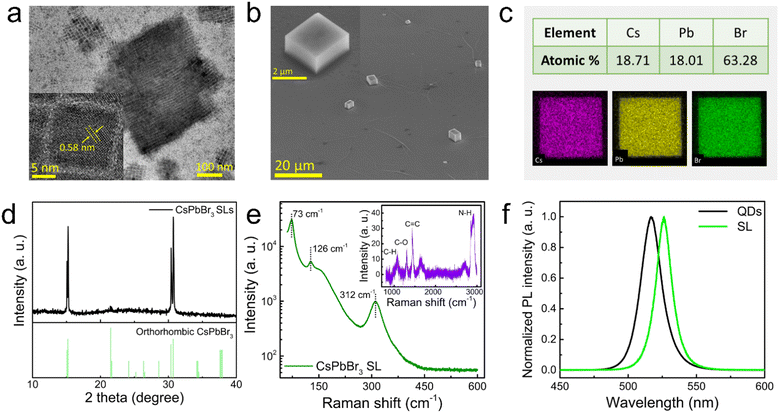
The CsPbBr3 SLs used in this study were prepared by the self-assembly of monodispersed QDs in hexane solution assisted by polar solvent (here acetone) (see Fig. S1(a), ESI†). The narrow size distribution of QDs and the surface ligands on QDs play a crucial role in the formation of CsPbBr3 SLs. Fig. 1(a) shows the high-resolution transmission electron microscope (HR-TEM) image of pristine monodispersed CsPbBr3 QDs with an average size of ∼10 nm, which were synthesized by using typical high temperature hot injection following the procedure reported in literature.12 As shown in the HR-TEM image, clear lattice fringes with an interplane spacing of ∼0.58 nm, which is assigned to the (110) plane in the orthorhombic crystal of CsPbBr3, indicate the good crystallinity of the QDs.30 Basically, the self-assembly of QDs is driven by the intermolecular forces between aliphatic ligands.31 Here, the ligand molecules decorated on the surfaces of QDs, which are extremely sensitive to polar solvent, are employed to fabricate CsPbBr3 SLs. The fabricated CsPbBr3 SLs appear as cuboids with micrometer edges, as shown in Fig. 1(b) (see also Fig. S1(c)–(e), ESI†). The elemental mapping of a typical CsPbBr3 SL based on energy dispersive spectrocopy (EDS) is presented in Fig. 1(c). It indicates uniform spatial distributions for each element. The atomic ratio of Cs![[thin space (1/6-em)]](https://www.rsc.org/images/entities/char_2009.gif) :
:![[thin space (1/6-em)]](https://www.rsc.org/images/entities/char_2009.gif) Pb
Pb![[thin space (1/6-em)]](https://www.rsc.org/images/entities/char_2009.gif) :
:![[thin space (1/6-em)]](https://www.rsc.org/images/entities/char_2009.gif) Br is very close to the stoichiometry of 1
Br is very close to the stoichiometry of 1![[thin space (1/6-em)]](https://www.rsc.org/images/entities/char_2009.gif) :
:![[thin space (1/6-em)]](https://www.rsc.org/images/entities/char_2009.gif) 1
1![[thin space (1/6-em)]](https://www.rsc.org/images/entities/char_2009.gif) :
:![[thin space (1/6-em)]](https://www.rsc.org/images/entities/char_2009.gif) 3 (the small deviation of the atomic ratio from the stoichiometry is caused by instrumental error). The X-ray diffraction (XRD) pattern of a CsPbBr3 SL, which is shown in Fig. 1(d), exhibits two double peaks located at ∼15.3° and ∼30.6°, which are assigned to the diffractions from the (110) and (220) faces of the orthorhombic CsPbBr3 (ICSD 97851), respectively.32 This feature, which is consistent with monodispersed QDs, confirms the existence of pure orthorhombic phase of CsPbBr3 in the CsPbBr3 SL. In order to characterize the local atomic environment and bonding configuration a CsPbBr3 SL, we also examined the Raman spectrum of a SL, as shown in the Fig. 1(e). The three Raman modes with small wave numbers revealed in the Raman spectrum (a strong mode at 73 cm−1 and two weak modes at 126 and 312 cm−1) are assigned to the vibrational modes of the CsPbBr3 sublattice.33 In addition, the four intense bands with large wave numbers, which are located at 1085, 1305, 1440, and 2915 cm−1, are assigned to the bending vibrations of C–H, C–O, C
3 (the small deviation of the atomic ratio from the stoichiometry is caused by instrumental error). The X-ray diffraction (XRD) pattern of a CsPbBr3 SL, which is shown in Fig. 1(d), exhibits two double peaks located at ∼15.3° and ∼30.6°, which are assigned to the diffractions from the (110) and (220) faces of the orthorhombic CsPbBr3 (ICSD 97851), respectively.32 This feature, which is consistent with monodispersed QDs, confirms the existence of pure orthorhombic phase of CsPbBr3 in the CsPbBr3 SL. In order to characterize the local atomic environment and bonding configuration a CsPbBr3 SL, we also examined the Raman spectrum of a SL, as shown in the Fig. 1(e). The three Raman modes with small wave numbers revealed in the Raman spectrum (a strong mode at 73 cm−1 and two weak modes at 126 and 312 cm−1) are assigned to the vibrational modes of the CsPbBr3 sublattice.33 In addition, the four intense bands with large wave numbers, which are located at 1085, 1305, 1440, and 2915 cm−1, are assigned to the bending vibrations of C–H, C–O, C![[double bond, length as m-dash]](https://www.rsc.org/images/entities/char_e001.gif) C, and N–H bonds, respectively.34–36 This result implies that oleic acid/oleylamine ligands are remained on the surfaces of QDs after the self-assembly. This unique feature renders CsPbBr3 SLs excellent environmental and structural stability. As compared with the PL spectrum of QDs, the PL spectrum of the SL exhibited a red shift of the PL peak from 513 to 526 nm, which is attributed to the electronic coupling of the wavefunctions in neighboring QDs of the SL.37 In addition, a reduction in the full width at half maximum (FWHM) (from 18 to 13 nm) was observed in the SL because of the narrow size distribution of QDs. Moreover, remarkable two-photon-induced photoluminescece (TPL) was observed for the SL excited by femtosecond laser pulses and the polarization dependence of the TPL is similar to that observed in single-crystal CsPbBr3 resulting from anisotropic crystal structure (Fig. S2, ESI†). All these results indicate clearly the good crystallinity of the fabricated CsPbBr3 SLs. More importantly, the PL from the CsPbBr3 SLs remains stable for more than 60 min at room temperature under the excitation of a 325-nm CW laser light with a power density of 140 W cm−2 (Fig. S3, ESI†).
C, and N–H bonds, respectively.34–36 This result implies that oleic acid/oleylamine ligands are remained on the surfaces of QDs after the self-assembly. This unique feature renders CsPbBr3 SLs excellent environmental and structural stability. As compared with the PL spectrum of QDs, the PL spectrum of the SL exhibited a red shift of the PL peak from 513 to 526 nm, which is attributed to the electronic coupling of the wavefunctions in neighboring QDs of the SL.37 In addition, a reduction in the full width at half maximum (FWHM) (from 18 to 13 nm) was observed in the SL because of the narrow size distribution of QDs. Moreover, remarkable two-photon-induced photoluminescece (TPL) was observed for the SL excited by femtosecond laser pulses and the polarization dependence of the TPL is similar to that observed in single-crystal CsPbBr3 resulting from anisotropic crystal structure (Fig. S2, ESI†). All these results indicate clearly the good crystallinity of the fabricated CsPbBr3 SLs. More importantly, the PL from the CsPbBr3 SLs remains stable for more than 60 min at room temperature under the excitation of a 325-nm CW laser light with a power density of 140 W cm−2 (Fig. S3, ESI†).
It is well known that the PL from CsPbBr3 QDs at room temperature originates from the radiative recombination of excitons with a high rate because the exciton binding energy in CsPbBr3 QDs (40–200 meV) is larger than the thermal energy (∼26 meV).38–40 In order to understand the recombination dynamics of CsPbBr3 SLs, we measured the PL spectra of a SL at different excitation power densities, as shown in Fig. 2(a). Physically, the dominant recombination process in a semiconductor with direct bandgap is generally revealed in the relationship between the integrated PL intensity (IPL) and the excitation power density (Iex), which is expressed as follows:41
| IPL ∼ Ikex. | (2) |
Here, k is a parameter associated with the dominant recombination process. In general, k < 1, 1 < k < 2, and k > 2 represent a free-to-bound recombination or donor–acceptor pair recombination, an excitonic recombination, and a free-carrier recombination, respectively. We examine the relationship between IPL and Iex for a typical SL plotted in a logarithmic coordinate, as shown in the inset of Fig. 2(a). In this case, the value of k extracted from the dependence of IPL on Iex slope is ∼1.29, indicating that excitonic recombination is the dominant recombination process in the SL. This value is larger than that observed in QDs (k ∼ 0.99, see Fig. S4, ESI†), implying an increased radiative excitonic recombination rate in the SLs. This conclusion is also supported by the longer PL lifetime observed in the SL (Fig. S5, ESI†). We also compared the temperature-dependent PL spectra of CsPbBr3 QDs and SLs, as shown in Fig. 2(b) and Fig. S6 (ESI†). Basically, the temperature-dependent PL intensity IPL(T) of a CsPbBr3 SL can be fitted by the Arrhenius equation expressed as follows:42
 | (3) |
Here, IPL(T0) is the PL intensity at 80 K, β is a constant, kB is the Boltzmann constant, and Eb is exciton binding energy, respectively. The exciton binding energy in the SL derived from the fitting of the experimental data by using with the Arrhenius equation is found to be Eb ∼ 76 meV. This value is larger than that observed in bulk CsPbBr3 microplatelets (Fig. S7, ESI†), implying a robust excitonic transition at room temperature. The large Eb is beneficial for the realization of population inversion and amplified stimulated emission (ASE) with a low threshold. As a result, we observed two-photon-pumped ASE (λ = ∼531 nm) with a low threshold (Pth ∼ 0.9 mJ cm−2) in a SL at room temperature under the excitation of 800-nm femtosecond laser pulses (1 kHz, 130 fs), as shown in Fig. S8(a) and (b) (ESI†). More interestingly, the intensity of ASE remains stable for more than 60 min under a pumping power density of 1.45Pth (Fig. S8(c), ESI†), implying a potential application for lasing application. In Fig. 2(d), we present the evolution of the PL peak with increasing temperature observed for CsPbBr3 QDs and SLs. Basically, the temperature-dependent PL peak (or bandgap energy) is related to the thermal expansion (TE) and exciton–phonon (EP) interaction and it can be described by the following equation:43,44
 | (4) |
Basically, a CsPbBr3 SL with a regular geometric shape and a moderate refractive index (n = 2.3) can serve as an optical microcavity that provides necessary optical feedback for light amplification. Previous studies have demonstrated that cavity-enhanced superfluorescence can be achieved in a CsPbBr3 SL assembled by from CsPbBr3 QDs.27 Thus, it would be interesting to find out whether the optical modes supported by a CsPbBr3 SL microcavity can be exploited to the enhance light–matter interaction. In Fig. S9 (ESI†), we show the optical resonances observed in the forward scattering spectra of SLs with different sizes placed on a SiO2 substrate by using polarized white light and analyzer with cross polarization. It was previously reported that the coupling between a narrow exciton resonance and a broader cavity mode lead to the formation of asymmetric Fano resonance, making it difficult to reveal the larger Rabi splitting from exciton–photon coupling.45 Based on previous studies, the light–matter interaction can be greatly enhanced by constructing a hybrid cavity composed of a CsPbBr3 particle and a thin metal film.46–48 In particular, a significantly larger electric field enhancement factor within all-dielectric microcavities was observed for the Ag/SiO2 substrate, attributed to the lower optical loss of Ag.29 In this work, we created hybrid microcavities by synthesizing CsPbBr3 SLs directly on a Ag/SiO2 substrate with the conventional spin coating method. In Fig. 3(a), we show the forward scattering spectra of CsPbBr3 SLs with different side length (L) placed on a Ag/SiO2 substrate. In each case, one can identify a scattering dip in the scattering spectrum at the exciton resonance, which is resulted from the coherent coupling between the exciton resonance and the high-order Mie resonances supported by the hybrid micro-cavity. As a result, the radiative recombination of excitons can be enhanced by the Purcell effect, leading to the enhancement in the PL intensity. The corresponding electric field distribution simulation in the XZ plane calculated for such a hybrid microcavity is shown in Fig. S10 (ESI†). Here, the L and thickness of the CsPbBr3 SL are assumed to be 2 μm and 0.5 μm, respectively. It is shown that the electric field distribution appears inside the CsPbBr3 SL as a standing wave in both the x and z directions. In our case, the optical modes supported by the hybrid microcavity may originate from the coherent interaction between the high-order Mie resonances and the WGMs or F–P resonances. It is found that the optical resonances become denser and sharper with increasing L of the SL because of the reduced optical loss, indicating that the reduced photonic mode volume for such hybrid micro-cavities. In general, the narrower linewidths or higher quality factors of the optical resonances are favorable for promoting strong coupling between the excitons and photons, creating exciton–photon polaritons. Therefore, it is expected that such hybrid micro-cavities are promising candidates for realizing CW-pumped pervoskite lasers with low thresholds.
In Fig. 3(b), we present the normalized PL spectra of the CsPbBr3 SLs with L ranging from 1 to 6 μm placed on an Ag/SiO2 substrate under the excitation of 405-nm CW laser light with a power density of 528 W cm−2. For the CsPbBr3 SLs with L < 2 μm, one can see only a narrow PL band without any resonant modes. In this case, the radiation loss of the micro-cavity is relatively large and the coupling between the excitons and photons is weak. As revealed in the scattering spectra of the SLs with small sizes (see Fig. 3(a)), the optical resonance is much broader than the linewidth of the exciton resonance. It is noticed that the multiple optical modes with narrow linewidths emerge in the scattering spectra with increasing size, indicating the enhanced exciton–photon coupling. In Fig. 3(c), we present the PL spectrum of a SL with L = 5 μm under the excitation of 405-nm CW laser light at room temperature. It is noticed that the emission peaks at the low-energy side of the PL spectrum can be well fitted by multiple Lorentz lineshapes. In addition, the free spectral range (FSR) decreases gradually when the photon energy approaches the exciton resonance (Ex), the. This feature is in good agreement with the exciton–photon polariton model, as demonstrated in previous reports.49 In each case, the dispersion of the polaritons in the in the lower polariton branch (LPB) can be analyzed by using the Hamiltonian for the exciton–photon coupling (see Fig. S11, ESI†).50 In Fig. 3(d), we show the fitting of the dispersion relation characterized by the dependence of the polariton energy (E) versus the in-plane wavevector (k‖). The Rabi splitting energy extracted from the fitting of the dispersion relation is estimated to be Ω ∼ 225 meV, which is larger than that reported in CsPbBr3 microplatelets.11 It implies the existence of strong exciton–photon coupling in such hybrid cavities. Moreover, the PL spectra modulated by cavities and the fitting results of Rabi splitting energies for the SLs with L ranging from 3 to 5.5 μm are shown in Fig. S12 and S13 (ESI†), respectively. As L is increased from 3 to 5.5 μm, the Rabi splitting energy is increased gradually from 192 to 237 meV. This indicates that the improvement of the quality factors of the optical resonances supported by a CsPbBr3 SL with increasing L facilitates the coupling with the exciton resonance. For a hybrid microcavity, the exciton–photon coupling strength depends on the exciton oscillator strength, the photonic mode volume, and the linewidth match between the optical mode and the exciton resonance. As compared with bulk CsPbBr3, the oscillator strength of the excitons in a CsPbBr3 SL is increased by the coherent coupling of excitons induced by the reduced interdot distance and EP interaction. As a result, the exciton–photon coupling is enhanced, resulting in a larger Rabi splitting energy. On the other hand, it is well known that the effective mode volume is inversely proportional to the quality factor of the photonic mode. The confinement of light in the microcavity is governed by the group refractive index ng (Fig. S14, ESI†). When L is increased from 2.5 to 4.25 μm, the group refractive index ng is increased from 15 to 26.4. It implies that the light confinement becomes more efficient in large SLs. Therefore, a stronger exciton–photon coupling can be achieved in CsPbBr3 SLs with large sizes (L > 2 μm), which is benefit for the realization of polariton lasing under the excitation of CW laser light.
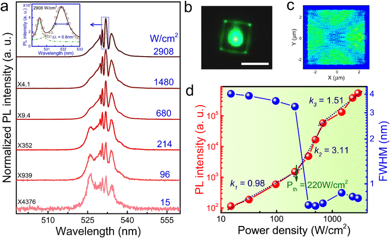
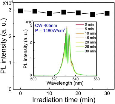
In Fig. 4(a), we show the PL spectra of a large CsPbBr3 SL with L = 4.5 μm (the thickness is 0.6 μm, see Fig. S15, ESI†) obtained at different excitation power densities. The CsPbBr3 SL was excited by using 405-nm CW laser light at 80 K. At a power density of 15 W cm−2, the PL spectrum is dominated by a broad SE peak centered at 526.5 nm with a FWHM of ∼4 nm. Meanwhile, several narrow oscillation peaks with nonidentical spacing, which are attributed to the optical modes selectively amplified by the optical gain in a square CsPbBr3 SL, appear on the low energy side of the SE peak. From the optical image of the SL (see Fig. 4(b)), one can see bright green light emission from four corners of the SL, suggesting that the dominant optical mode is the WGMs supported by the CsPbBr3 SL.51 This is consistent with the electric field distribution calculated for the hybrid microcavity, which shows notable leakage at the four corners (see Fig. 4(c)). In Fig. 4(d), one can see a rapid increase in the PL intensities of the oscillation peaks when the power density is further is increased. As the power density reaches 220 W cm−2, the dependence of the PL intensity on the excitation power density exhibits a transition from a sublinear to a superlinear relationship. Moreover, the linewidth of dominant emission peak is dramatically reduced to ∼0.8 nm, indicating clearly the appearance of multi-mode lasing (see the inset of Fig. 4(a)). In addition, it was found that the decay time drops rapidly to the order of picoseconds (∼380 ps) at a higher excitation fluence of 10 mJ cm−2 (see Fig. S16, ESI†), further confirming the lasing behavior. Thus, the lasing threshold is estimated to be Pth (∼220 W cm−2). To the best of our knowledge, the lasing threshold observed in this work is smaller than those reported in bulk CsPbBr3 polariton laser,10,11 indicating the important role of strong exciton–photon coupling achieved by using a hybrid microcavity composed of a CsPbBr3 SL and a Ag thin film. Furthermore, we have also verified that the lasing behavior observed in the CsPbBr3 SLs is reproducible (see Fig. S17, ESI†), indicating the stability of CsPbBr3 SLs under laser illumination. More interestingly, it was found that the PL intensity from such a hybrid microcavity remains stable under continuous irradiation of 405-nm CW laser with a power density of 1480 W cm−2 for at least 30 min (see Fig. 5). All these phenomena indicate that such CsPbBr3/Ag hybrid microcavities exhibit potential applications in realizing polariton lasers with low threshold and high stability. For comparison, we also examined the lasing behavior of a CsPbBr3 SL with a similar L but with a larger thickness of ∼1.1 μm (see Fig. S18, ESI†). As shown in Fig. S18(b) (ESI†), the lasing modes in the CsPbBr3 SL exhibit a highly degree of polarization (DOP = 0.66), which is close to that reported in single-crystal CsPbBr3 laser.11 It was found that the lasing threshold Pth is increased to ∼300 W cm−2 (see Fig. S18(c), ESI†). As shown in the optical image of the thicker CsPbBr3 SL (see the inset of Fig. S18(d), ESI†), light is emitted from the four corners and the centers of four edges of the CsPbBr3 SL. In the thicker CsPbBr3 SL, one need to consider the F–P cavity formed by the up and bottom facets. The interplay of the WGMs and the F–P modes may degrade the light confinement in the CsPbBr3 SL, leading to a higher lasing threshold.52
Conclusions
In summary, we have successfully synthesized CsPbBr3 SLs with outstanding optical properties by using acetone-assisted self-assembly. It was found that such SLs possess narrow emission linewidths, stable exciton emission and weaker EP coupling, which were identified by excitation-power and temperature-dependent PL spectra. This unique feature makes it possible to realize strong exciton–photon coupling in SLs. We proposed the use of a hybrid microcavity composed of a SL and an Ag thin film to greatly enhanced exciton–photon coupling, resulting in a Rabi splitting as large as ∼225 meV. We demonstrated polariton lasing in hybrid microcavities with a low threshold of ∼220 W cm−2 under the excitation of CW laser light. Our findings are helpful for understanding the strong exciton–photon coupling in CsPbX3 SLs and useful for designing polariton lasers with low thresholds.Author contributions
R. Huang and S. Lan revised paper; Z. Lin and S. Li engineered the experiments and wrote the first draft of the manuscript; H. Wu, J. Song, Z. Lin, Y. Zhang and H. Li prepared the samples and performed the photoluminescence measurements. M. Panmai and S. Li conducted numerical simulation. All authors contributed to the analysis of data and general discussion.Data availability
The authors declare that all data supporting the results reported in this study are available within the paper and the ESI.† Additional data used for the study are available from the corresponding authors upon reasonable request.Conflicts of interest
There are no conflicts to declare.Acknowledgements
The authors acknowledge the financial support from the National Natural Science Foundation of China (Grant No. 11874020, 12174123, 12374347 and 12104117), Guangdong Basic and Applied Basic Research Foundation (Grant No. 2022A1515010747), Start-Up Funding of Guangdong Polytechnic Normal University (2022SDKYA007), Project of Guangdong Province Key Discipline Scientific Research Level Improvement (2021ZDJS039, 2024ZDZX1026) and Program of Hanshan Normal University (XTP202402).Notes and references
- X. Wang, M. Shoaib, X. Wang, X. Zhang, M. He, Z. Luo, W. Zheng, H. Li, T. Yang, X. Zhu, L. Ma and A. Pan, ACS Nano, 2018, 12, 6170–6178 CrossRef CAS
.
- W. Du, S. Zhang, J. Shi, J. Chen, Z. Wu, Y. Mi, Z. Liu, Y. Li, X. Sui, R. Wang, X. Qiu, T. Wu, Y. Xiao, Q. Zhang and X. Liu, ACS Photonics, 2018, 5, 2051–2059 CrossRef CAS
.
- R. Su, A. Fieramosca, Q. Zhang, H. S. Nguyen, E. Deleporte, Z. Chen, D. Sanvitto, T. C. H. Liew and Q. Xiong, Nat. Mater., 2021, 20, 1315–1324 CrossRef CAS PubMed
.
- T. Byrnes, N. Y. Kim and Y. Yamamoto, Nat. Phys., 2014, 10, 803–813 Search PubMed
.
- H. Dong, C. Zhang, X. Liu, J. Yao and Y. S. Zhao, Chem. Soc. Rev., 2020, 49, 951–982 RSC
.
- R. Su, C. Diederichs, J. Wang, T. C. H. Liew, J. Zhao, S. Liu, W. Xu, Z. Chen and Q. Xiong, Nano Lett., 2017, 17, 3982–3988 CrossRef CAS
.
- Q. Han, J. Wang, J. Lu, L. Sun, F. Lyu, H. Wang, Z. Chen and Z. Wang, ACS Photonics, 2020, 7, 454–462 CrossRef CAS
.
- M. A. Masharin, A. K. Samusev, A. A. Bogdanov, I. V. Iorsh, H. V. Demir and S. V. Makarov, Adv. Funct. Mater., 2023, 33, 2215007 CrossRef CAS
.
- T. J. S. Evans, A. Schlaus, Y. Fu, X. Zhong, T. L. Atallah, M. S. Spencer, L. E. Brus, S. Jin and X. Y. Zhu, Adv. Opt. Mater., 2018, 6, 1700982 CrossRef
.
- Q. Shang, M. Li, L. Zhao, D. Chen, S. Zhang, S. Chen, P. Gao, C. Shen, J. Xing, G. Xing, B. Shen, X. Liu and Q. Zhang, Nano Lett., 2020, 20, 6636–6643 CrossRef CAS
.
- J. Song, Q. Shang, X. Deng, Y. Liang, C. Li, X. Liu, Q. Xiong and Q. Zhang, Adv. Mater., 2023, 35, 2302170 CrossRef CAS PubMed
.
- L. Protesescu, S. Yakunin, M. I. Bodnarchuk, F. Krieg, R. Caputo, C. H. Hendon, R. X. Yang, A. Walsh and M. V. Kovalenko, Nano Lett., 2015, 15, 3692–3696 CrossRef CAS
.
- S. Yakunin, L. Protesescu, F. Krieg, M. I. Bodnarchuk, G. Nedelcu, M. Humer, G. D. Luca, M. Fiebig, W. Heiss and M. V. Kovalenko, Nat. Commun., 2015, 6, 8056 CrossRef CAS
.
- J. Chen, W. Du, J. Shi, M. Li, Y. Wang, Q. Zhang and X. Liu, InfoMat, 2020, 2, 170–183 CrossRef CAS
.
- S. Zhang, Y. Zhong, F. Yang, Q. Cao, W. Du, J. Shi and X. Liu, Photonics Res., 2020, 8, A72–A90 CrossRef CAS
.
- C. Bujalance, L. Calio, D. N. Dirin, D. O. Tiede, J. F. G. López, J. Feist, F. J. G. Vidal, M. V. Kovalenko and H. Míguez, ACS Nano, 2024, 18, 4922 Search PubMed
.
- D. Mao, L. Chen, Z. Sun, M. Zhang, Z. Y. Shi, Y. Hu, L. Zhang, J. Wu, H. Dong, W. Xie and H. Xu, Light: Sci. Appl., 2024, 13, 34 Search PubMed
.
- L. K. Van Vugt, B. Piccione, C. H. Cho, P. Nukala and R. Agarwal, Proc. Natl. Acad. Sci. U. S. A., 2011, 108, 10050–10055 CrossRef CAS PubMed
.
- M. C. Yen, C. Jg Lee, Y. C. Yao, Y. L. Chen, S. C. Wu, H. C. Hsu, Y. Kajino, G. R. Lin, K. Tamada and Y. J. Lee, Adv. Opt. Mater., 2023, 11, 2202326 CrossRef CAS
.
- F. Krieg, P. C. Sercel, M. Burian, H. Andrusiv, M. I. Bodnarchuk, T. Stoferle, R. F. Mahrt, D. Naumenko, H. Amenitsch, G. Raino and M. V. Kovalenko, ACS Cent. Sci., 2020, 7, 135–144 CrossRef
.
- D. Baranov, A. Fieramosca, R. X. Yang, L. Polimeno, G. Lerario, S. Toso, C. Giansante, M. D. Giorgi, L. Z. Tan, D. Sanvitto and L. Manna, ACS Nano, 2020, 15, 650–664 CrossRef PubMed
.
- J. Liu, X. Zheng, O. F. Mohammed and O. M. Bakr, Acc. Chem. Res., 2022, 55, 262–274 CrossRef CAS
.
- L. Chen, B. Zhou, Y. Hu, H. Dong, G. Zhang, Y. Shi and L. Zhang, Adv. Opt. Mater., 2022, 10, 2200494 CrossRef CAS
.
- L. Chen, D. Mao, Y. Hu, H. Dong, Y. Zhong, W. Xie, N. Mou, X. Li and L. Zhang, Adv. Sci., 2023, 10, 2301589 CrossRef CAS
.
- T. V. Sekh, I. Cherniukh, E. Kobiyama, T. J. Sheehan, A. Manoli, C. Zhu, M. Athanasiou, M. Sergides, O. Ortikova, M. D. Rossell, F. Bertolotti, A. Guagliardi, N. Masciocchi, R. Erni, A. Othonos, G. Itskos, W. A. Tisdale, T. Stöferle, G. Raino, M. I. Bodnarchuk and M. V. Kovalenko, ACS Nano, 2024, 18, 8423 CrossRef CAS PubMed
.
- G. Rainò, M. A. Becker, M. I. Bodnarchuk, R. F. Mahrt, M. V. Kovalenko and T. Stöferle, Nature, 2018, 563, 671–675 CrossRef
.
- C. Zhou, Y. Zhong, H. Dong, W. Zheng, J. Tan, Q. Jie, A. Pan, L. Zhang and W. Xie, Nat. Commun., 2020, 11, 329 CrossRef CAS PubMed
.
- W. Du, S. Zhang, Q. Zhang and X. Liu, Adv. Mater., 2019, 31, 1804894 CrossRef CAS
.
- Z. Lin, R. Huang, S. Li, S. Liu, J. Song, M. Panmai and S. Lan, J. Phys. Chem. Lett., 2022, 13, 9967 CrossRef CAS PubMed
.
- M. Zhang, Z. Zheng, Q. Fu, Z. Chen, J. He, S. Zhang, L. Yan, Y. Hu and W. Luo, CrystEngComm, 2017, 19, 6797–6803 RSC
.
- L. Chen, Y. Hu, B. Zhou, H. Dong, N. Mou, J. He, Z. Yang, J. Li, Z. Zhu and L. Zhang, Adv. Mater. Interfaces, 2022, 9, 2200187 CrossRef CAS
.
- D. Zhang, S. W. Eaton, Y. Yu, L. Dou and P. Yang, J. Am. Chem. Soc., 2015, 137, 9230–9233 CrossRef CAS
.
- M. Liu, J. Zhao, Z. Luo, Z. Sun, N. Pan, H. Ding and X. Wang, Chem. Mater., 2018, 30, 5846–5852 CrossRef CAS
.
- Y. E. Mendili, F. Grasset, N. Randrianantoandro, N. Nerambourg, J. M. Greneche and J. F. Bardeau, J. Phys. Chem. C, 2015, 119, 10662–10668 CrossRef
.
- D. Baranov, M. J. Lynch, A. C. Curtis, A. R. Carollo, C. R. Douglass, A. M. M. Tejada and D. M. Jonas, Chem. Mater., 2019, 31, 1223–1230 CrossRef CAS
.
- J. Qiu, H. Y. Hou, N. T. Huyen, I. S. Yang and X. B. Chen, Appl. Sci., 2019, 9, 2807 CrossRef CAS
.
- C. Zhou, J. M. Pina, T. Zhu, D. H. Parmar, H. Chang, J. Yu, F. Yuan, G. Bappi, Y. Hou, X. Zheng, J. Abed, H. Chen, J. Zhang, Y. Gao, B. Chen, Y.-K. Wang, H. Chen, T. Zhang, S. Hoogland, M. I. Saidaminov, L. Sun, O. M. Bakr, H. Dong, L. Zhang and E. H. Sargent, Adv. Sci., 2021, 8, 2101125 CrossRef CAS
.
- D. Rossi, H. Wang, Y. Dong, T. Qiao, X. Qian and D. H. Son, ACS Nano, 2018, 12, 12436–12443 CrossRef CAS PubMed
.
- B. R. C. Vale, E. Socie, A. B. Caminal, J. Bettini, M. A. Schiavon and J. E. Moser, J. Phys. Chem. Lett., 2019, 11, 387–394 CrossRef
.
- Q. Li, Y. Yang, W. Que and T. Lian, Nano Lett., 2019, 19, 5620–5627 CrossRef CAS
.
- Z. Y. Wu, J. H. Zhuang, Y. T. Lin, Y. H. Chou, P. C. Wu, C. L. Wu, P. Chen and H. C. Hsu, ACS Nano, 2021, 15, 19613–19620 CrossRef CAS
.
- X. Li, Y. Wu, S. Zhang, B. Cai, Y. Gu, J. Song and H. Zeng, Adv. Funct. Mater., 2016, 26, 2435–2445 CrossRef CAS
.
- Z. Liu, Q. Shang, C. Li, L. Zhao, Y. Gao, Q. Li, J. Chen, S. Zhang, X. Liu, Y. Fu and Q. Zhang, Appl. Phys. Lett., 2019, 114, 101902 CrossRef
.
- S. Wang, J. Ma, W. Li, J. Wang, H. Wang, H. Shen, J. Li, J. Wang, H. Luo and D. Li, J. Phys. Chem. Lett., 2019, 10, 2546–2553 CrossRef CAS PubMed
.
- E. Y. Tiguntseva, D. G. Baranov, A. P. Pushkarev, B. Munkhbat, F. Komissarenko, M. Franckevicius, A. A. Zakhidov, T. Shegai, Y. S. Kivshar and S. V. Makarov, Nano Lett., 2018, 18, 5522–5529 CrossRef CAS PubMed
.
- H. Li, Y. Xu, J. Xiang, X. F. Li, C. Y. Zhang, S. L. Tie and S. Lan, Nanoscale, 2016, 8, 18963–18971 RSC
.
- S. Kushida, D. Okada, F. Sasaki, Z. H. Lin, J. S. Huang and Y. Yamamoto, Adv. Opt. Mater., 2017, 5, 1700123 CrossRef
.
- S. Li, M. Yuan, W. Zhuang, X. Zhao, S. Tie, J. Xiang and S. Lan, Laser Photonics Rev., 2021, 15, 2000480 CrossRef CAS
.
- K. Park, J. W. Lee, J. D. Kim, N. S. Han, D. M. Jang, S. Jeong, J. Park and J. K. Song, J. Phys. Chem. Lett., 2016, 7, 3703 CrossRef CAS
.
- S. Makarov, A. Furasova, E. Tiguntseva, A. Hemmetter, A. Berestennikov, A. Pushkarev, A. Zakhidov and Y. Kivshar, Adv. Opt. Mater., 2019, 7, 1800784 CrossRef
.
- Q. Zhang, R. Su, X. Liu, J. Xing, T. C. Sum and Q. Xiong, Adv. Funct. Mater., 2016, 26, 6238–6245 CrossRef CAS
.
- Q. Li, C. Li, Q. Shang, L. Zhao, S. Zhang, Y. Gao, X. Liu, X. Wang and Q. Zhang, J. Chem. Phys., 2019, 151, 211101 CrossRef
.
Footnote |
| † Electronic supplementary information (ESI) available. See DOI: https://doi.org/10.1039/d5tc00226e |
| This journal is © The Royal Society of Chemistry 2025 |


