
 Open Access Article
 This Open Access Article is licensed under a Creative Commons Attribution-Non Commercial 3.0 Unported Licence
A highly selective fluorescent sensor for glucosamine†
Tam Minh
Tran
,
Yuksel
Alan
and
Timothy Edward
Glass
*
Department of Chemistry, University of Missouri, Columbia, MO 65211-7600, USA. E-mail: glasst@missouri.edu
First published on 1st April 2015
Abstract
A new fluorescent chemical sensor for glucosamine is reported. The sensor is based on a boronic acid-containing coumarin aldehyde and shows excellent selectivity for glucosamine by forming a boronic ester with the sugar diol as well as an iminium ion with the amine group of glucosamine. The sensor successfully discriminates glucosamine over other similar biomolecules in terms of both fluorescence intensity and binding affinity. This method provides a new concept for the design and synthesis of very selective turn-on optical sensors for selective detection of multi-functional biomolecules.
Glucosamine is one of the most popular non-prescription nutriceutical products on the market and has been used for years as an over the counter dietary supplement for the treatment of osteoarthritis, though its pharmacokinetics and pharmacodynamics are unclear.1–5 Many contradictory reports have been published about its efficiency at treating related diseases, such as rheumatoid arthritis, gastric ulcers and hepatitis.5–11 The normal cellular concentration of glucosamine is 1–2 μM, but can reach 10 μM when taken orally. Recently, high concentrations of glucosamine and its derivatives have shown growth inhibitory effects against certain cancers.12–16 This important investigation could lead to potential development of new agents for cancer therapy. For these reasons, a fluorescent sensor that can selectively detect glucosamine in very complex media, such as the cell environment, would be an effective tool to support on-going research with glucosamine.
Significant effort has been made in developing fluorescent sensors for the discrimination of amine-containing biomolecules, including glucosamine. However, the high structural similarity of these biological amines, in addition to the complexity of cellular media, makes this endeavour difficult. To our knowledge, only a few sensors for glucosamine have been reported.17,18 Typically, sensors which bind ammonium ions use crown ethers as a recognition group.17 Although crown ethers can have good affinity toward ammonium cations, they would likely not suffice for cellular use due to competition from the very high concentration of sodium ion. As a result, a turn-on fluorescent sensor that can bind to glucosamine with high selectivity remains challenging.
Our group has been developing a coumarin–aldehyde system for fluorescent sensing of amines.19–22 Some time ago, we introduced a fluorescent sensor (sensor 1, Scheme 1) for norepinephrine and dopamine by appending a phenyl boronic acid via a flexible linker to the coumarin aldehyde.20 Although, the sensor operated in a turn-off mode due to the quenching nature of catecholamines, the sensor showed good selectivity for norepinephrine and dopamine over many similar molecules including glucosamine. It was surprising that sensor 1 did not bind glucosamine well (Ka = 5.0 M−1) since both dopamine and glucosamine have a diol and an amine in their structures. At the time, we speculated that sensor 1 had a cavity that was too large to accommodate glucosamine well, though admittedly, the cavity appeared to be quite flexible. In this report, we describe the design and properties of a new sensor that has a smaller binding cavity with a view toward binding glucosamine selectively.
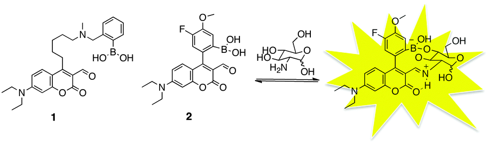 | ||
| Scheme 1 A sensor for dopamine/norepinephrine (1); a sensor for glucosamine (2). | ||
To target glucosamine, sensor 2 was designed with a boronic acid group in the closest possible position to the aldehyde, creating a small pocket suited for the amino-sugar analyte, while also potentially excluding larger analytes. We have found that aryl-substituted coumarin–aldehydes perform very well as fluorescent sensors,22 so an ortho-phenylboronic acid was chosen for the diol-binding unit. A simple phenyl boronic acid has a pKa that is too high to operate properly at neutral pH. Thus, most phenyl boronic acid-based receptors utilize an aminomethyl substituent (e.g., sensor 1) to maintain the proper pKa. For sensor 2, a fluoro substituent was used to achieve the optimal pKa.23
The synthesis of the sensor 2 is outlined in Scheme 2. Acetophenone derivative 3 was converted to ketoester 4 by Claisen condensation,24 which also resulted an SNAr substitution of the fluoro group para to the ketone with liberated methoxide. A Pechman reaction gave derivative 5.25 The chlorine substituent on intermediate 5 was converted to a boronic ester under palladium catalysis in moderate yield.26,27 The sensor (as the boronate ester) was obtained via Vilsmeier–Haack formylation reaction under carefully controlled conditions due to the acid sensitivity of the boronate ester.28 For these studies, compound 7 was used to prepare stock solutions of sensor 2 as the boronate ester hydrolysed in the dilute, aqueous media used for the titration experiments.
 | ||
| Scheme 2 Synthesis of sensor 2. | ||
Sensor 2 was titrated with various primary amines to determine its selectivity profile (Table 1). The aldehyde group of sensor 2 can bind reversibly to the primary amines to form an iminium ion.19 The iminium ion enhances the internal charge transfer (ICT) of the coumarin, leading to a large red shift in the excitation spectra (Fig. 1a). By exciting the sensor at the red excitation wavelength, only the bound form is excited resulting in a large increase in fluorescence upon binding (expressed as Isat/I0 where Isat is the fluorescence of the sensor at saturation). When excited at 488 nm (a convenient wavelength) the sensor emission increases and shifts from 520 nm to 568 nm upon addition of glucosamine (Fig. 2b). Comparing results from dicarboxylate-containing guests such as glutamate and aspartate with the mono-carboxylate glycine and butylamine (no carboxylates) shows that the binding constants for such simple analytes are similar but the fluorescent response is stronger for analytes with more carboxylate groups. This effect stems from the fact that the carboxylates raise the pKa of the formed imine, producing more iminium ion, which is key to the sensor response.19 Secondary amines produce no response from the sensor.
| Guest | λ ab (nm) | λ em (nm) | K a (M−1) | I sat/I0b | |
|---|---|---|---|---|---|
| a K a measured by fluorescence spectroscopy, errors are ±5% based on triplicate titration; titrations performed with sensor 2 (10−5 M in 120 mM NaCl, 25 mM HEPES, pH = 7.4). b I sat: fluorescence intensity at saturation from a fit to the binding isotherm; λex = 488 nm. c Determined by absorption changes at 488 nm. | |||||
| D-Glucosamine |

|
485 | 568 | 4100 | 32 |
| Norepinephrine |

|
487 | NA | 25c | 0 |
| D-Glucose |
|
452 | NA | 35 | 0 |
| L-Glutamic acid |

|
492 | 572 | 105 | 41 |
| L-Aspartic acid |

|
492 | 575 | 107 | 41 |
| Glycine |

|
484 | 565 | 93 | 22 |
| N-Butylamine |

|
455 | 550 | 73 | 13 |
| Diethylamine |

|
— | — | — | — |
 | ||
| Fig. 1 (a) UV-vis absorption titration of sensor 2 with glucosamine ([2] = 10−5 M in 120 mM NaCl, 25 mM HEPES, pH = 7.4). (b) Fluorescence titration of sensor 2 with glucosamine (λex = 488 nm). Inset is a fit of the fluorescence data to a one-site binding isotherm. | ||
 | ||
| Fig. 2 (a) Fluorescence response of sensor 2 (10−5 M) with addition of various analytes at concentration 1 mM, (λex = 488 nm). (b) Binding constants of sensor 2 with various analytes. | ||
Interestingly, not only did sensor 2 exhibit a very strong turn-on fluorescence for glucosamine compared all other primary amines, but also an excellent binding constant (Ka = 4100 M−1). This binding constant is nearly two orders of magnitude higher than that of the simple amino acids. The strong binding indicates glucosamine interacts with both the boronic acid and the aldehyde in a cooperative fashion (Scheme 1).29–33 It should be noted that boronic acids are well known to interact with the furanose form of glucose,34 and indeed, it may be possible that glucosamine adopts this form in solution as well. However, the formation of glucofuranose-boronic acid complexes is driven by the stability of the boronate ester of the 1,2 diol of the glucofuranose, which is not possible for glucosamine.
It appears from the titration data that norepinephrine, which also possesses both a primary amine and a diol, elicited a poor response from sensor 2, both in terms of binding constant and fluorescence response (Table 1). Apparently, the small cavity between the boronic acid and the aldehyde in the sensor 2 is not suitable for the extended catechol system in norepinephrine. The observed absorption changes indicate that the iminium ion is formed, however cooperative binding of the catechol was not observed. Furthermore, the electron rich catechol engages in PET quenching with the electron poor coumarin giving an overall decrease in emission. This result stands in contrast to the high binding constant (Ka = 6500 M−1) achieved with norepinephrine and sensor 1.20 Thus, the cavity size of sensor 2 provides excellent discrimination between glucosamine and chatecholamines. In addition, glucose itself gives only a small change upon binding sensor 2 since it lacks an amine groups, and the binding constant is quite low, as expected.
To demonstrate the selectivity of sensor 2, the sensor was mixed with equal concentration of analytes in buffer (Fig. 2a). A concentration of 1 mM which is similar to therapeutic concentration was selected.35 Sensor 2 gave a much stronger fluorescence response to glucosamine than any other analyte. Interestingly, the emission behaviour of this sensor is quite different than other sensors in this series. For sensors such as 1, the emission band stays constant at about 520 nm upon binding analytes, however it is clear from Fig. 2a that the emission band of sensor 2 shifts almost 50 nm upon binding glucosamine. This shift gives sensor 2 a Stokes shift of 83 nm in the bound state. Such large Stokes shifts are helpful for overcoming background fluorescence. This shift in emission upon binding results from the binding geometry that increases the π-overlap of the aromatic substituent with the fluorophore, leading to higher wavelengths of both excitation and emission.36 Taken together, these results indicate that sensor 2 is a very selective sensor for glucosamine both in terms of fluorescent response and binding constant as graphically illustrated in Fig. 2b.
In conclusion, a new turn-on fluorescent sensor was developed that shows excellent affinity and selectivity to glucosamine over various amines and diol-containing analytes. The suitable geometrical arrangement of multiple functional groups within the sensor produces an ideal binding cavity for glucosamine under physiological conditions of salt and pH. This probe will greatly benefit on-going research on the pharmaceutical effects of glucosamine. In addition, this observed fluorescence responses may inspire research toward highly sensitive and selective fluorescent sensors for other complex biomolecules.
We wish to acknowledge NSF for financial support (CHE-1112194) and would like to thank the United States Government, National Academy of Sciences and Vietnam Education Foundation for a graduate fellowship (T.M.T.).
Notes and references
- O. Bruyere, K. Pavelka, L. C. Rovita, R. Deroisy, M. Olejarova, J. Gatterova, G. Giacovelli and J. Y. Reginster, Menopause, 2004, 11, 138–143 CrossRef.
- S. Christgau, Y. Henrotin, L. B. Tanko, L. C. Rovati, J. Collette, O. Bruyere, R. Deroisy and J. Y. Reginster, Clin. Exp. Rheumatol., 2004, 22, 36–42 CAS.
- H. Gray, P. Hucheson and R. Slacin, J. Allergy Clin. Immunol., 2004, 114, 459–460 CrossRef PubMed.
- J. Erickson and T. Messer, J. Hand. Surg. Am., 2013, 38, 1638–1640 CrossRef PubMed.
- C. T. Vangsness, W. Spiker and J. Erickson, Arthroscopy, 2009, 25, 86–94 CrossRef PubMed.
- J. B. Houpt, R. MacMillan, C. Wein and S. D. Paget-Dellio, J. Rheumatol., 1999, 26, 2423–2430 CAS; J. Y. Reginster, A. Neuprez, M. P. Lecart and N. Sarlet, J. Rheumatol., 2012, 32, 2959–2967 Search PubMed.
- M. J. Pouwels, J. Jacobs, P. Span, J. Lutterman, P. Smits and C. Tack, J. Clin. Endocrinol. Metab., 2001, 86, 2099–2103 CAS.
- F. Richy, O. Bruyere, O. Ethgen, M. Cucherat, Y. Herotin and J. Y. Reginster, Arch. Intern. Med., 2004, 163, 1514–1522 CrossRef PubMed.
- T. E. Toweed, L. Maxwell and T. P. Anastassiades, Cochrane Database Syst. Rev., 2005, 2, CD002946 Search PubMed.
- S. Wandel, P. Juni, B. Tendal, E. Nuesch, P. M. Villiger, N. J. Welton, S. Reichenbach and S. Trelle, BMJ., 2010, 314, c4675 CrossRef PubMed.
- A. D. Sawitzke, H. Shi, M. F. Finco, D. D. Dunlop, C. O. Bingham, C. L. Harris, N. G. Singer, J. D. Bradley, D. Silver, C. G. Jackson, N. E. Lane, C. V. Oddis, F. Wolfe, J. Lisse, D. E. Furst, D. J. Reda, R. W. Moskowwits, H. J. Williams and D. O. Clegg, Arthritis Rheum., 2008, 58, 3183–3191 CrossRef CAS PubMed.
- J. H. Quastel and A. Cantero, Nature, 1953, 171, 252–254 CrossRef CAS; B. Q. Liu, X. Meng, C. Li, Y. Y. Gao, N. Li, X. F. Niu, Y. Guan and H. Q. Wang, Exp. Mol. Med., 2011, 43, 487–493 CrossRef PubMed.
- J. Bekesi, Z. Molnar and R. Winzler, Cancer Res., 1969, 29, 353–359 CAS.
- J. Bekesi and R. Winzler, Cancer Res., 1970, 30, 2905–2912 CAS.
- S. Friedman and P. Skehan, Proc. Natl. Acad. Sci. U. S. A., 1980, 77, 1172–1176 CrossRef CAS.
- E. Krug and A. Zweibaum, Biochem. J., 1984, 217, 701–708 CAS.
- C. Cooper and T. James, J. Chem. Soc., Perkin Trans. 1, 2000, 963–969 RSC.
- R. Cheng, Y. Liu, S. Ou, Y. Pan, S. Zhang, H. Chen, L. Dai and J. Qu, Anal. Chem., 2012, 84, 5641–5644 CrossRef CAS PubMed.
- E. Feuster and T. Glass, J. Am. Chem. Soc., 2003, 125, 16174–16175 CrossRef CAS PubMed.
- K. Secor and T. Glass, Org. Lett., 2004, 6, 3727–3730 CrossRef CAS PubMed.
- J. Klockow and T. Glass, Org. Lett., 2013, 15, 235–237 CrossRef CAS PubMed.
- K. Hettie, X. Liu, K. Gillis and T. Glass, ACS Chem. Neurosci., 2013, 4, 918–923 CrossRef CAS PubMed.
- J. Yan, G. Springsteen, S. Deeter and B. Wang, Tetrahedron, 2004, 60, 11205–11209 CrossRef CAS PubMed.
- C. L. Ni, H. Q. Wang and H. Yan, Youji Huaxue, 2006, 26, 357–359 CAS.
- J. Madhav, B. Kuarm, P. Someshwar, B. Rajitha, Y. Reddy and P. Crooks, J. Chem. Res., 2008, 4, 232–234 CrossRef.
- N. Kornblum and A. Lurie, J. Am. Chem. Soc., 1959, 81, 2705–2715 CrossRef CAS.
- T. Ishiyama, M. Murata and N. Miyaura, J. Org. Chem., 1995, 60, 7508–7510 CrossRef CAS.
- K. Rajanna, F. Solomon and M. Ali, Int. J. Chem. Kinet., 1996, 28, 865–872 CrossRef CAS.
- J. P. Lorand and J. O. Edwards, J. Org. Chem., 1959, 24, 769–774 CrossRef CAS.
- T. Kimura, S. Arimori, M. Takeuchi, T. Nagasaki and S. Shinkai, J. Chem. Soc., Perkin Trans. 2, 1995, 1889–1894 RSC.
- J. Yoon and A. W. Czarnink, J. Am. Chem. Soc., 1992, 114, 5874–5875 CrossRef CAS.
- L. K. Mohler and A. W. Crarnik, J. Am. Chem. Soc., 1993, 115, 2998–2999 CrossRef CAS.
- T. D. James, K. R. A. S. Sandanayake and S. Shinkai, Nature, 1995, 374, 345–347 CrossRef CAS.
- J. C. Norrild and H. Eggert, J. Am. Chem. Soc., 1995, 117, 1479–1484 CrossRef CAS.
- S. J. Friedman and P. Skehan, Proc. Natl. Acad. Sci. U. S. A., 1980, 77, 1172–1176 CrossRef CAS.
- K. S. Hettie and T. E. Glass, Chem. – Eur. J., 2014, 20, 17488–17499 CrossRef CAS PubMed.
Footnote |
| † Electronic supplementary information (ESI) available: Synthesis, characterization of materials, and additional details. See DOI: 10.1039/c5cc00415b |
| This journal is © The Royal Society of Chemistry 2015 |