Supramolecular hydrogels for wound repair and hemostasis
Shaowen
Zhuo
a,
Yongping
Liang
 a,
Zhengying
Wu
a,
Xin
Zhao
a,
Zhengying
Wu
a,
Xin
Zhao
 *a,
Yong
Han
 *ab and
Baolin
Guo
 *abc
*abc
aState Key Laboratory for Mechanical Behavior of Materials, and Frontier Institute of Science and Technology, Xi’an Jiaotong University, Xi’an, 710049, China. E-mail: zhaoxinbio@mail.xjtu.edu.cn; yonghan@mail.xjtu.edu.cn; baoling@mail.xjtu.edu.cn
bDepartment of Orthopaedics, The First Affiliated Hospital of Xi’an Jiaotong University, Xi’an, 710061, China
cKey Laboratory of Shaanxi Province for Craniofacial Precision Medicine Research, College of Stomatology, Xi’an Jiaotong University, Xi’an, 710049, China
First published on 27th October 2023
Abstract
The unique network characteristics and stimuli responsiveness of supramolecular hydrogels have rendered them highly advantageous in the field of wound dressings, showcasing unprecedented potential. However, there are few reports on a comprehensive review of supramolecular hydrogel dressings for wound repair and hemostasis. This review first introduces the major cross-linking methods for supramolecular hydrogels, which includes hydrogen bonding, electrostatic interactions, hydrophobic interactions, host–guest interactions, metal ligand coordination and some other interactions. Then, we review the advanced materials reported in recent years and then summarize the basic principles of each cross-linking method. Next, we classify the network structures of supramolecular hydrogels before outlining their forming process and propose their potential future directions. Furthermore, we also discuss the raw materials, structural design principles, and material characteristics used to achieve the advanced functions of supramolecular hydrogels, such as antibacterial function, tissue adhesion, substance delivery, anti-inflammatory and antioxidant functions, cell behavior regulation, angiogenesis promotion, hemostasis and other innovative functions in recent years. Finally, the existing problems as well as future development directions of the cross-linking strategy, network design, and functions in wound repair and hemostasis of supramolecular hydrogels are discussed. This review is proposed to stimulate further exploration of supramolecular hydrogels on wound repair and hemostasis by researchers in the future.
Wider impactOver the past few years, supramolecular hydrogels have gained increasing attention in the field of hemostasis and wound repair due to their unique cross-linking and high designability, making them significantly different from traditional chemical hydrogels. However, the relationship between the network design of supramolecular hydrogel dressings and their unique biomedical functions, as well as the status and future trends of supramolecular hydrogel dressings, has not been systematically summarized. In this review, we provide a comprehensive summary of the main cross-linking methods and their cross-linking principles, and elaborate the relationship between various network structures and the properties of supramolecular hydrogels. Additionally, we summarize the functions of supramolecular hydrogels in promoting hemostasis and wound repair, and discuss the methods for realizing each specific function. At the end of the paper, we put forward the current challenges and feasible countermeasures related to supramolecular hydrogels, and forecast the future development trends of supramolecular hydrogels, encompassing network design, wound healing and hemostatic applications. We hope that this review will help researchers to gain a deeper understanding of supramolecular hydrogels and their applications in hemostasis and wound repair, paving the way for the emergence of better supramolecular hydrogel dressings in the future. |
1 Introduction
The skin is one of the most vital organs of the human body, which can make direct contact with the external environment, detect external stimuli, regulate body temperature, and protect the body from external damages. As a result, skin is one of the most vulnerable tissues. Hemostasis, inflammation, proliferation, and remodeling are the four stages of skin wound healing.1 Excessive bleeding can lead to symptoms of anemia and insufficient blood volume in patients, or hemorrhagic shock and even death in severe cases such as car accidents, on the battlefield, operation or in patients with blood clotting disorders. Timely hemostasis is necessary, as it can prevent the occurrence of shock and enable the progression to the next stage of wound healing.2,3 The inflammation, proliferation, and remodeling are also important parts of the wound healing process. Although most common skin injuries can heal, various factors may cause a wide range of wound healing problems (including producing scar tissue and hindering the regeneration of appendages),4–7 such as excessive inflammation, burns, full-thickness skin defects, infection and diabetic wounds, which lead to difficulties in wound repair. Therefore, it is necessary to develop wound dressings to facilitate the wound healing process including hemostasis and subsequent high quality wound repair.Ideal wound dressings should satisfy the following requirements to accelerate wound healing: good biocompatibility and moisture retention, adequate mechanical strength, and suitable biochemical properties.8,9 Hydrogels are widely used as soft materials that can absorb large amounts of water while maintaining their integrity. Hydrogels have high water content and good flexibility and possess a number of properties similar to those of soft tissues in the human body. Hydrogels are often biocompatible and can be designed to show a good anti-adhesion effect against proteins in body fluids. In addition, hydrogels can be easily designed to show responsiveness to various stimuli. These properties enable hydrogels to be used in critical biomedical applications such as use as adhesives, dressings, drug/cell delivery systems, and scaffolds.10,11 Thus, hydrogels are the most competitive dressing materials and there has been an increasing use of hydrogels as wound dressings in recent years.12–15
Chemically cross-linked hydrogels with permanent covalent bonds are relatively stable, possessing high stiffness and resistance to rapid degradation. However, when the hydrogel networks are broken, their 3D structures are irreversibly broken.16–18 In contrast, supramolecular hydrogels are formed through reversible non-covalent interactions (hydrogen bonds, electrostatic interactions, hydrophobic interactions, host–guest interactions, metal ligand coordination or other interactions) between macromolecules and/or small molecules. These non-covalent interactions are usually reversible and can respond to external stimuli. When the external environment changes or artificial stimuli occur, the non-covalent bonds will respond by breaking or recombining, showing the properties of reversible expansion, gel–sol transformation, injectable capacity under environmental stimulation, self-healing, shape memory or other properties.19 Moreover, they are biocompatible and easily degradable. More importantly, these reversible properties provide the foundation for the application of supramolecular hydrogels as delivery systems and tissue engineering scaffolds.20–22 Therefore, supramolecular hydrogels have gained popularity in the field of wound dressings, and nowadays a variety of functions (antibacterial properties, tissue adhesion, anti-inflammatory function, antioxidation, and functions of cell control) are introduced in supramolecular hydrogels, leading to the development of more multi-functional supramolecular hydrogel dressings.
Recently, research on supramolecular hydrogels has gained significant momentum, and the number of reviews on supramolecular hydrogels and related fields has also been increasing year by year. These reviews mostly focus on the introduction of different kinds of supramolecular interactions,23 comparisons between supramolecular hydrogels and covalent macromolecular hydrogels,24 recent technologies of various characterization methods25 and so on. However, there has never been a thorough analysis of supramolecular hydrogels for hemostasis and wound repair. In this review, we discuss the cross-linking interactions and network structures of supramolecular hydrogels in detail, and focus on their applications in wound repair and hemostasis. We have outlined the specific functions of supramolecular hydrogels that have emerged over the last decade in wound repair and hemostasis, and highlighted their structural features, and the underlying mechanisms that enable hydrogels to achieve these functions. Specifically, the functions related to promoting wound repair are divided into antibacterial activity, tissue adhesion, substance delivery, anti-inflammatory and antioxidant functions, control of cell behaviors, pro-angiogenesis function and other functions. We subsequently classified the functions of hemostasis into physical hemostasis and coagulation promotion. In addition, the design of supramolecular hydrogel dressings and the challenges they face in wound repair and hemostasis are discussed, and more detailed prospects are presented to stimulate further research in this field.
2 Cross-linking and characteristics of supramolecular hydrogels
The 3D structure of hydrogel is formed by cross-linking the polymer chains (one or more). The effect of one or more non-covalent cross-linking interactions on the properties and even the functions of supramolecular hydrogels cannot be ignored. In this section, the cross-linking methods and properties of supramolecular hydrogels will be described in six parts (hydrogen bonds, electrostatic interactions, hydrophobic interactions, host–guest interactions, metal ligand coordination and other interactions). Table 1 summarizes supramolecular hydrogels with different cross-linking interactions and their characteristics.| Cross-linking interaction | Hydrogel compositions | Functional groups\molecules that provide cross-linking | Characteristics | Ref. |
|---|---|---|---|---|
| Hydrogen bonds | Polyacrylamide, sodium alginate (SA) and diethylene triamine pentaacetic acid (DTPA) | Amino, amide and carboxyl | Good mechanical properties; tissue adhesion | 26 |
| Mono-(6-ethylenediamine-6-deoxy)-β-cyclodextrin (EDA-CD) and hydroxyethyl cellulose (HEC) | Amino and hydroxyl; EDA-CD molecule | Serving as a drug carrier; self-healing properties | 27 | |
| Deferoxamine grafted alginate (SA-DFA) | Carbonyl, hydroxyl and amino | Self-healing properties; structural stability | 28 | |
| Silk fibroin (SF) and carboxymethyl chitosan (CMCS) | Amino, hydroxyl and carboxyl | pH- and temperature-sensitive swelling behavior | 29 | |
| Humic acid (HA) and polyvinylpyrrolidone (PVP) | Carboxyl; PVP | Injectability; good flexibility; self-healing properties | 30 | |
| Dopamine-modified γ-PGA and tannic acid (TA) | Carboxyl, catechol; polyphenol | Enhanced wet tissue adhesion; high stretchability | 31 | |
| Poly(N-hydroxyethyl acrylamide) (PHEAA) and TA | Amide and hydroxyl; polyphenol | Self-healing properties; strong adhesion | 32 | |
| 2-amino-2′-fluoro-2′-deoxyadenosine (2-FA) | Amino and hydroxyl; 2′-F atom | High-strength; injectability | 33 | |
| Carboxymethyl agarose (CMA) | Hydroxyl | pH-responsiveness; temperature-responsiveness | 34 | |
| N-Acryloyl 2-glycine monomer | Amide and carboxyl | High tensile strength (∼1.3 MPa); large stretchability (up to 2300%); high compressive strength (∼10.8 MPa); good toughness (∼1000 J m−2) | 35 | |
| Aramid nanofibers (ANFs), PVA and TA | Polyphenol, amide and alcohol hydroxyl | Excellent mechanical properties | 36 | |
| PVA, HA, TA and polyethylene glycol (PEG) | Polyphenol, ether, carboxyl and alcohol hydroxyl | Shape memory behavior; self-healing properties | 37 | |
| Casein micelles (CEs) and PVA | PVA and glycerol; H2O and glycerol | Excellent mechanical properties; self-healing properties | 38 | |
| Chitosan (CS) and poly(D,L-lactide)–poly(ethylene glycol)–poly(D,L-lactide) (PPP) | Amino, hydroxyl, amide, carboxyl, carbonyl and ether | Good fluidity; strong adhesion | 39 | |
| Fenugreek gum and cellulose | Hydroxyl | Excellent mechanical properties; thermal stability | 40 | |
| PVA and CS | Hydroxyl and amino | Superior compressive (60%–230 kPa), tensile (152 kPa–360%); recover ability (90.77% after 5 cycles); anti-swelling properties | 41 | |
| Hydrophilic synthetic polyacrylic acid Carbomer 940 (CBM940) and CMCS | Hydroxy, amino and carboxyl | Elastic properties; water retention | 42 | |
| CS and PEC | Amino, carboxyl and hydroxy | Good printability; dimensional integrity | 43 | |
| TA and CS | Phenol hydroxyl, amino and hydroxy | High swelling rate | 44 | |
| TA and imidazolidinyl urea reinforced polyurethane (PMI) | Phenol hydroxyl, ether and urethane; imidazolidiny urea | Excellent mechanical strength (break strength: 0.28–0.64 MPa; elongation at break: 650–930%) | 45 | |
| Poly(N-acryloyl 2-glycine) (PACG) | Amide and carboxyl | High tensile strength (up to 1.1 MPa); outstanding compressive strength (up to 12.4 MPa); large Young's modulus (up to 320 kPa); high compression modulus (up to 837 kPa) | 46 | |
| Boron nitrogen nanosheet (f-BNNS), PSS and PNIPAM | Amide, sulfonic acid group, siloxane group and boron hydroxyl | Enhanced mechanical properties; self-healing properties | 47 | |
| Agarose (AG) | Hydroxy | Good mechanical properties; superior processing performance | 48 | |
| TA and SF | Phenol hydroxyl and amide | High extensibility (i.e., up to 32![[thin space (1/6-em)]](https://www.rsc.org/images/entities/char_2009.gif) 000%); real-time self-healing properties 000%); real-time self-healing properties |
49 | |
| TA and thioctic acid | Polyphenol and carboxyl | Self-healing property; injectability | 50 | |
| Hydrophilic PVA and certain hydrophobic compounds | Hydroxyl and phenolic hydroxyl | Good plasticity; self-healing properties | 51 | |
| Ureido-pyrimidinone (UPy) | UPy | Self-healing properties; injectability | 52 | |
| Supramolecular branched polymer HDU, SF | UPy | Self-healing properties; injectability | 53 | |
| SA and CMCS | Hydroxyl, carboxyl and amino | Self-healing properties; controlled swelling properties | 54 | |
| Orotic acid (OA) modified chitosan and 2,6-diaminopurine (DAP) | OA and DAP molecules | Self-healing properties; injectability | 55 | |
| Poly(acrylic acid) (PAA), lauryl methacrylate (LMA), cetyltrimethylammonium bromide (CTAB) | Carboxyl | Superior tensile strength (≈1.6 MPa), large stretchability (>900%), rapid self-recovery (≈3 min at 100% strain) | 56 | |
| TA, PVP, basic fibroblast growth factor (bFGF) | Polyphenol; PVP | Self-healing properties; injectability | 57 | |
| PVA, poly(sodium acrylate) (PAANa), polyallylamine (PAH) | Hydroxyl, carboxyl and amino | Excellent mechanical properties (fracture strain: 768% and toughness: 2.25 MJ m−3); recovery capabilities (recovery ratio of maximum stress: 90.5%) | 58 | |
| Electrostatic interaction | Fmoc-FFpY peptide and PAH | –NH3+ and phosphate | Superior mechanical properties; injectability | 59 |
| Polysaccharides salecan and CMCS | Polysaccharides salecan and CMCS | Good swelling properties; adsorbability | 60 | |
| Residue of CaCO3 and CS | Ca2+ and CS chain | Excellent mechanical properties; low gelation temperature | 61 | |
| Poly(hexamethylene guanidine) hydrochloride and CMCS | Poly(hexamethylene guanidine) hydrochloride and CMCS | Shear thinning; self-healing properties | 62 | |
| Lignin and CS | Phenoxide and ammonium | Decontamination; environmental protection | 63 | |
| CS and carmellose (CM) | Amino and carboxyl | Adjustable swelling properties and erosion susceptibility | 64 | |
| Poly[3-[2-(methacryloyloxy)ethyl](dimethyl)-ammonio]-1-propanesulfonate (PSBMA) | Quaternary ammonium salts and sulfonates | Degradability | 65 | |
| Poly(acrylic acid) (PAA), lauryl methacrylate (LMA), cetyltrimethylammonium bromide (CTAB) | Carboxy and trimethyl ammonium bromide | Superior tensile strength (≈1.6 MPa), large stretchability (>900%), rapid room-temperature self-recovery (≈3 min at 100% strain) | 56 | |
| PVA, poly(sodium acrylate) (PAANa), polyallylamine (PAH) | Amino and carboxyl | Excellent mechanical properties (fracture strain: 768% and toughness: 2.25 MJ m−3); recovery capabilities (recovery ratio of maximum stress: 90.5%). | 58 | |
| Hydrophobic interaction | Methylcellulose (MC) and gallic acid (GA) | Aromatic ring of GA and methyl groups of MC | Dynamic viscoelasticity | 66 |
| Dihydrophenonthrolin-4-one-3-carboxylic acid (DPCA) and polyethylene glycol (PEG) | DPCA | Shear thinning; injectability | 67 | |
| Hydrophobic monomer stearyl methacrylate C18M and an amphiphilic regenerated silk fibroin (RSF) | C18M and RSF | Excellent mechanical properties; self-healing properties | 68 | |
| Poly(acrylic acid) (PAA), lauryl methacrylate (LMA), cetyltrimethylammonium bromide (CTAB) | LMA and CTAB | Superior tensile strength (≈1.6 MPa), large stretchability (>900%), rapid room-temperature self-recovery (≈3 min at 100% strain) | 56 | |
| Host–guest interaction | Acrylamide, a host–guest macrocross-linker assembled from poly(β-cyclodextrin) nanogel and azobenzeneacrylamide | Host: β-CD | Adjustable self-healing properties | 69 |
| Guest: azobenzeneacrylamide | ||||
| Tripeptide Phe-Gly-Gly ester derivative (FGG-EA) and cucurbit[8]uril (CB[8]) | Host: CB[8] | Softness and elasticity | 70 | |
| Guest: FGG-EA | ||||
| Well-defined star [Ad-(PNIPAM-Ad)4] and linear [Ad-(PNIPAM-βCD)2] | Host: linear [Ad-(PNIPAM-βCD)2] | Temperature sensitivity; self-healing properties | 71 | |
| Guest: well-defined star [Ad-(PNIPAM-Ad)4] | ||||
| Quaternized chitosan-graft-cyclodextrin (QCS–CD), quaternized chitosan-graft-adamantane (QCS–AD) and graphene oxide-graft-cyclodextrin (GO–CD) | Host: QCS–CD and GO–CD | Fast self-healing properties; conductivity; photo-thermal properties; injectability | 72 | |
| Guest: QCS–AD | ||||
| β-Cyclodextrin modified hyaluronic acid (HA-CD) and adamantane modified 4-arm-PEG (4-arm-PEG-Ad) | Host: HA-CD | Self-healing property; adjustable profiles in strength and swelling property | 73 | |
| Guest: 4-arm-PEG-Ad | ||||
| Acryloyl-β-cyclodextrin (Ac-CD), 2-hydroxyethyl acrylate (HEA) and SF | Host: Ac-CD | Long-term stability; rapid self-healing properties; injectability | 14 | |
| Guest: aromatic amino acid | ||||
| γ-Cyclodextrin dimer and tetraaniline and poly(ethylene glycol) | Host: γCD2 | Injectability; conductivity | 74 | |
| Guest: AT | ||||
| Multivalent host molecule HCD and multivalent gest molecule 8PEG-Ada | Host: HCD | Acid-triggered gelation | 75 | |
| Guest: 8PEG-Ada | ||||
| β-CD units and adamantyl groups | Host: β-CD | High strength; self-healing properties; injectability; tissue adhesion | 76 | |
| Guest: adamantyl | ||||
| Octa-cyclodextrin polyhedral oligomeric silsesquioxane (OCDPOSS) and acrylamide-modified adamantane (Ad-AAm) | Host: OCDPOSS | Super-stretchability; self-healing properties and injectability | 77 | |
| Guest: Ad | ||||
| DAS@SCD, NIPAZO | Host: CD | Injectability | 78 | |
| Guest: azobenzene derivative | ||||
| Cucurbit[6]urils (CB[6]), 1,4,5,8-naphthalenediimide (NDI)-bridged CDs, Paxle | Host: cucurbit[6]urils (CB[6]), 1,4,5,8-naphthalenediimide (NDI)-bridged CDs | Excellent mechanical behaviors, showing a Young's modulus of up to 1.01 MPa, toughness of up to 3.80 MJ m−3, maximum stress of up to 1.12 MPa, and stretchability up to 1966% | 79 | |
| Guest: Axle-1 | ||||
| Hyaluronic acid modified with cyclodextrin (HA-CD), hyaluronic acid modified with adamantane (Ad-HA) | Host: CD | Self-healing properties and injectability | 80 | |
| Guest: Ad | ||||
| γ-CD and anthracene dimer | Host: γ-CD | Self-healing properties; injectability | 81 | |
| Guest: anthracene dimer | ||||
| Alginate-g-DA-g-β-CD, PAAM-g-Ad | Host: β-CD | Self-healing properties; injectability | 82 | |
| Guest: Ad | ||||
| γ-CD and 1-(4-carboxybenzyl)-4,4′-bipyridinium chloride (1+·Cl−) | Host: γ-CD | Self-healing properties | 83 | |
| Guest: 1-(4-carboxybenzyl)-4,4′-bipyridinium chloride (1+·Cl−) | ||||
| Metal ligand coordination | SA, metal ions (Gd3+), and bisphosphate functionalized polycitrate | Ligand: carboxyl, diphosphate | Injectability; self-healing properties | 84 |
| Metal ion: Gd3+ | ||||
| Ag+ and the solution of HA containing BP groups (HA-BP) | Ligand: carboxyl, diphosphate | Shear-thinning; self-healing properties | 85 | |
| Metal ion: Ag+ | ||||
| Fe3+, Al3+ and CMCS | Ligand: carboxyl from CMCS | Self-healing properties; adaptive characteristics | 86 | |
| Metal ion: Fe3+, Al3+ | ||||
| Fe3+ and catechol | Ligand: catechol | Self-healing properties; injectability | 52 | |
| Metal ion: Fe3+ | ||||
| TA, PVP, basic fibroblast growth factor (bFGF) | Ligand: phenolic hydroxyl | Self-healing properties; injectability | 57 | |
| Metal ion: Fe3+ | ||||
2.1 Hydrogen bonds
Hydrogen bonding is a widely studied supramolecular interaction with reversibility, directionality and sensitivity.87 It widely exists between hydrogen atoms of different groups, such as hydroxyl, amino, amide, and strong electronegative atoms with lone pair electrons (such as O, N, and F).23,87 The hydrogen atom will acquire a positive charge when bonding with the extremely electronegative atoms mentioned above, while the electronegative atom will have a negative charge.23,88 Hydrogen bonding is crucial to many chemical systems and biological processes in nature, and it is also an important driving force for the construction of hydrogels.87 Although a single hydrogen bond is weaker than a single covalent or ionic bond, the concerted hydrogen bond interactions can improve the cross-linking strength,33 and their collective properties can promote the formation of hydrogels, especially supramolecular hydrogels.23,87,89Reversible cross-linking can be established between different polymer chains through hydrogen bonds. The hydrogen bonds dissociate at high temperatures while recombine at low temperatures.88,89 Thus, hydrogen bonds can be used in the preparation of self-healing hydrogels. For example, the hydroxyl groups on polyvinyl alcohol (PVA) can form intermolecular hydrogen bonds, leading to the formation of self-healing hydrogels following repeated freezing–thawing operations.89,90 Zhao et al.91 prepared a self-healing supramolecular hydrogel through hydrogen bonds between tertiary amide groups in polyvinylpyrrolidone (PVP) and phenol hydroxyl groups in gallic acid (GA) (Fig. 1A(a)). The presence of hydrogen bonds imparts the hydrogel with good tensile properties and injectability (Fig. 1A(b) and (c)). Hydrogen bonds can also be combined with other supramolecular interactions to prepare hydrogels with multiple cross-linking interactions or multi-networks. For example, we previously developed a heat shrinkable hydrogel by combining hydrogen bonds and host–guest interaction. In our hydrogel, the dual non-covalent cross-linking strategy, especially the hydrogen bonds, imparts it with good elasticity and self-healing properties.92 The similar synergistic strategies of multi-interaction will be demonstrated in more detail later.
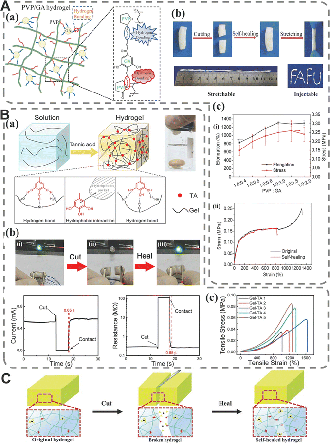 | ||
Fig. 1 Supramolecular hydrogels cross-linked by hydrogen bonds and their characteristics. (A) (a) Schematic diagram of hydrogen bonds between PVP and GA in PVP/GA composite hydrogel; (b) self-healing process of PVP/GA composite hydrogel after recovery, optical demonstration of the stretchability, and injecting PVP/GA composite hydrogel to a “FAFU” shape through a syringe; (c) (i) elongation (black square, indicated by a black arrow) and stress (red circles, indicated by a red arrow) of the PVP/GA composite hydrogels and (ii) stress–strain curves of original hydrogel and self-healed hydrogel with PVP![[thin space (1/6-em)]](https://www.rsc.org/images/entities/char_2009.gif) : :![[thin space (1/6-em)]](https://www.rsc.org/images/entities/char_2009.gif) GA of 1.0 GA of 1.0![[thin space (1/6-em)]](https://www.rsc.org/images/entities/char_2009.gif) : :![[thin space (1/6-em)]](https://www.rsc.org/images/entities/char_2009.gif) 1.0. Copyright 2020 Elsevier.91 (B) (a) Formation process and mechanism of gel-TA hydrogel formation; (b) self-healing tests and photographs of the self-healing process. Circuit containing self-healing gel-TA-Ag NW 4 hydrogel connected in a series with a green LED: (i) original, (ii) completely severed (open circuit), and (iii) healed. Time evolution of the self-healing process for the conductive gel-TA-Ag NW 4 hydrogel: real-time current and resistance; (c) typical tensile stress–strain curves of gel-TA hydrogels at 25 °C. Copyright 2019 American Chemical Society.93 (C) Schematic diagram of the self-healing mechanism. Copyright 2020 Wiley-VCH GmbH.52 1.0. Copyright 2020 Elsevier.91 (B) (a) Formation process and mechanism of gel-TA hydrogel formation; (b) self-healing tests and photographs of the self-healing process. Circuit containing self-healing gel-TA-Ag NW 4 hydrogel connected in a series with a green LED: (i) original, (ii) completely severed (open circuit), and (iii) healed. Time evolution of the self-healing process for the conductive gel-TA-Ag NW 4 hydrogel: real-time current and resistance; (c) typical tensile stress–strain curves of gel-TA hydrogels at 25 °C. Copyright 2019 American Chemical Society.93 (C) Schematic diagram of the self-healing mechanism. Copyright 2020 Wiley-VCH GmbH.52 | ||
Self-healing speed and self-healing efficiency are also important parameters of hydrogen bond-based self-healing hydrogels. Numerous studies have been conducted to study the self-healing properties of hydrogels based on these two parameters. Wang et al.93 prepared a supramolecular hydrogel (Fig. 1B(a) and (c)) via hydrogen bonds between phenol hydroxyl groups on TA and carbonyl, hydroxyl and amino groups of hydrogel chains. The water in the hydrogel network can improve the fluidity of TA molecules and unsaturated hydrogen bonds, thus greatly improving the speed of self-healing (indicates a healing efficacy of 95%) (Fig. 1B(b)). Zhao et al.94 prepared semi-interpenetrating polymer network hydrogels using sodium polyacrylamide (PAM) and alginate (SA). Based on the hydrogen bonds between PAM and SA molecules, the hydrogel had a rare self-healing ratio of 99% at room temperature.
In the field of hydrogels, the stimuli-responsive property has been a hot topic, because this property is closely related to many applications of hydrogels. Hydrogen bonds in supramolecular hydrogels can act as thermo-triggered regulators of mechanical behaviors, thus imparting shape memory properties to the hydrogels.88,95,96 For example, Zhu et al.97 prepared a fluorescent hydrogel with good shape memory properties and toughness. The dense and strong hydrogen bonds between carboxylic acid and imidazole groups make the hydrogel glassy at room temperature with moderate water content. In addition, hydrogen-bonded supramolecular hydrogels with stimuli-responsive expansion properties have also been reported. For example, Xu et al.54 prepared a polysaccharide supramolecular hydrogel with self-healing and dual-responsive swelling properties by adding hymexazol into a mixed solution of alginate and carboxymethyl chitosan. Results showed that the increasing concentration of PBS could delay the water adsorption of hydrogels. Moreover, the swelling of the hydrogel increased with the pH value in a 0.1 M PBS solution.
Hydrogen bonds can also achieve adjustable self-healing performance and shape adaptivity by responding to external stimuli. Our team designed an injectable DN (double network) hydrogel, which consisted of catechol-Fe3+ coordination cross-linked poly(glycerol sebacate)-co-poly(ethylene glycol)-g-catechol (PEGSD) and quadruple hydrogen bonds cross-linked ureido-pyrimidinone modified gelatin (GTU). The tests confirmed the good self-healing efficiency of the hydrogels, partly due to quadruple hydrogen bonds between UPy motifs from GTU. Apart from injectability and self-adaptability, the quadruple hydrogen bonds and Fe3+-catechol coordination can also respond to heat (resulting in decreased cross-linking density) and NIR irradiation (with the rapidly rising temperature, fluidity of the hydrogel network increased rapidly, thereby significantly fastening the self-healing property)52 (Fig. 1C).
Hydrogen bonds can also be used as a method of strengthening hydrogel systems to endow hydrogels with a variety of other properties at the same time. For example, natural polymer TA can have hydrogen bond interaction with various polymers, such as PVA, polyethylene glycol (PEG) and gelatin.44,98,99 Hydrogen bonds can also drive specific phase separation processes. Kamakshi Bankoti et al.100 prepared a macroporous hydrogel scaffold under room temperature drying conditions. The intermolecular and intramolecular hydrogen bonds between chitosan and polyurethane diol dispersion (PUDs) are used to induce the phase separation process in the system. They studied the effects of phase separation on scaffold's degradation and mechanical properties, and concluded that it was suitable for wound healing (showing increased wound contraction, higher collagen synthesis and vascularization).100,101 The applications of supramolecular hydrogels in wound repair can be found in Section 4.
In conclusion, since the mechanical properties of supramolecular hydrogels cross-linked by a single hydrogen bond are poor, the majority of research has concentrated on their exceptional reversible properties, such as self-healing, shape memory, and stimulus response. In contrast, supramolecular hydrogels cross-linked by multiple hydrogen bonds have better mechanical properties and can be combined with other non-covalent interactions. The theory of their reversible properties needs further study.
2.2 Electrostatic interaction
Electrostatic interaction is a force resulting from electrostatic attraction between ions or groups with opposite charges. Although electrostatic interaction is the reason for the formation of chemical bonds such as ionic bonds and metal ion coordination bonds, as a kind of non-covalent interaction, its bond energy is quite different from that of some common chemical bonds. Ions can interact with dipoles (polar molecules or groups), or induce nonpolar molecules to become polarized (induced dipoles). The electrostatic attraction between oppositely charged monomers, the interaction between cations (such as protonated amines) and anions (such as carboxylates and sulfates) in polymers, and the interaction between charged polymers and oppositely charged ions can all be driving forces for the formation of hydrogels.23,89The electrostatic interactions between polycations and polyanions can be used to form hydrogels.102 Numerous studies have shown that chitosan (CS), a positively charged natural cationic polymer, can form polyelectrolyte complexes with negatively charged polymers like polysaccharides (such as hyaluronic acid), proteins (such as gelatin) and synthetic polymers (such as polyacrylic acid).23,103 For example, Zhang et al.104 constructed a hydrogel using polycation CS, polyanion γ-PGA and heparin, and loaded superoxide dismutase (SOD) in the hydrogel to impart it with antioxidant properties. The hydrogel shows a good and connected porous sponge structure, which is conducive to the transfer of nutrients and the elimination of cell metabolites. With the increase in heparin content, the electrostatic interaction and cross-linking degree increased while the porosity of materials reduced.102,104 Similarly, Lai et al.64 developed a biodegradable polyelectrolyte hydrogel through the electrostatic interaction between CS (positively charged) and carmellose (negatively charged). Electrostatic interactions made the hydrogel preparation process simpler and more convenient by allowing the mixing of the two polymer solutions before application (Fig. 2A).
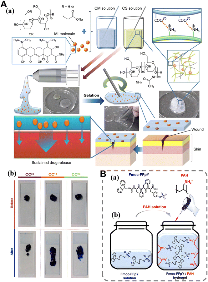 | ||
| Fig. 2 Supramolecular hydrogels cross-linked by electrostatic interaction and their characteristics. (A) (a) A schematic diagram depicting the generation and use of the dressing in wound treatment. Abbreviation: MI, minocycline hydrochloride; (b) photos of MB-loaded CC33, CC11, and CC03 dressings before and after spreading. Copyright 2019 Elsevier.64 (B) Representation of the PAH/Fmoc-FFpY supramolecular hydrogel: (a) electrostatic interaction between Fmoc-FFpY and PAH, leading to the (b) formation of a PAH/Fmoc-FFpY hydrogel by simple mixing of PAH and Fmoc-FFpY solutions. Copyright 2020 American Chemical Society.59 | ||
The researchers also explored the methods to improve the preparation of electrostatic interaction cross-linked hydrogels. Generally, the self-assembly of low-molecular-weight hydrogelators is triggered by a sol–gel process. External stimuli (such as temperature, pH, solvent change and chemical reactions) are able to adjust their solubility. Due to their weak solubility in water, peptides are typically induced to undergo self-assembly by first dissolving them in dimethyl sulfoxide (DMSO), followed by a subsequent dilution step in water and a change in pH, or alternatively, by heating the solution to enhance its solubility and subsequently cooling it. Miryam et al.59 introduced a new preparation strategy of supramolecular hydrogel in their work, where they triggered and controlled the self-assembly of Fmoc FFpY peptides through direct electrostatic interaction with polycations, thus avoiding the dephosphorylation of peptides (Fig. 2B). Compared with the Fmoc FFpY hydrogel induced by enzymatic dephosphorylation, the obtained hydrogel showed enhanced mechanical properties on a large scale.
The phenolic group in polyphenols is partially ionized when the pH is higher than pKa, resulting in a large number of negatively charged groups.105 Based on this principle, polyphenols and cationic polymers (such as poly(dimethyldiallylamide) (PDDA)) and PNIPAm can be physically self-assembled in a layer-by-layer fashion to prepare supramolecular hydrogels or engineering scaffolds.106,107 This process is the assembly of molecules with opposite charges on the substrate through electrostatic interactions between their surfaces.108 The number of relevant studies in this field still accounts for a small proportion in the entire field of supramolecular hydrogel dressings. Although the preparation process is relatively simple, the conditions for selecting raw materials are quite strict: they must have a certain charge and good biocompatibility. In addition, charged polymers are sensitive to the surrounding environment (e.g., pH and ions), so reaction conditions are a key factor to consider.109,110 Finally, a thorough assessment of the effect of electrical charge on the wound and its surrounding tissues is necessary to help understand the potential effects that these dressings face during actual use. With further research, more mature supramolecular hydrogel dressings based on electrostatic interactions will be developed. The application of electrostatic interaction cross-linked supramolecular hydrogels in hemostasis and wound healing is discussed Sections 5 and 6.
2.3 Hydrophobic interaction
Hydrophobic interaction plays a crucial role in the process of biological structures’ formation (such as the tertiary structure of proteins). The strength of this interaction surpasses that of hydrogen bonds and van der Waals interactions. The non-covalent hydrophobic interaction between non-polar hydrophobic groups can be used to drive the reversible hydrogel network.23,111 For instance, copolymerization of acrylamide with n-alkyl acrylamide or n-alkyl methacrylate having alkyl chains ranging from 4 to 12 carbon atoms yields a tough hydrogel.112,113The hydrogel network formed through hydrophobic interactions typically consists of both hydrophobic and hydrophilic components. The amphiphilic molecular chain folds in water in such a way that the hydrophobic regions become the core while polar groups are exposed to surrounding water. When the minimum gelling concentration is reached, the molecules aggregate and form micelles, finally leading the formation of a hydrogel.114,115 To this end, the commonly used micelle self-assembly technique can be used to incorporate hydrophobic sequences into amphiphilic polymer micelles. For example, our group prepared an injectable micellar-reinforced hydrogel with self-healing properties. The cross-linking agent PF127-CHO can self-assemble into micelles, and then cross-link with quaternary ammonium chitosan (QCS), resulting in a double-cross-linked hydrogel through dynamic Schiff base bonds and micellar cross-linking.116 Besides, the hydrophobic domains’ size, number and geometry will affect the mechanical properties of hydrophobic interaction cross-linked hydrogels, and their properties can also be modified by adding surfactants or salts. For instance, the water solubility of stearyl methacrylate (C18M) and alkyl acrylate (C22) is relatively low. However, their copolymerization with acrylamide in a micelle solution can be facilitated by the introduction of salt.112,114
The reestablishment of hydrophobic interaction contributes to the exceptional self-healing performance of these hydrogels.115,117 For example, Lei et al.68 prepared a hydrogel based on silk fibroin through hydrophobic interaction (Fig. 3A(a)). The hydrogel achieved self-healing performance through reversible hydrophobic interaction between C18M and regenerated silk fibroin (RSF) at room temperature without external stimulation. The results of mechanical tests and rheological measurements showed that the hydrophobic interaction, acting as sacrificial bonds in the system, broke before the alginate–Ca2+ ion network under an external load, dissipating a lot of energy, and enhancing the mechanical elongation, strength and toughness of the hydrogel. In addition, due to the existence of hydrophobic interaction, the hydrogel can also quickly restore its structure after injection (Fig. 3A(b) and (c)).89 However, the degradation rate of the hydrogel will decrease with the increase in C18M concentration, which may be an obstacle to the degradation process of the hydrogel.
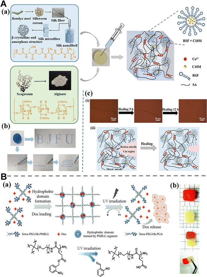 | ||
| Fig. 3 Supramolecular hydrogels cross-linked by hydrophobic interactions and their characteristics. (A) (a) Schematic illustration of the preparation process of the sodium alginate-regenerated silk fibroin (SA-RSF) hydrogels; (b) injection behavior of the SA-RSF-C18M hydrogels (stained with methylene blue); (c) (i) optical images of the self-healing process of SA-RSF-C18M-30 hydrogels; (ii) hydrogel region before and after healing. Copyright 2019 American Chemical Society.68 (B) (a) Schematic illustration of the design and application of the photo-responsive injectable hydrogel; (b) separated pieces of hydrogel (stained with eosin Y and without dye, respectively) were brought together and then the self-healing process occurred within 5 min, and the healed hydrogel could support its own weight. Copyright 2018 Royal Society of Chemistry.118 | ||
Recently, Zhang et al.118 reported an injectable and self-healing hydrogel cross-linked by hydrophobic interactions (Fig. 3B(a) and (b)). The o-nitribenzyl ester group is cleaved under UV irradiation with the hydrophobic domain transforming into a hydrophilic one, leading to the effective release of doxycycline (DOX).89,118 Both examples mentioned above are based on strong hydrophobic interactions, but weak hydrophobic interactions can also be applied to hydrogel structures. Jing et al.67 prepared a PEG-DPCA supramolecular hydrogel. As a structure directing agent and therapeutic agent, DPCA (dihydrophenonthrolin-4-one-3-carboxylic acid) exhibits reversible weak hydrophobic interactions between its domains, which enables the hydrogel to flow under external shear stress, and fully recover to the gelled state immediately after the stress is relieved.67,119
In addition, hydrogels formed through hydrophobic interactions usually exhibit a negative thermal response behavior (the rise of temperature leads to the hydrogel formation process). Generally speaking, the hydrophilic groups of macromolecules will undergo solvation by water molecules below the phase transition temperature, establishing hydrogen bonds with the surrounding water molecules. When the solution is heated, the mobility of water molecules increases, which destroys the solvated polymer chains and further promotes the formation of hydrogels.89,120,121
In conclusion, hydrophobic interaction is an intermolecular interaction that occurs universally in the human body, and supramolecular hydrogels cross-linked by this reversible interaction can have injectable or self-healing properties. Besides, the hydrophobic domain in hydrogels based on hydrophobic interactions can be used to load drugs or active substances and it will be discussed in Section 4.1.3.
2.4 Host–guest interaction
The host–guest interaction is realized through the mutual recognition of “host” and “guest” molecules. It is based on the formation of a selective inclusion complex between the cavities of host molecules and smaller guest molecules (such as adamantine).23,87 The shape and size complementarity between the host cavity and the guest molecules is crucial in this interaction. In this way, the host can pair with a variety of guest molecules which not only show a degree of inertness, but also respond to stimuli. In the hydrogel structure, the formation of the host–guest structure is relatively easy, which provides greater repeatability for the hydrogel performance. The reversible nature of host–guest interactions imparts hydrogels with the self-healing and shear thinning properties.122,123The host group encompasses a diverse array of naturally derived and synthesized macrocycles, as well as their derivatives. Cyclodextrin (CD) is the most prominent representative of hosts; it has a series of water-soluble and low-toxicity cyclic oligomers with 6, 7 and 8 truncated glucose units, which are called α-CDs, β-CDs and γ-CDs, respectively.23,124 CDs have hydrophilic outer shells and relatively hydrophobic inner cavities, and the hydrophobic inner cavity can form inclusion complexes with various types of guests23 like adamantane, azobenzene, ferrocene, cholesterol and polyethylene glycol.103,125 Besides, CD units can also be grafted onto other polymer chains, such as alginate, HA and PEG. The guest molecule is subsequently linked to another polymer chain, thereby facilitating the formation of supramolecular hydrogel.123,126 For example, Li et al.75 developed a new strategy for the preparation of hydrogels: developing convertible nanoparticles through the self-assembly of hydrophobic and proton sensitive host materials (HCD) and hydrophilic guest macromolecules (8PEG-Ada). Hydrophilic host molecules released from hydrolyzed nanoparticles under acidic conditions can spontaneously assemble with guest molecules through enhanced host–guest interactions, thus producing hydrogels (Fig. 4A(a)). It is noteworthy that orally administered transformable nanoparticles can effectively mitigate ethanol or drug-induced gastric injury in mice by forming an in situ hydrogel barrier (Fig. 4A(b)). As mentioned in the first three sections of this chapter, supramolecular interactions can exist in hydrogels as standalone interaction segments, or they can be combined with a variety of other supramolecular interactions to achieve synergistic effects. Moreover, self-healing, injectable, good mechanical properties and other properties are usually associated with many functions, and the host–guest supramolecular hydrogels described in this section are no exception. Previously, our group developed a series of self-healing and injectable supramolecular hydrogels by utilizing quaternized chitosan-graft-cyclodextrin (QCS–CD), quaternized chitosan-graft-adamantane (QCS–AD), and graphene oxide-graft-cyclodextrin (GO–CD) as building blocks. This approach combines the excellent antibacterial activity of QCS with the photothermal properties of reduced graphene oxide (rGO) (Fig. 4B(c)). The hydrogels exhibit rapid recovery within 14 seconds, owing to a diverse range of dynamic non-covalent interactions including host–guest interaction and hydrogen bonding among QCS–CD, QCS–AD, and GO–CD (Fig. 4B(b)).72 In addition, we also prepared a supramolecular hydrogel, which is composed of silk fibroin (SF), acryloyl-β-cyclodextrin (Ac-CD) and 2-hydroxyethyl acrylate through photopolymerization. Host–guest interactions and the hydrophobic β-sheet configuration are used to achieve a dual cross-linked hydrogel with good mechanical properties, rapid self-healing, injectability and good biocompatibility, which is conducive to wound healing (Fig. 5A(a) and (b)). It is worth noting that the self-healing process of our hydrogels can be completed quickly without any additional rest time, which makes them highly competitive among other physical hydrogels, because it usually takes several minutes or hours for traditional physical hydrogels to restore their original properties.127
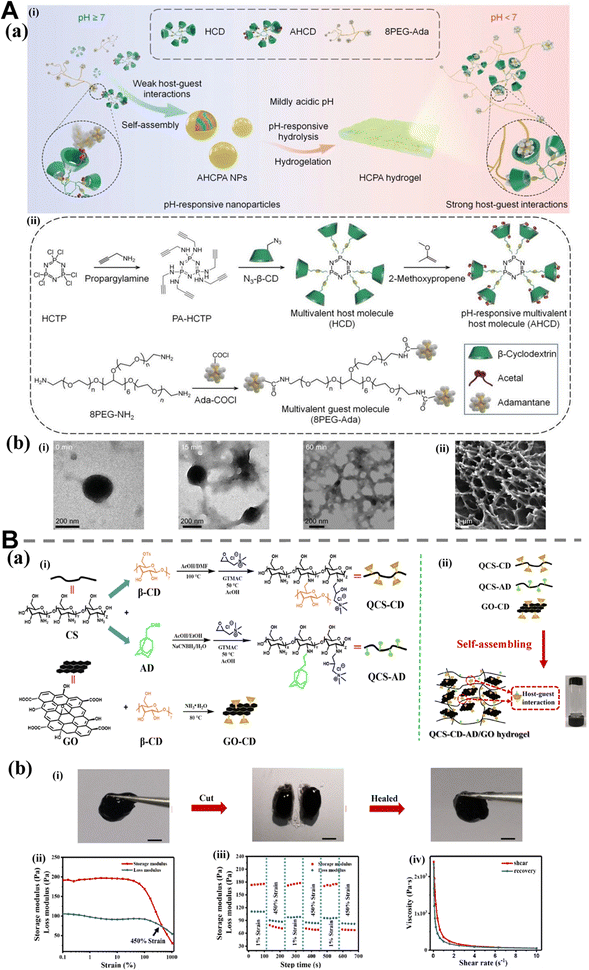 | ||
| Fig. 4 Supramolecular hydrogels cross-linked by host–guest interactions and their characteristics. (A) (a) Design and synthesis of pH-triggerable hydrogel-transforming nanoparticles based on host–guest recognition. (i) Schematic illustration of host–guest interaction mediated self-assembly of pH-responsive nanoparticles and acid-triggerable hydrogel transformation; (ii) schematics showing the chemical structure and synthesis of multivalent host and guest molecules. Abbreviation: HCTP, cyclic hexachlorocyclotriphosphazene; PA-HCTP, propargylamine-conjugated HCTP; N3-β-CD, mono-6-azido-β-cyclodextrin; HCD, β-cyclodextrin-conjugated HCTP; AHCD, acetalated HCD; 8PEG-NH2, 8-arm poly(ethylene glycol)amine; Ada-COCl, 1-adamantanecarbonyl chloride; 8PEG-Ada, 8-arm poly(ethylene glycol) conjugated with adamantane; AHCPA NPs, nanoparticles assembled by AHCD and 8PEG-Ada; HCPA hydrogel, host–guest hydrogel formed by pH-triggered transformation of AHCPA NPs; (b) (i) STEM images of AHCPA NPs after incubation at pH 5 for 0, 15, or 60 min; and (ii) a typical SEM image shows the cross-section structure of hydrogel transformed from AHCPA NPs at pH 5 after freeze-drying. Copyright 2022 Wiley-VCH GmbH.75 (B) (a) Schematic representation of QCS–CD–AD/GO supramolecular hydrogels preparation. (i) Preparation scheme of QCS–CD, QCS–AD and GO–CD polymers and (ii) QCS–CD–AD/GO supramolecular hydrogel; (b) (i) photographs taken during the self-healing process of hydrogel; (ii) G′ and G′′ of the supramolecular hydrogels on strain sweep test; (iii) the rheological property of the QCS–CD–AD/GO hydrogels when alternate step strain was switched from 1% to 450%; and (iv) viscosity dependence on the shear rate of the QCS–CD–AD/GO4 hydrogel. Scale bar: 5 mm. Copyright 2020 Elsevier.72 | ||
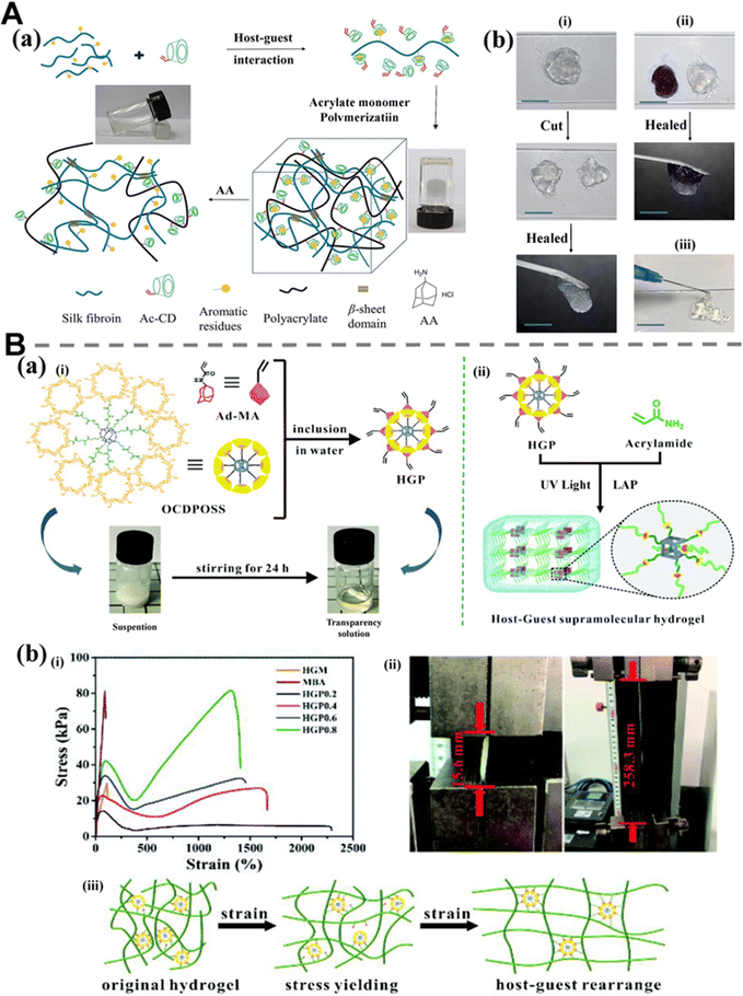 | ||
| Fig. 5 Supramolecular hydrogels cross-linked by host–guest interactions and their characteristics. (A) (a) Schematic illustration of the fabrication of self-healing SF/Ac-CD hydrogel; (b) photographs of the self-healing behavior (i) and (ii) and injectable behavior (iii) of SF/Ac-CD hydrogel dyed with crystal violet. Scale bar: 1 cm. Copyright 2021 Elsevier.127 (B) (a) Preparation of the HGP (POSS, polyhedral oligomeric silsesquioxan) supramolecular cross-linker and the HGP hydrogel. (i) The synthesis of HGP, where the solution can be observed by the transformation from suspension to transparent solution after inclusion complexation; and (ii) schematic illustration of the preparation of the HGP hydrogel via reversible host–guest interactions. Supramolecular cross-linker (HGP), acrylamide (AAm), and photo-initiator (LAP) were mixed in water and the supramolecular hydrogel was formed by UV-initiated cross-linking; (b) mechanical properties of the HGP hydrogels. (i) Stress–strain curves of the hydrogels (MBA, HGM, HGP0.2, HGP0.4, HGP0.6, and HGP0.8) subjected to tensile tests; (ii) photographs of the HGP0.8 hydrogel under tensile testing that extended over 15 fold to its initial length; and (iii) schematic of the possible mechanism involved in the fatigue resistance and stress yield mechanical behavior of the HGP hydrogels. Copyright 2020 Royal Society of Chemistry.77 | ||
As an indispensable cross-linking medium in traditional covalent hydrogel networks, chemical cross-linking agents are generally difficult to use in the cross-linking process of supramolecular hydrogels.128 In recent years, the emergence of a new type of cross-linking agent, the supramolecular cross-linking agent, has paved the way for scientific researchers, and has been gradually applied in the development of supramolecular hydrogels.129 Yang et al.69 prepared a self-healing supramolecular hydrogel by cross-linking acrylamide with a cross-linking agent assembled from poly(β-cyclodextrin)nanogel and azo phenylacrylamide. Photoisomerization of azobenzene can change its relationship with β-cyclodextrins, so that the cross-linking density and rheological properties of hydrogels can be adjusted by external light stimulation. It is also worth mentioning that damaged hydrogels lose their self-healing ability under ultraviolet irradiation, but can restore their self-healing behavior under visible light irradiation. The switchable self-healing properties are innovative, combining the properties of supramolecular networks with the concept of stimulus response.69,127,130 Similarly, Zhou et al.77 prepared a supramolecular hydrogel with self-healing properties and injectability by using a “supramolecular cross-linker” method (Fig. 5B(a)). The star supramolecular cross-linking agent is formed through the host–guest interaction between octa-cyclodextrin polyhedral oligomeric silsesquioxane (OCDPOSS) and acrylamide-modified adamantane (Ad-AAm). Subsequently, the host–guest interaction in the cross-linking agent became the actual cross-linking mechanism for hydrogels by means of co-polymerization. The multivalent host–guest interactions in the hydrogel improves its ductility, rapid self-healing and injectability. Simultaneously, the incorporation of rigid polyhedral oligomeric silsesquioxane as a supramolecular cross-linker endows supramolecular hydrogels with remarkable mechanical properties (Fig. 5B(b)).
In essence, supramolecular cross-linkers function as “intermediaries” in non-covalent cross-linking, combining the concept of “cross-linkers” in chemical hydrogels with non-covalent interactions. The emergence of supramolecular cross-linkers mitigates the adverse effects of toxic cross-linkers to a great extent, and offers a fresh approach for the creation of supramolecular hydrogels.
Thanks to the stability of the main part of the host–guest structure, its excellent molecular recognition ability and the ease of binding with guest molecules, researchers have utilized host–guest interactions to prepare hydrogels with self-healing, adhesion, and excellent biocompatibility, which have found widespread application in the field of wound dressings. However, many challenges and unmet needs remain. Most host–guest supramolecular hydrogels are produced at the laboratory level. The synthetic routes and post-processing procedures of materials are usually quite complex, and the reproducibility of experimental steps is poor, which greatly hinders their clinical conversion.131,132 On the other hand, the in vivo long-term toxicity of various host or guest chemical agents in supramolecular hydrogels is also a key issue that needs attention at this stage and is closely related to dressing removal (avoiding mechanical removal of dressing) and application safety (preventing the accumulation of toxic components over time).133 On the basis of the summary of previous research results and in-depth future studies, the application of host–guest supramolecular hydrogels in wound repair and hemostasis can be constantly improved by optimizing the structural design ideas, identifying and investigating potential problems.
2.5 Metal–ligand coordination
Chelation is a special interaction, which is based on two or more independent binding sites of a central atom on the same ligand. This chemical entity is called a chelate, and this interaction is called chelation interaction.134 In the coordination bond, multiple donor groups (ligands) may participate in the process of bonding with the central metal ion. Because it offers the full structure of metalloproteins and the catalytic centers of many enzymes, coordination chemistry is essential to biological systems. The strength of coordination bonds varies widely, and it can be equal to or even higher than that of covalent bonds, depending on the type of ligand and the properties of metal ions.134 But at the same time, metal coordination bonds are dynamic and reversible compared with covalent bonds. Various organic or inorganic ligands can combine with metal ions to form linear, branched or star-shaped complexes.23,134 Compared with other non-covalent bonds135 or dynamic covalent bonds, metal ligand coordination binding is unique in that biomaterials prepared through this interaction can have multiple wound healing functions when introducing inorganic particles and ions with inherent physical and chemical properties (such as antibacterial and anti-inflammatory function).136,137 The function of promoting wound repair associated with metal coordination will be discussed in detail in Sections 4 and 5.Hydrogels based on the coordination between metal ions and ligands are mainly obtained by functionalizing the polymer skeleton through chelating ligands.134,138 There are many kinds of ligands used to prepare supramolecular adhesion hydrogels, such as iron ion and catechol ligands.139 The complex is found in the adhesion protein of mussels, and it provides strong adhesion for different wet surfaces. For example, our group prepared a DN supramolecular hydrogel under physiological conditions, and endowed it with functions such as resistance to drug-resistant bacteria and promotion of skin repair. The first network of hydrogels is formed by catechol–Fe3+ coordination cross-linking. In addition to dynamic cross-linking, the network can also provide a variety of functions such as stimulus response, photothermal ability, tissue adhesion and free radical scavenging ability.52
Some modified products of chitosan, such as quaternary ammonium chitosan (QCS) and carboxymethyl chitosan (CMCS), are often used as matrix substances to construct metal coordination bonds. For instance, our group has previously developed an injectable and self-healing Mg-QCS/PF composite hydrogel capable of sustaining the release of Mg2+. Within the hydrogel structure, the hydroxyl groups and amino groups on the QCS main chain can form metal coordination bonds with divalent magnesium ions. The self-healing properties of QCS/PF hydrogels enhance the safety profile for their application in tendon sheath repair. Furthermore, it is noteworthy that this hydrogel exhibits improved stability at the tendon–bone interface due to its adhesive characteristics.116,140 CMCS has abundant coordination groups and groups which can generate hydrogen bonds. It has a class of naturally occurring ligands that can coordinate with metal ions, and has significant potential in various fields. For example, in the research conducted by Cao et al.,86 Fe3+ and Al3+ can form coordination bonds with CMCS that is uniformly dispersed in the alkali/urea aqueous solution for an ultrafine gelation process (Fig. 6A(a)). Due to the dynamic and reversible properties of metal coordination bonds, hydrogels have self-healing, adaptive and thermal response capabilities (Fig. 6A(b)).86 In addition, owing to a series of excellent characteristics of metal ligand coordination, it is often applied in multi-cross-linked and multi-network supramolecular hydrogels. Yan et al.141 presented the concept of multiple metal ions participating in coordination (Fig. 6B(a)). They successfully prepared a DN supramolecular hydrogel through strong coordination interaction between CMCS and various metal ions (Cu2+, Zn2+, Ni2+, Co2+, Fe3+ and Cr3+) (Fig. 6B(b)). Hydrogen bonds and metal coordination interaction are functionally independent and reversible, which makes the programming of hydrogels multifunctional, including the pH regulated shape memory behavior and multi-stage self-healing properties. Wu et al.142 prepared a tough double non-covalent cross-linking network hydrogel (Fig. 6C(a)). The rigid CS-ionic network is used as the first network while the pectin network cross-linked by Fe3+ is used as the second network of the hydrogel. The experimental results demonstrate the hydrogel's exceptional toughness (1.04–11.20 MJ m−3) and adjustable mechanical properties (tensile strength: 0.97–1.61 MPa, elongation: 133–1346%, elastic modulus: 0.30–2.20 MPa) (Fig. 6C(b)). Zhang et al.143 thought that in situ polymerization of conductive precursors into a polymer matrix can be a simple and effective method to improve the uniformity of silly putty-like materials. Consequently, a series of supramolecular hydrogels exhibiting excellent self-healing capabilities and photothermal conversion properties were synthesized through the utilization of metal–ligand interactions and hydrogen bonds. The incorporation of coordination bonds between Fe3+ and phytate polyvinyl alcohol (PPVA) imparts unique rheological characteristics to the hydrogels, including shear thinning behavior (Fig. 6D(a) and (b)).
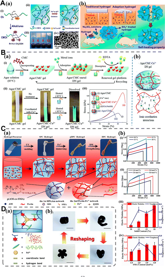 | ||
| Fig. 6 Supramolecular hydrogels cross-linked by metal ligand coordination and their characteristics. (A) (a) (i) Schematic illustration of CMCS; (ii) the structure of CMCS-Fe/Al hydrogels; (iii) photographs of CMCS-Fe 3.0–1.0 hydrogels prepared using CMCS with different degrees of substitution (from left to right, the hydrogels were prepared using CMCS-3, 6, 9, and 12); (iv) SEM image of the CMCS-Fe 3.0–1.0 hydrogel; (b) characteristics of the CMCS-Fe hydrogels applied in wound healing. Copyright 2021 American Chemical Society.86 (B) (a) (i) Concept of the two-step synthesis process of Agar/CMCS-metal DN hydrogels and erasable structure/properties; (ii) the copper ion-induced changes in CMCS in the optical and thermal behaviors of Agar/CMCS hydrogel; (iii) mechanical properties of Agar/CMCS hydrogels were varied in response to the metal-CMCS coordination interactions; (b) scheme of the supramolecular complexation of the metal ions and CMCS chains within the agar gel matrix. Copyright 2020 Royal Society of Chemistry.141 (C) (a) Digital images of the formation process of the pectin-based hydrogel precursor for DPC (double non-covalent cross-linking) and corresponding gelation mechanisms of SPC hydrogel, pDPC hydrogel, and DPC hydrogel; (b) mechanical properties of the pectin-based DPC hydrogel with different solid contents and SMA (stearyl methacrylate) contents. (i) Typical tensile stress–strain curves and (iii) elastic modulus (left) and toughness (right) of DPC hydrogels with different solid contents. (ii) Typical tensile stress–strain curves and (iv) elastic modulus (left) and toughness (right) of DPC hydrogels with different SMA contents. Copyright 2020 Elsevier.142 (D) (a) A schematic diagram showing the bonding interactions between every component inside the PPFe hydrogel; (b) photographs of the PPFe4 hydrogel cyclic remolding into different geometrical shapes, scale bar = 1 cm. Copyright 2020 Royal Society of Chemistry.143 | ||
Metal ligand-based supramolecular hydrogel dressings are still in their infancy. Due to the presence of metal ions and reversible coordination, these hydrogels can exhibit inherent antibacterial properties, electrical conductivity, and the ability to deliver active substances. Although they have been used as wound dressings or hemostatic materials, they also face many challenges. For example, the toxicity of metal ions should be considered when introducing them, and the commonly used metal ions are usually more toxic. We believe that following are the possible solutions to this problem: (1) change their forms (such as transforming them into metal nanoparticles), which will reduce the toxicity of metal ions in hydrogels and (2) select less toxic metal ions or biologically active metal ions (such as Mg2+ and Zn2+).144 On the other hand, when designing hydrogels, researchers mainly use natural polymers (such as gelatin and hyaluronic acid) or biocompatible synthetic polymers (such as polyethylene glycol). However, predicting the biocompatibility following the introduction of chelating ligands poses challenges due to the involvement of macromolecules and essential metal ions in biochemical reactions within the body, and their coordination with ligands may potentially impact endogenous biochemical processes.145 Therefore, for application in wound repair and hemostasis, the supramolecular hydrogels based on metal ligand coordination still have a long way to go and need further in-depth study.
2.6 Other interactions
In addition to the five common non-covalent cross-linking interactions described above, several uncommon non-covalent interactions have also been used in the cross-linking strategies of supramolecular hydrogels. Cation–π interaction is a non-covalent interaction arising from the mutual attraction between a cation and an aromatic system, and it is stronger than other π interactions (such as π–π interactions), so we can consider this interaction as a new method to control the polymer region and stereoselectivity. Many studies have proved this feature, and it exhibits synergistic effects with some substances, which is particularly important for the structural design of hydrogels with self-healing and injectable properties.146–148 For example, inspired by barnacle bone cement protein, Ni et al.149 combined cationic structure modified polyphosphazene with polyvinyl alcohol to prepare a series of dynamic viscous hydrogels. The cation–π interaction in the hydrogel further enhances the inherent interfacial toughness and cohesion of polymer chains, enabling stable adhesion to various wet tissue surfaces. In addition, π–π stacking (existing in the poly(N,N-dimethylethylenediamine-g-3-bromomethylphenylboronic acid)phosphazene chain) and cation–π interactions also enable the hydrogel to undergo rapid self-repair when damaged.Protein–protein interaction generally refers to the specific contact between proteins through molecular docking in a specific biological system. It can be divided into two types: direct contact between protein molecules, and complexation among proteins and other intermediates. It is worth noting that the stability of protein is not the same at different pH values due to its pH charge dependent ionizable groups, which makes it a key factor to consider in practical applications.150–153
In general, hydrogels cross-linked by multiple hydrogen bonds have mechanical properties comparable to those of covalent cross-linked hydrogels. Most supramolecular hydrogels constructed through hydrogen bonds have self-healing characteristics and some of them can also acquire shape memory properties; electrostatic interactions can be used to prepare self-healing supramolecular hydrogels, achieve drug delivery functions and improve the cross-linking degree of hydrogels; the hydrophobic domain in hydrogels based on hydrophobic interactions can be used to load drugs or active substances. Supramolecular hydrogels based on hydrophobic interactions can exhibit self-healing, stimulus response and other relative characteristics. They can also be used to enhance the mechanical properties of hydrogels, such as mechanical elongation, strength and toughness; the supramolecular hydrogel prepared via host–guest interaction has the characteristics of self-healing and shear thinning, which can also be used to increase the cross-link density of the hydrogel and correspondingly improve some mechanical properties; metal ligand coordination can be used to fabricate self-healing and stimuli sensitive supramolecular hydrogels, and can combine hydrogels with other excellent properties, such as building drug controlled release systems according to different intrinsic properties of various coordination bonds.
With advancements in research on supramolecular hydrogels, researchers have gained a deeper understanding of existing non-covalent cross-linking methods, and more and more non-covalent interactions will be introduced into the structure of supramolecular hydrogels. We expect that future work will facilitate the design of better supramolecular hydrogels. Some properties of hydrogels (rheological properties and mechanical properties) will be improved by regulating the non-covalent force and the cross-linking structure of hydrogels, and these hydrogels can be endowed with more intelligence.
3 Network structures of supramolecular hydrogels
It is well known that the structure of a material determines its properties, which is also suitable for hydrogels. The network structure design of a hydrogel affects a series of properties, so different properties of hydrogels are attributed to different types of network structure. This section will commence with an exploration of the network design of supramolecular hydrogels, aiming to elucidate the intricate relationship between the network structures and the properties exhibited by these hydrogels. We divide supramolecular hydrogel network structures into single, double and triple network structures, and they are described and discussed in detail, aiming to stimulate further thinking among readers. Table 2 lists three different network types of supramolecular hydrogels.| Network type | Cross-linking interaction | Acting substance | Characteristics | Ref. |
|---|---|---|---|---|
| Multi-cross-linked single network | Hydrogen bonds; electrostatic interactions | CS and CS; cordycepin (CY) and CS | Shear-thinning behavior | 154 |
| Hydrogen bonds; metal ligand coordination | CS and TA; Fe3+ and TA | Strongly hygroscopic | 155 | |
| Electrostatic interaction; hydrophobic interaction | Methacrylate sulfonated betaine (PSBMA) and PSBMA; hydroxybutyl chitosan (HBC) and HBC | Self-healing properties | 156 | |
| Hydrogen bonds; electrostatic interaction | CS and CS; CS and heparin sodium (HS) | Superior strength and toughness (8.53 MPa and 11.32 kJ m−2); excellent extensibility (133.7%); good self-recoverability | 157 | |
| Electrostatic interaction; metal ligand coordination | Carboxyl and guanidine groups; peptide conjugate (Pept-1) and Ca2+, alginate (ALG) and Ca2+ | Self-healing properties (enables the hydrogel to fill irregularly shaped wound defects without premolding); injectability | 158 | |
| Host–guest interaction; electrostatic interaction | MPEG-grafted poly(ethylene argininylaspartate diglyceride) (MPEG-g-PEAD) and α-cyclodextrin; MPEG-g-PEAD and heparin | — | 159 | |
| Host–guest interaction; hydrogen bonds | Chitosan-graft-β-cyclodextrin and PNIPAM; adenine and adenine | Superior stretchability; good elasticity and resilience; rapid autonomous self-healing properties | 92 | |
| Host–guest interaction; hydrophobic interaction | SF and acryloyl-β-cyclodextrin (Ac-CD); SF | Injectability (After the hydrogel was extruded through a 26-gauge needle, G′ of hydrogel showed a slight increase to ∼ 72 Pa) | 14 | |
| Hydrogen bonds; metal ligand coordination | PVA and TA, CS and TA; TA and Fe3+ | Injectability; self-healing properties | 160 | |
| Hydrogen bonds; metal ligand coordination | Phytate polyvinyl alcohol and polypyrrole; phytate polyvinyl alcohol and Fe3+ | Self-healing properties; unique solid–liquid viscoelasticity | 143 | |
| Hydrogen bonds; hydrophobic interaction | UPy; alkyl chain | Compressive strength (4 MPa); rapid self-healing properties; injectability | 161 | |
| Hydrogen bonds; hydrophobic interaction | PAAM and P(S-AA) core-shell nanoparticles; SMA chain | Excellent mechanical properties (the tensile strength and elongation at break after swelling reached 71 kPa and 978%, respectively); self-healing properties | 162 | |
| Hydrogen bonds; electrostatic interaction; metal ligand coordination | Acrylamide and acrylic acid (PAM); CATNFC and carboxyl; Al3+ and alginate-dopamine (Alg-DA) | Self-healing properties (the healed hydrogel could be bent or stretched, and easily lift a load of 500 g without breaking) | 163 | |
| Hydrogen bonds; hydrophobic interaction; metal ligand coordination | Betamethasone phosphate; betamethasone phosphate; betamethasone phosphate and Ca2+ | Injectability, nanofiber hydrogel, durable drug release | 164 | |
| Hydrogen bonds; π–π stacking | Orotic acid modified chitosan (OACS) and 2,6-diaminopurine (DAP); OACS and DAP | Self-healing properties; injectability | 55 | |
| Hydrogen bonds; metal ligand coordination | PVP and TA; TA and Fe3+ | Self-healing properties; injectability | 57 | |
| Hydrogen bonds; electrostatic interaction; hydrophobic interaction | AA and AA; positively charged CTAB head group and negatively charged carboxy groups on the AA; CTAB | Superior tensile strength (≈1.6 MPa), large stretchability (>900%), rapid room-temperature self-recovery (≈3 min at 100% strain) | 56 | |
| Hydrogen bonds; host–guest interaction | Paxle polymer and Cucurbit[6]urils (CB[6]); Paxle polymer and 1,4,5,8-naphthalenediimide (NDI)-bridged CDs | Excellent mechanical behaviors, showing a Young's modulus of up to 1.01 MPa, toughness of up to 3.80 MJ m−3, maximum stress of up to 1.12 MPa, and stretchability up to 1966% | 79 | |
| Double network | Hydrogen bonds; π–π stacking; hydrophobic interaction | Aldehyde of FPMA; benzene ring of FPMA; poly{[FPMA(4-formylphenyl methacrylate)-co-DEGMA[di(ethylene- glycol) methyl ether methacrylate]-b-MPC(2-methacry-loyloxyethyl phosphorylcholine)-b-(FPMA-co-DEGMA)} | Excellent injectability; repeatable self-healing properties | 165 |
| Hydrogen bonds; electrostatic interaction | Hydroxyl of AAC; CS and citrate ions, SBMA | Highly stretchability, anti-fatigue property; self-healing properties | 166 | |
| Hydrogen bonds; metal ligand coordination | Hydroxyl of agar; CMCS and metal ions (Cu2+, Zn2+, Ni2+, Co2+, Fe3+, and Cr3+) | Multi-staged self-healing properties | 141 | |
| Metal ligand coordination; electrostatic interaction | SMA and Fe3+; PSMA chain | Tunable mechanical properties | 142 | |
| Metal ligand coordination; electrostatic interaction | SA and Ca2+; CS and SA | Excellent mechanical properties (with the maximum tensile strength of 0.190 MPa) | 167 | |
| Hydrogen bonds; electrostatic interaction | PAA; PANI and the incorporated tannic acid coated cellulose nanocrystals (TA@CNCs) | Excellent mechanical and electrical self-healing properties | 168 | |
| Hydrogen bonds; electrostatic interaction; hydrophobic interaction | CS and AAM chain of p(AAm-co-LMA); carboxyl-functionalized multi-walled carbon nanotubes (c-MWCNTs) and CS; LMA and SDS | Excellent flexibility, puncture resistance and self-healing capability | 169 | |
| Hydrogen bonds; hydrophobic interaction | Hydroxyl of PVA and phenolic hydroxyl of TA, amide of BSA and phenolic hydroxyl of TA; benzene ring of TA and hydrophobic pocket of BSA | Ultrahigh tensile strength | 170 | |
| Hydrogen bonds; electrostatic interaction | Hydroxyl of PVA, hydroxyl of PVA and carboxyl of PAANa, hydroxyl of PVA and PAH; carboxyl of PAANa and PAH | Excellent mechanical properties (fracture strain: 768% and toughness: 2.25 MJ m−3); recovery capabilities (recovery ratio of maximum stress: 90.5%) | 58 | |
| Triple network | Hydrogen bonds and metal ligand coordination | Gelatin, PVA and PVA; PAA and Fe3+ | Superior toughness, strength and recovery capacity | 171 |
| Hydrogen bonds, electrostatic interaction and metal ligand coordination | Al3+ and CMCS NPs, Al3+ and PAA; CMCS NPs and PAA; CMCS NPs and PAA | Good extensibility (1930%) and high mechanical strength (190.9 kPa); self-healing properties (HE of stress is 68–80%, HE of strain is 87–96%, 25 °C, 24 h of healing) | 172 | |
3.1 Single network supramolecular hydrogels
A hydrogel which is connected by a cross-linking agent or relies on only one cross-linking interaction and has one single 3D network is called a single cross-linked single network hydrogel.173 When the cross-linking in these hydrogels is based on noncovalent interactions, we call them single cross-linked single network supramolecular hydrogels in the review. Compared with traditional chemically cross-linked hydrogels, single cross-linked single network supramolecular hydrogels exhibit superior performance in some aspects (such as self-healing, injectability, shape memory properties and stimulus responsiveness). For example, supramolecular hydrogels prepared by Jing et al.67 through hydrophobic interactions promote tissue regeneration while possessing many attractive properties: simple injection, shear thinning and high drug-carrying. However, because of their single cross-linking mode and network structure, non-covalent cross-linked hydrogels may also exhibit disadvantages (poor mechanical properties). With the introduction of the concept of dual networks and multi-networks, and the development of multifunctional, multi-crosslinked and composite hydrogels, some defects of single cross-linked single network supramolecular hydrogels can be addressed.When macromolecules are connected by two or more than two cross-linking interactions (or cross-linking agents), the network structure is called a multi-cross-linked network structure.173 A double cross-linked single polymer supramolecular network is mainly a single network structure containing two different, dynamic and noncovalent cross-linking interactions. This allows them to compensate multiple stimuli, and provides better mechanical properties than single cross-linked polymers. Even so, their mechanical properties can still be improved a lot, which will be discussed in comparison with other structures later. Double cross-linked single network supramolecular hydrogels can be constructed through any combination which consists of two kinds of supramolecular interactions, and the two types of noncovalent cross-linking interactions may exhibit sensitivity to two different stimuli.173,174 For example, Song and his colleagues154 developed a self-healing cordycepin (CY)/CS supramolecular hydrogel dressing, which was prepared through hydrogen bonds and electrostatic interactions without any cross-linking agents. The hydrogel has an appropriate swelling ratio and self-healing properties, and can fill irregularly shaped wound defects without the need for preforming, due to the network's synergy between hydrogen bonds and electrostatic interactions. Similarly, Azadikhah et al.160 prepared a double cross-linked supramolecular hydrogel by using the freeze–thaw method based on hydrogen bonds and metal coordination. TA molecules act as a cross-linking agent by establishing multiple hydrogen bonds between polymer chains and coordinating with Fe3+ to further connect them (Fig. 7A(a) and (b)). The hydrogel has a double cross-linked network structure, which endows it with good mechanical properties, injectability, self-healing and shear thinning properties.
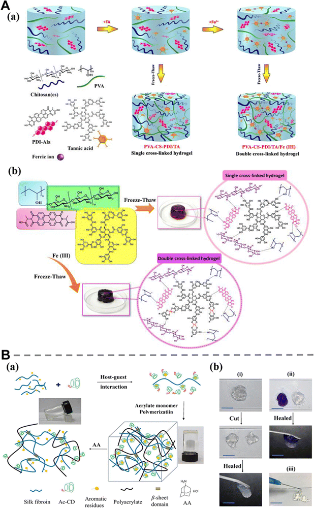 | ||
| Fig. 7 Multi-cross-linked single network supramolecular hydrogels and their characteristics. (A) (a) Schematic illustration of the formation of single cross-linked and double cross-linked supramolecular hydrogels; (b) preparation of PVA-CS-PDI/TA (single cross-linked) and PVA-CS-PDI/TA/Fe(III) (double cross-linked) hydrogels and possible interactions involving hydrogen bonds and metal-catechol coordination bonds. Copyright 2022 Elsevier.160 (B) (a) Schematic illustration of the fabrication of self-healing SF/Ac-CD hydrogel; (b) photographs of self-healing behavior (i) and (ii) and injectable behavior (iii) of SF/Ac-CD hydrogel dyed with crystal violet. Scale bar: 1 cm. Copyright 2021 Elsevier.14 | ||
In view of the limitations of silk fibroin hydrogel, such as long gelling time, high temperature and lack of self-healing, our team prepared a supramolecular hydrogel using 2-hydroxyethyl acrylate, acryloyl-β-cyclodextrin (Ac-CD) and SF. Unlike other methods of grafting CD onto SF through complex steps, this method can integrate the advantages of SF, Ac-CD and Cur into one system by simply modifying the original SF and CD. Ac-CD was selected as a host molecule while the aromatic amino acids of SF was selected as a guest molecule, and the second polymer skeleton was formed by the polymerization of Ac-CD and 2-hydroxyethyl acrylate (HEA) (Fig. 7B(a)). Double cross-linked hydrogels through host–guest interactions and a hydrophobic β-sheet configuration (hydrophobic interaction) exhibit long-term stability, rapid self-healing properties (Fig. 7B(b)), injectability and good biocompatibility, which was conducive to its application as a wound dressing.14
A multi-cross-linked single supramolecular network is usually a single network structure formed by more than two different non-covalent cross-linking interactions. The concept of multi-cross-linking is based on single cross-linking and double cross-linking, so that the types of cross-linking in the structure of multi-cross-linked networks are relatively more complex, which indirectly leads to the complexity and flexibility of the synergies between these cross-linking modes.175 For example, Qi et al.56 prepared a tough supramolecular hydrogel (Fig. 8A(a)) by modifying hydrophilic poly(acrylic acid) (PAA) with a little of hydrophobic lauryl methacrylate (LMA) in the presence of cetyltrimethylammonium bromide (CTAB). Due to the synergistic effects of electrostatic and hydrophobic interactions between CTAB and the P(AA-co-LMA) copolymer, the hydrogel exhibits exceptional resistance to swelling with a water content of 58.5 wt%. Additionally, it demonstrates remarkable tensile strength (approximately 1.6 MPa), high stretchability (>900%), and rapid self-recovery at room temperature (around 3 minutes at 100% strain) (Fig. 8A(b)). Wang et al.176 prepared SPU-PAA/Zn self-healing hydrogel films (Fig. 8B(a)). The synergistic effect of coordination interaction (between sulfonate groups of SPU and Zn2+, carboxylic acid groups of PAA and Zn2+) and hydrogen bonds (between carboxylic acid groups of PAA and urea as well as urethane groups of SPU) in the network and the enhancement effect of hydrophobic domains formed in situ greatly improve the mechanical properties of the hydrogel. Meanwhile, these multiple non-covalent cross-linkages also impart it with self-healing and elastic properties (Fig. 8B(b)). Deng et al.163 developed a supramolecular hydrogel with antibacterial properties using Al3+ and alginate–dopamine (Alg-DA) chains to cross-link with the copolymer chains of acrylamide and acrylic acid (PAM) through triple dynamic noncovalent interactions (hydrogen bonds, metal coordination, and electrostatic interaction). It is worth noting that the hydrogel can be easily recycled in a pollution-free manner, thanks to the synergy of three non-covalent interactions and setting it apart from other hydrogels with a similar structure. Furthermore, the hydrogel exhibits remarkable advantages in terms of cellular and tissue affinity as compared to other hard and antibacterial hydrogels, which is typically harmful to cell growth, owing to the presence of Alg-DA, so that it has high application potential in repairing soft tissue and preventing bacterial infection of wounds. The application of multi-cross-linked single network hydrogels in wound repair and hemostasis will be discussed in Chapters 4 and 5.
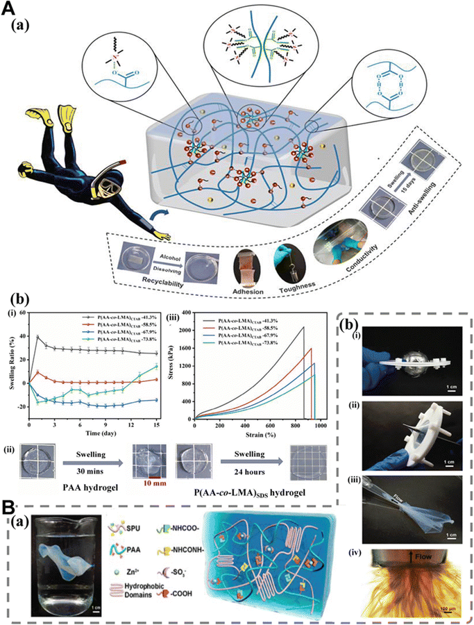 | ||
| Fig. 8 Multi-cross-linked single network supramolecular hydrogels and their characteristics. (A) (a) Schematic illustration of the fabrication of the tough, anti-swelling, and conductive supramolecular hydrogel for underwater sensors. (b) Swelling behaviors of the hydrogels: (i) swelling curves of different P(AA-co-LMA)CTAB hydrogels in deionized water at 25 °C for 15 days. (ii) Photographs of the swelling behaviors of the PAA hydrogel and P(AA-co-LMA)SDS hydrogel in deionized water at 25 °C. (iii) Typical stress–strain curves of different P(AA-co-LMA)CTAB hydrogels. Copyright 2022 American Chemical Society.56 (B) (a) Photographs of a piece of SPU-PAA/Zn hydrogel membrane suspended in water and schematic illustration of the structure of the SPU-PAA/Zn hydrogel membrane. (b) Photographs and optical micrograph to demonstrate the excellent mechanical properties of the SPU-PAA/Zn hydrogel membrane. Hydrogel membrane can withstand (i) the weight of a 200 g steel ball and (ii) the stab of tweezers with a sharp tip. (iii) Process of sucking a SPU-PAA/Zn hydrogel membrane with an area of ∼4 × 6 cm2 into a micropipette with an inner diameter of the tip being approximately 800 μm. (iv) Close-up optical micrograph around the tip during the sucking process of the hydrogel membrane. Copyright 2021 American Chemical Society.176 | ||
3.2 Double network supramolecular hydrogels
The traditional double network (DN) hydrogel consists of interpenetrating networks (IPNs) synthesized and infiltrated by two different polymer components in turn to form the whole system.177,178 The first network is a highly cross-linked polyelectrolyte that serves as a rigid support to maintain its shape, while the second network is a loosely cross-linked flexible neutral polymer used as a filler to absorb external stress. Therefore, DN hydrogel showed both rigidity and softness.177,179 Under conditions of large deformation, the rigid network dissipates energy through cross-linked fracture, while the flexible network remains relatively intact, which endows the hydrogel with the ability to recover from deformation to a certain extent. Therefore, the combination of these two different types of networks enhances the mechanical properties, and the resulting hydrogels also show different types of mechanical response properties.180–182 Compared with DN hydrogels synthesized using two covalent cross-linking networks,178,181,183 the DN hydrogels prepared with partial or complete non-covalent cross-linking exhibit greater toughness.178,184 The rupture of the non-covalent bonds absorbs mechanical energy, improve toughness, and the broken bond can also be restored to rebuild the structure of the hydrogel.185,186Although biomedical applications require high strength, high toughness and self-healing properties, traditional concepts hinder the possibility of coexistence of these different properties in DN hydrogels. Moreover, the use of toxic initiators and acrylamide-based monomers is not feasible. In addition, when considering the operation of hydrogel in tissue engineering, the injectability of DN hydrogel is also an important factor affecting the performance of hydrogel.187 Different from the multi-cross-linked single network supramolecular hydrogels introduced in the previous section, the two networks in DN supramolecular hydrogels can not only synergistically enhance the performance of hydrogels, but also exist independently, each dominating different performance of hydrogels and being endowed with corresponding functions.188 For example, Xia et al.169 fabricated a DN multi-functional supramolecular hydrogel for wearable strain sensors (Fig. 9A(a)). The polyacrylamide (PAAm) network is cross-linked by hydrophobic interactions, and the CS network is cross-linked by the electrostatic interaction of carboxyl-functionalized multi-walled carbon nanotubes (c-MWCNTs). The interconnection between these two networks is further reinforced through physical entanglement and the formation of hydrogen bonds. These cross-links work together to endow the hydrogel with excellent stretch and puncture resistance. In the CS network, the presence of CS endows hydrogels with enhanced self-adhesion behavior, and the introduction of c-MWCNT endows the hydrogels with superior strain-sensitive conductivity (Fig. 9A(b)). Similarly, Zhao et al.165 reported a DN supramolecular hydrogel with injectability and self-healing properties (Fig. 9B(a) and (b)). When the temperature is above the critical gel temperature, the heat responsive polymer segment drives dehydration and the hydrophobic interaction, causing the polymer chain to become entangled with the first network. At the same time, non-covalent interactions between benzaldehyde groups will be formed, including hydrogen bonds, hydrophobic interactions and π–π interactions as the second network. When the benzaldehyde content increases from 0 to 8.2 mol%, the critical gel temperature of the hydrogel decreases significantly from 35.5 °C to 19.9 °C, while concurrently exhibiting a substantial increase in the storage modulus from 21 Pa to 1411 Pa, which indicates that the critical gel temperature and storage modulus of hydrogels are only controlled by the second network in the structure. In order to enhance the therapeutic effect of hydrogels on damaged tissues, Dong et al.156 fabricated a series of composite hydrogels based on hydroxybutyl chitosan (HBC) and methacrylate sulfonated betaine with heat sensitivity, self-healing properties, and antibacterial activity (Fig. 9C(a)). The methacrylate sulfonated betaine network and the hydrophobic interaction cross-linking HBC network are cross-linked by electrostatic interaction to form a fully non-covalently cross-linked double network structure. The existence of the methacrylate sulfonated betaine network can promote the sol–gel transition of HBC. The HBC network has good killing activities against very few bacteria adhering to the surface of the material. The synergistic effect between anti-protein adhesion ability of the methacrylate sulfonated betaine network and the bactericidal ability of the HBC network makes hydrogels contributes to the good synergistic antibacterial performance of hydrogels. In addition, hydrogels do not need contain any cross-linking agents (Fig. 9C(b)).
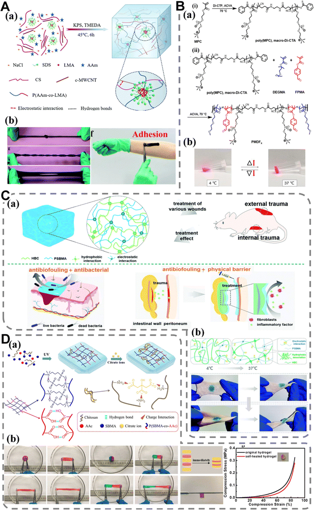 | ||
| Fig. 9 Double network supramolecular hydrogels and their characteristics. (A) (a) Formation of the HPAAm/CS-c-MWCNT hybrid hydrogel. (b) The HPAAm/CS-c-MWCNT hybrid hydrogel exhibits extraordinary mechanical properties: ordinary stretching with knotting, twisting and holing; photographs of the HPAAm/CS-c-MWCNT hybrid hydrogel adhering to arm. Copyright 2019 American Chemical Society.169 (B) (a) Synthesis of (i) difunctional macro-CTA poly(MPC) and (ii) triblock copolymer PMDFx. (b) Temperature-triggered sol–gel transition of the GMDF40 hydrogel. Copyright 2022 American Chemical Society.165 (C) (a) Schematic diagram of the hydrogel and its biomedical application. (b) Schematic diagram of the state of composite hydrogel at different temperatures and composite hydrogel can accommodate complex deformations (the hydrogel was dyed blue with methylene blue or pink with rhodamine B). Copyright 2022 Royal Society of Chemistry.156 (D) (a) Schematic diagram of the preparation of P(SBMA-co-AAc)/CS-Cit DN hydrogels. (b) Tensile properties of the P(SBMA-co-AAc)/CS-Cit DN hydrogel; self-healing properties of the hydrogel; assembly properties of the hydrogel; compression performance of the original and self-healed hydrogel. Copyright 2020 Elsevier.166 | ||
Supramolecular double-network structures are also commonly used by researchers as basic structures to better enshrine the corresponding functions of hydrogels. DN supramolecular hydrogel sensors are a good example. Zhang et al.166 fabricated a supramolecular hydrogel of poly(sulfobetaine-co-acrylicacid)/chitosan-citrate (P(SBMA-co-AAc)/CS-Cit) through the utilization of electrostatic interaction and hydrogen bonds between polymer chains (Fig. 9D(a)). The hydrogel exhibits high tensile strength, transparency, fatigue resistance, self-adhesive and good self-healing performance (Fig. 9D(b)). The self-healing efficiency reaches an impressive 95.4%. Furthermore, the hydrogel exhibits high sensitivity to a wide range of strains, making it suitable for use as a strain sensor capable of detecting joint movements such as bending and swallowing.
Thermosensitive hydrogels have always been the key objects for the functionalization of supramolecular hydrogels, especially multi-network supramolecular hydrogels, because of their characteristic of responding to external temperature changes. However, the majority of temperature-sensitive hydrogels reported to date are based on synthetic amphiphilic copolymers. Nevertheless, these synthesized copolymers lack biological activity, while agarose hydrogel exhibits a high melting temperature and chitosan can only be dissolved in acidic solvents. Moreover, physical gelatin hydrogel is unstable at body temperature and demonstrates low strength and poor stability similar to other single network supramolecular hydrogels. These problems greatly hinder the application of sports-related skin tissue healing.189,190 In view of the above restrictions, our team utilized poly(glycerol sebacate)-co-poly(ethylene glycol)-g-catechol (PEGSD) and ureido pyrimidinone-hexamethylene diisocyanate (UPy-HDI) modified gelatin (GTU) as raw materials in 2020. By exploiting the catechol–Fe3+ coordination between PEGSD and Fe3+, along with the quadrupole hydrogen bonds of GTU, we successfully fabricated a supramolecular DN hydrogel exhibiting rapid shape adaptability, swift self-healing, and responsiveness to near-infrared/pH stimuli. As a kind of synthetic branched macromolecule, PEGSD can endow hydrogel with good flexibility, while the catechol groups on it can coordinate with Fe3+, resulting in dynamic cross-linking, NIR/pH-responsiveness, photothermal capacity, tissue adhesiveness and free radical scavenging. The gelatin (GT) network can promote tissue regeneration, and the UPy groups on the GT backbone can provide the second non-covalent network. Furthermore, the hydrogen bonds and the catechol–Fe3+ coordination can respond to photothermal and/or acidity. When exposed to NIR radiation and/or the acid solution wash, the hydrogel dressing can be easily removed.52
Overall, after the mechanical properties of DN supramolecular hydrogels are enhanced, a variety of functions can be incorporated into them. In contrast to previous research, our work has endowed the hydrogel with a number of excellent reversible properties (on-demand removable tissue adhesion, dual stimulation response, rapid shape adaptation and self-healing) and multiple functions of promoting wound-healing (antibacterial, antioxidant, and angiogenesis promotion function), all while satisfying the high strength of the hydrogel network.
3.3 Triple network supramolecular hydrogels
Triple network hydrogels are a kind of multi-network hydrogel. They are similar to DN hydrogels, but show a more complex relationship among their networks. In previous studies, multi-network technology has been widely used to prepare hydrogels with excellent performance.171,191 A variety of non-covalent interactions described in Chapter 2 can be introduced into the structure to construct triple network compound hydrogels. For example, Dai et al.192 designed a triple network hydrogel with physical and chemical double cross-linking. The first non-covalent network is constructed by utilizing the hydrogen bonds and related entanglement between the hydroxyl groups of PVA chain as the cross-linking point. Then, the PVA chain reacts with borax to form reversible diol-borax coordination bonds, forming the second network. Finally, the PVA crystal domain generated through the freeze–thaw cycle is used to form the third network. The experimental results show that the triple cross-linked network structure provides reliable and stable recovery performance and self-healing ability without any external stimulation.192,193 Moreover, it is still difficult to achieve a shape memory hydrogel with multi-networks, multi-stimuli response and multiple shape memory effects with three-dimensional deformation at the same time. Wang et al.194 developed a simple method to prepare a triple-network SMH. Their hydrogel consists of a stable chemically cross-linked PVA network, a reversible ion cross-linked CMCS–Fe3+ network and a hydrogen bonded PVA network. The shape memory behavior triggered by Fe3+, heat and near-infrared light was realized by using the coordination interaction between CMCS and Fe3+ and the stable temporary shape of reversible hydrogen bonds formed by PVA-OH.However, due to the existence of chemical cross-linking in the structure, although the composite triple network hydrogels listed above have better mechanical properties and stability, their dynamic properties such as self-healing effects and injectivity may be weakened. Because chemical cross-linking networks are more likely to interact with the surrounding material, the bonds formed tend to be strong. The emergence of triple network supramolecular hydrogels provides a feasible solution to this problem.
Triple network supramolecular hydrogels add a new network to the foundation of DN supramolecular hydrogels, enhancing their designability, but the relationships and interactions between networks become more complex at the same time. Thanks to the strength and stability of the triple network itself, the triple network supramolecular hydrogels can even have the obvious advantages of single network and DN supramolecular hydrogels in some aspects without the need for any reinforcement means. For example, Pan et al.172 utilized Al3+ and carboxymethyl chitosan nanoparticles to cross-link PAA chains via metal coordination interaction, hydrogen bonds, and electrostatic interaction, resulting in the preparation of supramolecular hydrogels with noncytotoxic properties (Fig. 10(a) and (b)). Benefiting from the strong triple noncovalent cross-linking network and the dynamic nature of non-covalent bonds, the hydrogel shows ideal mechanical properties (Fig. 10(d)) (a breaking strength of 190.9 kPa and a breaking elongation of 1930%) and excellent self-healing properties (Fig. 10(c)) (a stress healing efficiency of 92.9% and a strain of 98.8%, 25 °C, healing for 24 h), and will not be significantly disturbed by surface aging or erosion (such as acid, alkali and salt solutions). What is striking is that due to the complete non-covalent cross-linking structure, the prepared hydrogel is also easily recycled in a pollution-free manner. Similarly, Jing et al.171 synthesized a triple network supramolecular hydrogel. The first network comprises a PAA-Fe3+ hydrogel, which is formed through metal coordination between carboxyl groups and Fe3+. The second and third networks consist of a gelatin hydrogel and PVA hydrogel, respectively, formed via hydrogen bonds. The non-covalent cross-linking networks can provide PAA-Fe3+/gelatin/PVA with good extensibility and shape recovery properties (Fig. 11(a)). Compared with single network and DN hydrogels, PAA-Fe3+/gelatin/PVA triple network hydrogels have better toughness, strength and recovery capacity, and their mechanical properties can be dynamically changed by adjusting their composition (Fig. 11(b)). The triple network supramolecular hydrogel exhibits excellent self-healing performance, with a remarkable healing efficiency exceeding 95% (Fig. 11(c) and (d)).
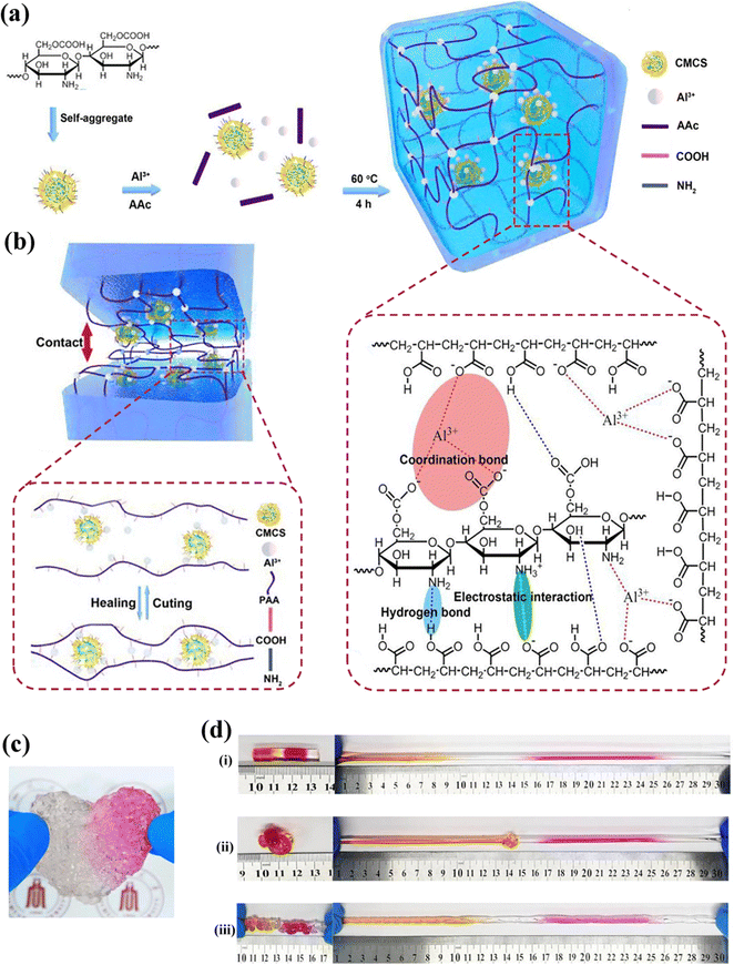 | ||
| Fig. 10 Triple network supramolecular hydrogels and their characteristics. (a) Schematic illustration of the preparation and structure of PAA-Al3+-CMCS hydrogels; (b) schematic illustration of the self-healing process of PAA-Al3+-CMCS hydrogels; (c) self-gluing behavior of the PAA-Al3+-CMCS1.3 hydrogel; (d) the four pieces of rod-shaped specimens from the PAA-Al3+-CMCS1.3 hydrogel were healed into a complete hydrogel that can sustain (i) stretching, (ii) knotting, and (iii) twisting. Copyright 2019 Elsevier.172 | ||
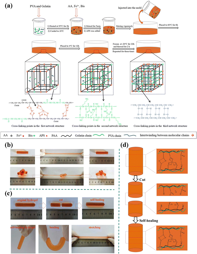 | ||
| Fig. 11 Triple network supramolecular hydrogels and their characteristics. (a) Synthesis of PAA-Fe3+/gelatin/PVA triple-network supramolecular hydrogels; (b) photographs of the synthesized triple-network hydrogel (PAA-Fe3+(0.20)/gelatin3%/PVA15%) under different conditions demonstrating excellent mechanical deformation and processability. Hydrogel showing ability to withstand stretching and knotting; (c) macroscopic self-healing behavior of the synthesized triple-network hydrogel (PAA-Fe3+(0.20)/gelatin3%/PVA15%). (d) Schematic of the self-healing mechanism of PAA-Fe3+/gelatin/PVA triple-network hydrogels. Copyright 2019 Society of Chemical Industry.171 | ||
At this point, we can sum up the traits of various network structures found in supramolecular hydrogels and the connections between them as follows. Single network supramolecular hydrogels typically have poor mechanical characteristics, but these characteristics can be improved. Most single network supramolecular hydrogels have very good reversible properties due to their relatively weak cross-linking. Multi-cross-linked single network hydrogels, on the other hand, have cross-linking combinations that are more varied and flexible, allowing them to respond to various stimuli while also having better mechanical properties. The mechanical properties of DN hydrogels have been somewhat enhanced (double three-dimensional structure) compared to single network supramolecular hydrogels. The designability of the structure has been greatly enhanced by the ability of the two networks to either work together synergistically or separately. Finally, triple network supramolecular hydrogels are among the best in terms of mechanical properties, designability and reversibility; however, the relationships and interactions between the networks have grown more complex, raising the bar for research in cross-linking, network design, and raw material selection.
4 Application of supramolecular hydrogels in wound repair and hemostasis
4.1 Application of supramolecular hydrogels in wound repair
The situation of the wound and its surroundings is very complex, and the wound healing process is affected by many internal and external factors.8 As a unique member of the hydrogel family, supramolecular hydrogels have shown unprecedented potential and advantages in the field of wound dressings. In this section, the application of supramolecular hydrogels in wound healing will be discussed in detail from six aspects: antibacterial ability (Table 3), tissue adhesion (Table 4), substance delivery, anti-inflammatory and antioxidant function, function of cell control, angiogenesis promotion (Table 5) and other functions. Specifically, starting from the mechanism behind each function, we will classify and discuss the common function implementation methods, and then evaluate and summarize them.| Antibacterial strategies | Functional components | Antibacterial mechanism | Ref. |
|---|---|---|---|
| Using substances with inherent antibacterial properties | TA | Inhibiting extracellular microbial enzymes, depriving microorganisms of the substrate they need to grow | 31,32,36,44,45,57,160 |
| CY | — | 154 | |
| GA | Inhibiting the synthesis of polysaccharides on bacterial biofilms | 195,196 | |
| Using metal particles/ions | Ag+, Cu2+, Zn2+, Fe3+, Al3+ | Damage to cell wall function, damage to cell enzymes or proteins, damage to cell DNA structure | 85,141,157,197,198 |
| Polycationic antibacterial strategy | CS, CMCS, HBC | Cationic functional groups are adsorbed to negatively charged bacterial surfaces by Coulomb force. The insertion of lipophilic alkyl chains into the phospholipid bilayer helps destroy the bacterial membrane structure | 29,39,156,160 |
| QCS | Quaternary ammonium groups disrupt the bacteria cell membrane and further cause the death of bacterial | 199 | |
| Photothermal antibacterial strategy | AT | Converting light into heat, the high temperature inactivates enzymes in bacterial cells, thus achieving the effect of sterilization | 200 |
| PPy NTs | — | 199 | |
| rGO | The stronger contact between rGO's sharp nanowalls and bacterial cell membranes, as well as better charge transfer between bacteria and nanowalls lead to bacterial cell membrane damage | 72 | |
| Synergetic antibacterial strategy | Alginate–dopamine (Alg-DA); Al3+; cationized nanofibrillated cellulose (CATNFC) | The Alg-DA can provide the hydrogel with stronger adhesion, which will facilitate the contact with bacteria at the wound site, and improve the antibacterial effectiveness of CATNFC | 163 |
| CS, TA and Fe3+ | — | 155 | |
| CS and Ag+ | — | 201 |
| Different adhesion functions | Functional components | Adhesion mechanism | Ref. |
|---|---|---|---|
| Ordinary adhesion | TA | TA supplies free pyrogallol, which can bind to tissue surface by forming reversible noncovalent or irreversible covalent interactions | 31,32,37 |
| CS | The positively charged amino groups of chitosan adhered to the negatively charged acidic groups in human tissues through electrostatic interaction | 202 | |
| UPy, aldehyde group modified PEG | Hydroxyl groups on UPy form hydrogen bonds with groups on the tissue surface; the aldehyde group on PEG forms an imine bond with the amino group on the tissue surface | 161 | |
| Repeatable adhesion | CEs | Hydrogen bonds between micelles hair layer and molecules of various interface | 38 |
| Catechol | The catechol-mediated non-covalent interactions between the catechol-functionalized hydrogel and soft tissue (including π–π and cation–π interactions, electrostatic interaction, and hydrogen bonds) | 203 | |
| Shape adaptive adhesion | HA, PVP | Hydrogen bonds between hydrogel and tissue (The softy of hydrogel makes it easy to fill the irregular wound bed and increase the contact area between the matrix and itself) | 30 |
| Functions of promoting wound repair | Functional classification | Functional components | Action mechanism | Ref. |
|---|---|---|---|---|
| Function of substance delivery | Drug delivery | Menthol, lidocaine hydrochloride (LDC), curcumin, minocycline hydrochloride, doxycycline (DOX), 1,4-dihydrophe-nonthrolin-4-one-3-carboxylic acid (DPCA), ofloxacin, dexamethasone sodium phosphate (Dexp), dexamethasone, diclofenac sodium (DS), quercetin (QUE), stimulator of interferon genes (STING) agonist-4 (SA), metformin (MET), hymexazol (Hy), paclitaxel (PTX) | — | 27,43,51,54,62,64,66,67,70,73,76–78,204–209 |
| Bioactive substance delivery | Fibroblast growth factor 2, vascular endothelial growth factor (VEGF), basic fibroblast growth factor (bFGF) | — | 14,159,210 | |
| APD1 antibody, IL15 cytokine and STING agonist (CDA) | — | 211 | ||
| — | — | 212 | ||
| A kind of antimicrobial peptide: KRGCGALGLPCKRIVKRIKKWLR, C-C cyclic | — | 80 | ||
| Functional gas delivery | Nitric oxide (NO) | — | 213 | |
| Anti-inflammatory function | Using natural or synthetic polymers | GA | Activate the inflammatory stage earlier by secreting a large number of chemokines and accelerating epithelialization | 195 |
| AT | — | 200 | ||
| Loading anti-inflammatory drugs | Dexamethasone (DEX) | Inhibit the aggregation of inflammatory cells at the inflammatory site, and inhibit the phagocytosis, the release of lysosomal enzymes, and the synthesis and release of inflammatory chemical mediators | 75 | |
| Heparin | The binding effect to inflammatory chemokines | 204 | ||
| SN50(a specific inhibitor of the NF-κB signaling pathway); betamethasone phosphate (BetP); pinocembrin (PNCB) | — | 164,214 | ||
| Antioxidant function | Using natural or synthetic polymers | TA | Phenolic hydroxyl groups of TA transferring electrons or donating hydrogen atoms to ABTS+ free radicals, and the inherent anti-inflammatory activity of TA | 37 |
| 44 | ||||
| Loading relevant substances | Superoxide dismutase (an enzyme) | Remove harmful superoxide anion free radicals | 104 | |
| PCA | Superoxide dismutase (SOD) and Nrf2 induced by PCA treatment increase | 215 | ||
| MET | — | 209 | ||
| Using metal ions | Ag+ | Stimulates immune functions to produce a large number of white blood cells in the early phase of the wound healing process, leading to synergistic antibacterial activity, and then quickly reduces the number of white blood cells, shorten the time of inflammation | 34 | |
| Function of cell control | Promotion of cell adhesion | HA | hold the cells within the matrix over the stroma and then allowing them to settle over time | 216 |
| rGO | rGO supports the adhesion and proliferation of human fibroblasts | 72 | ||
| Mg2+ | Interaction between Mg2+and the integrins | 140 | ||
| Promotion of cell proliferation | Anti-bacterial chitosan | Provide an environment conducive to cell interaction | 100 | |
| SF | — | 217 | ||
| Pectin | — | 142 | ||
| Epidermal growth factor (EGF) | Binding to EGF receptor, resulting in dimerization and activation of the receptor as well as phosphorylation of tyrosine from downstream proteins | 217 | ||
| Promotion of cell differentiation | PDGF-BB protein | 218 | ||
| Cell migration promotion | Cell adhesive peptide conjugate (Pept-1) and ALG | RGD moieties accelerated cell migration through integrin αvβ3 receptors | 158 | |
| Platelet-derived growth factor (PDGF) | — | 219 | ||
| Function of promoting angiogenesis | Fenugreek gum | Promote the expression of VEGF and TGF-β | 40 | |
| Gelatin | Promote the expression of VEGF | 220 | ||
| PDGF mimetic peptide | Promote the expression of vascular endothelial-specifc marker CD31 | 219 | ||
| Fibroblast growth factor | The sustained-release of fibroblast growth factor | 221 |
Some natural polymers with biocompatibility are widely used in the preparation of inherently antibacterial supramolecular hydrogels because of their universal or conditional antibacterial properties. Among them, several polyphenolic compounds (tannic acid and catechol) and natural polysaccharides (chitosan and agarose) are the most popular.227 Tannic acid (TA) is a natural nontoxic polyphenol compound with good antibacterial, anti-inflammatory, antioxidant and hemostatic properties. TA also has the characteristics of interacting with polymers and molecules through hydrogen bonds and hydrophobic interaction under neutral conditions. So, TA can be cross-linked with hydrophilic polymers (PVA and PEG) to form a supramolecular hydrogel network through non-covalent interactions, conferring it with certain antibacterial activity.32,36,228 Usually, a series of bacteria such as S. aureus, E. coli and Str. pyogenes are used in bacterial experiments. For example, Zhang et al.32 prepared a self-healing, antibacterial and adhesive supramolecular hydrogel (TAHE hydrogel) through multiple hydrogen bonds between TA and poly(N-hydroxyethyl acrylamide) (Fig. 12A(a)); the resulting hydrogel was called TAHE-x–y, where x is PHEAA concentration in percentage by weight, and y is TA concentration in percentage by weight. The results of the study on antibacterial properties revealed that TAHE-2–50 and TAHE-2–100 hydrogels exhibited bactericidal activity against approximately 80% of S. aureus and Str. pyogenes. Following incubation for 12 hours, no colony growth was observed on the agar plate when the diluted LB medium containing treated bacteria was plated (Fig. 12A(b)). Guo et al.36 constructed a new PVA/ANFs/TA DN supramolecular hydrogel (PAT hydrogel) based on several different strength hydrogen bonds, and tested the antibacterial activity of PVA, PA and PAT hydrogels against E. coli, S. aureus and P. aeruginosa. The results showed that the inhibition rate of the PAT hydrogel against the above three bacteria was 100%. TA in hydrogel could inhibit the formation of S. aureus biofilms through inhibition of gene expression and destruction of peptidoglycan. In addition, TA has a strong binding ability to metal ions. Therefore, it complexes or electrostatically reacts with metal ions, causing microorganisms to lose their substrate, thus inhibiting the proliferation of bacteria.
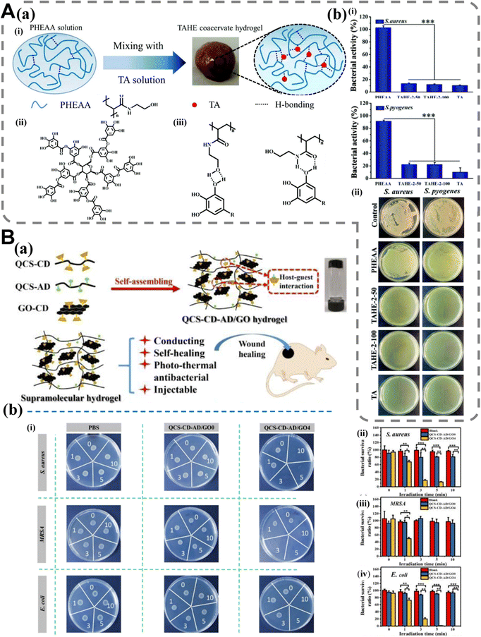 | ||
| Fig. 12 Supramolecular hydrogel dressings with antibacterial function. (A) (a) (i) Schematic diagram of the preparation process of the TAHE hydrogel. (ii) The molecular structure of TA; (iii) the possible hydrogen bonds between the TA and PHEAA polymer; (b) comparison of the bactericidal activity of PHEAA, TAHE-2–50, and TAHE-2–100 hydrogels with TA against S. aureus and S. pyogenes (i) and digital images of surviving bacterial clones on agar plates after coming into contact with hydrogels (ii). Data values correspond to mean ± standard deviation (SD), n = 5. Error bars represent SD. ***P < 0.001, one-way analysis of variance (ANOVA). Copyright 2020 Royal Society of Chemistry.32 (B) (a) QCS–CD–AD/GO supramolecular hydrogel, characteristics of QCS–CD–AD/GO hydrogel and application in wound healing; (b) photo-thermal antibacterial behavior of PBS, QCS–CD–AD/GO0 and QCS–CD–AD/GO4 hydrogels. (i) Images of NIR-induced photo-thermal antibacterial activities of QCS–CD–AD/GO hydrogel against S. aureus, MRSA and E. coli, with 0, 1, 3, 5 and 10 representing different irradiation times (min). The bacterial survival ratios of S. aureus (ii), MRSA (iii) and E. coli (iv) as a function of NIR irradiation time for the QCS–CD–AD/GO4 hydrogels. Copyright 2020 Elsevier.72 | ||
Chitosan (CS)229 has positive charges in an acidic environment, and it can kill bacterial cells by changing the permeability of the cell membrane.39,157 Moreover, the amino group present in chitosan readily undergoes protonation, thereby conferring an inherent bactericidal effect to the polymer.230 For example, in the work carried out by Song et al.,154 CY/CS supramolecular hydrogel showed good antibacterial activity against S. aureus and E. coli. The CS-TA@CeO2 supramolecular hydrogel developed by Teng et al.44 showed good antibacterial activity against Gram-positive bacteria and Gram-negative bacteria. This work adopts the strategy of introducing both TA and CS to endow the corresponding hydrogel with antibacterial properties. However, the development of chitosan in antibacterial hydrogels is limited due to its poor antibacterial performance and solubility. By introducing quaternary ammonium, carboxymethyl or hydroxybutyl groups into CS, quaternized chitosan (QCS), carboxymethyl chitosan (CMCS) and hydroxybutyl chitosan (HBC) with enhanced antibacterial ability can be synthesized.231,232 In 2020, our team successfully developed a series of supramolecular hydrogels with enhanced antibacterial properties, injectability, self-healing capability, and conductivity through the utilization of host–guest interactions (Fig. 12B(a)). After being in contact with three kinds of bacteria at 37 °C for 2 hours, all hydrogel groups were able to kill S. aureus and MRSA completely and 95% of E. coli. The antibacterial properties of the hydrogel are mainly attributed to QCS (Fig. 12B(b)).72 Compared to previous work, we combined the antibacterial activity of QCS with photothermal properties and introduced a new photothermal antibacterial strategy into the supramolecular hydrogels prepared in another work of our group in 2022. The hydrogel showed good antibacterial activity, and the killing ratio of both bacteria (E. coli and MRSA) was higher than 95%. Therefore, these hydrogels can be highly valuable for wound disinfection and protection from bacterial invasion.92 In addition to the natural polymers with inherent antibacterial activity described above, other natural polymers, such as cellulose, do not have antibacterial ability in essence. Therefore, researchers have employed the cationic method to synthesize antibacterial nano-cellulose; however, it is crucial to consider the impact of quaternary ammonium compound (QAC) molecular weight on the performance of these antibacterial materials. In summary, this approach presents a novel opportunity for developing hydrogel materials with inherent antibacterial activity, thereby circumventing the need for potentially toxic substances such as antibiotics or heavy metal particles.233–235
Antibacterial supramolecular hydrogels based on CMCS and HBC have also been reported. For example, the hydrogen bond cross-linked SF/CMCS supramolecular hydrogel prepared by Chen et al.29 maintains a suitable water environment for wound healing, and shows unique characteristics in terms of antibacterial activity against E. coli and S. aureus. Dong et al.156 constructed a composite supramolecular hydrogel by using the electrostatic interaction between polymethyl methacrylate sulfonated betaine molecules and the hydrophobic interaction between HBC molecules (Fig. 13A(b)). The hydrogel maintained strong antibacterial activities against E. coli and S. aureus (at least 87.3% and 92.2%, respectively), because the antifouling network can effectively prevent the adhesion of proteins and bacteria, while the HBC network can kill the attached bacteria via the positive charge (Fig. 13A(a) and A(c)). The synergetic antibacterial system is innovative in its design. Deng et al.163 developed a new type of supramolecular hydrogel by using Al3+ ligand coordination, electrostatic interaction and hydrogen bonds (Fig. 13B(a)). The experimental results demonstrated that the Alg-DA-CATNFC-PAM-Al3+ hydrogel exhibited inhibitory effects against both Gram-positive and Gram-negative bacteria (Fig. 13B(b)). The broad-spectrum antibacterial ability of CATNFC (cationized nanofibrillated cellulose) provides it with good antibacterial performance, while the activity of Al3+ only slightly improves this antibacterial performance.
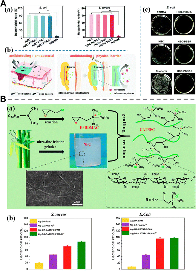 | ||
| Fig. 13 Supramolecular hydrogel dressings with antibacterial function. (A) (a) Quantitative statistics of the bactericidal effect of E. coli and S. aureus after being co-cultured for 12 h with hydrogels; (b) schematic diagram of the hydrogel and its biomedical application; (c) optical photographs of the antibacterial effect. *p < 0.05 and **p < 0.01. Copyright 2022 Royal Society of Chemistry.156 (B) (a) The preparation principle of CATNFC; (b) bactericidal ratios on S. aureus and bactericidal ratios on E. coli. Copyright 2021 American Chemical Society.163 | ||
Inorganic metal nanoparticles/ions, including silver, copper, gold, zinc and other metal-containing nanoparticles, have been extensively employed for investigating potential antibacterial applications.236 Silver demonstrates a wide range of antibacterial activity and compatibility with mammalian tissues. Numerous strategies have been developed to incorporate silver into wound dressings, aiming to achieve bactericidal properties. Initially, silver nanoparticles were primarily physically coated onto the surfaces of hydrogels composed of a range of natural or synthetic polymers such as CS, gelatin, cellulose, xylitol, PVA and polyvinyl pyrrolidone (PVP). In addition to its antibacterial properties alone, Ag can also be combined or used together with various other substances (such as antibacterial drug and other metals) to achieve better antibacterial effects.8,236 For example, Wang et al.206 prepared a supramolecular hydrogel through simple Ag+ complexation, and successfully loaded the anionic drug diclofenac sodium (DS) using electrostatic interaction and host–guest interaction. The evaluation of the wound healing model in vivo showed that the hydrogel had excellent antibacterial activity. In addition to silver, Cu2+, as an important material for promoting wound healing, has also been widely studied. However, high concentration of Cu2+ may lead to heavy metal poisoning and serious toxic side effects. The method of loading Cu2+ onto nanocarriers has been proven to significantly reduce its toxicity and side effects, while showing excellent antibacterial activity.237,238 For example, Gao and his colleagues198 synthesized a supramolecular hydrogel for wound healing by using two physical processes: Cu2+ ligand coordination and hydrogen bonding. Choline–glycolate can promote the release of Cu2+ and generate hydroxyl radicals (˙OH) in the wound through chemodynamic therapy to kill drug-resistant bacteria. The survival rate of the two bacteria (Gram-positive methicillin-resistant S. aureus and drug-resistant Gram-negative E. coli) decreased significantly due to the presence of Cu2+.
In recent years, with the increasing demand for antibacterial properties of materials in the biomedical field, the reports of supramolecular hydrogels based on synergistic antibacterial strategies have also increased year by year. In addition, the combination of NIR and strong light absorption materials has been widely developed, especially for killing multi-drug resistant bacteria.239 For example, Yu et al.155 prepared a multifunctional supramolecular hydrogel wound dressing through one step mixing of CS, SF, TA and Fe3+via freeze-drying. The amine groups on CS have limited antibacterial properties. At the same time, TA and Fe3+ possess good NIR-responsive photothermal activity and could inhibit the growth of bacteria. The synergistic action of these substances provides hydrogels with excellent antibacterial properties. The antimicrobial efficiency of the CSTFe (3) hydrogel remained consistently high at 99.99% even after undergoing four cycles, which can be attributed to its repeatable photothermal properties and excellent UV-resistant photothermal stability. This remarkable stability is primarily due to the abundance of polyphenols in TA, which effectively capture UV-induced free radicals.
The introduction of photothermal effects into thermosensitive hydrogels is a reliable antibacterial design strategy. When the photothermal material is introduced into the temperature-sensitive hydrogel, severe infection can be overcome by photothermal sterilization, and the hydrogel can be removed on request by NIR radiation.240 For example, our team has prepared supramolecular hydrogels with rapid self-healing, tissue adhesion, antioxidant activity, photothermal antibacterial and near-infrared/pH stimulus responsiveness through Fe3+ ligand coordination and quadruple hydrogen bonds. The results demonstrate the effective near-infrared assisted photothermal antibacterial activity of our hydrogel against both Gram-positive and Gram-negative bacteria, rendering it a promising photothermal antibacterial adhesive dressing for in vivo treatment of mouse skin full-thickness defect. The exceptional photothermal properties of the hydrogels primarily stem from the efficient near-infrared absorption and thermal conversion facilitated by catechol–Fe3+ coordination, as well as the contributions from catechol groups and UPy motifs.52
However, one of the fatal drawbacks of photothermal antibacterial strategies is that most of them can only achieve a topical bactericidal effect. Therefore, photothermal antibacterial strategies combined with synergistic effects have been developed to fill this gap. For instance, our team has successfully developed a series of supramolecular hydrogels with promising characteristics by employing dual dynamic bond cross-linking involving Fe3+, QCS, and protocatechualdehyde (PA) containing catechol and aldehyde groups. The Schiff bases incorporated in these hydrogels possess an aromatic ring that exhibits notable antibacterial activity, enabling effective pathogen inactivation through electrostatic interactions between positively charged QCS and the negatively charged bacterial cell membrane. Additionally, the catechol–Fe3+ coordination cross-linked QCS exhibits a moderate photothermal capacity, enabling the hydrogel to efficiently absorb and convert near-infrared (NIR) light into heat for bactericidal activity.241
Issues such as dressing falling off under external force and inaccurate adhesion during use often appear in the actual application process of adhesive hydrogels. The adhesion process with repeatability and fault tolerance (mispositioned hydrogel can be removed and repositioned without any loss of tissue adhesion250) can effectively solve the above problems. Supramolecular hydrogels are one of the best choices for reusable adhesive hydrogels because of their non-covalent dynamic properties. Repeatable adhesion can be realized either through a single non-covalent interaction or through the synergy of multiple non-covalent interactions. For example, a new type of organic supramolecular hydrogel synthesized by Wang et al.38 exhibited enhanced mechanical properties and good adhesion ability through the unique “load sharing” effect (Fig. 14(a) and (b)). The hydrogel exhibits fast adhesion formation and good adhesion repeatability, and it is suitable for use as a wound dressing for major surface injuries. The adhesive properties mainly arise from the hydrogen bonds present between the micelles hair layer and various interfacial molecules (Fig. 14(c) and (d)). The adhesion repeatability test demonstrated that even after undergoing 10 cycles of adhesion and detachment, the interfacial toughness remained 153.46 J m−2, representing a retention rate of 74% compared to the initial application. Similarly, Wei et al.203 (Fig. 15A(a)) confirmed that the adhesion strength of the prepared hydrogel could be restored after separation, and repeated application did not have a significant effect on adhesion strength, which was due to π–π and cation–π interactions, electrostatic interaction and hydrogen bonds between hydrogel and soft tissue (Fig. 15A(b)).
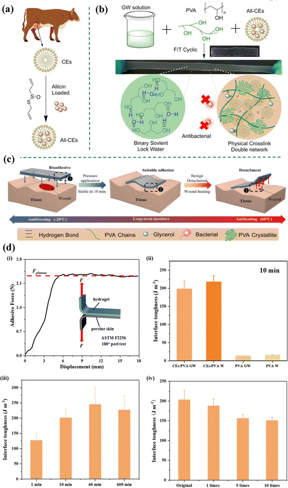 | ||
| Fig. 14 Supramolecular hydrogel dressings with adhesive properties. (a) Casein mainly originated from bovine milk and had a unique micelle structure, which was designed to load allicin; (b) the schematic of the formation and mechanism of CEs (casein micelles)/PVA GW (glycerol–water) and its images in tensile tests; (c) the tissue adhesion and skin matching mechanical properties of CEs/PVA GW remain stable even in hot (60 °C) and cold (−20 °C) environments, making CEs/PVA GW a potential candidate for use as a wound dressing; (d) the adhesion properties of the hydrogel system. (i) The scheme illustration of the method of interfacial toughness, following ASTM F2256; (ii) the interfacial toughness of hydrogel samples (PVA W, PVA GW, CEs/PVA W and CEs/PVA GW) after applying external pressure for 10 min; (iii) the interfacial toughness of CEs/PVA GW with external pressure applied at different time points (1 min, 10 min, 100 min and 600 min, respectively); (iv) the interfacial toughness of CEs/PVA GW after 0, 1, 5 and 10 cyclic adhesion and detachment. Copyright 2022 Elsevier.38 | ||
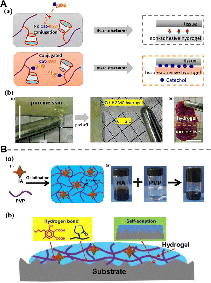 | ||
| Fig. 15 Supramolecular hydrogels with tissue adhesive properties. (A) (a) Schematic illustration of the unsuccessful functionalization of the control HGMC hydrogel and the successful functionalization of TU-HGMC hydrogels with Cat-RGD; (b) (i) tissue adhesion of the activated TU-HGMC hydrogels demonstrated by stretching the TU-hydrogels adhered to porcine skin; (ii) tissue adhesion of the TU-HGMC hydrogels demonstrated by gluing two pieces of the porcine liver together with the TU-HGMC hydrogel. Copyright 2019 American Chemical Society.203 (B) (a) (i) Schematic illustration of HPC hydrogels cross-linked by HA in an ultrafast process; (ii) images showing the preparation process of the HPC hydrogels; (b) the adhesion mechanism of the HPC hydrogels. Copyright from an open access journal of Frontiers in Bioengineering and Biotechnology.30 | ||
In order to better fill wounds of different shapes and maximize the effect of hydrogel dressings, injectable and adaptable hydrogels have been developed. In addition, certain situations require that hydrogels only work for certain periods of time and can be easily removed to avoid the additional damage thought to be caused by removal. For example, Yu et al.30 developed a typical supramolecular hydrogel cross-linked by hydrogen bonds in order to meet some special requirements for hydrogel based biomaterials, in which hyaluronic acid (HA) forms hydrogen bonds with polyvinylpyrrolidone (PVP) as a non-covalent cross-linking means. First, the hydrogen bonds incorporated within the hydrogel network act as cross-linkers, effectively enhancing cohesion. Additionally, at the interface between the hydrogel and matrix, interactions are further strengthened through the establishment of hydrogen bonds with functional groups. Second, the soft nature endows the hydrogel with good environmental adaptability, and makes it easy to fill the irregular wound bed, thus increasing the contact area between the matrix and itself.
Adhesion performance can also be linked to other functions, such as adhesion performance as a way to achieve hemostasis, through the physical closure of the injured site for rapid and effective hemostatic treatment. This part will be further discussed in 4.2.1.
As far as drug loading is concerned, chemical bonding between drugs and carriers is one of the commonly used methods in hydrogel systems.256 Compared with chemical bonding, physical encapsulation causes less chemical interference in cell activity and drug action, and can reduce the structural changes introduced into the loaded drug. In addition, physically encapsulated drug loading does not involve any chemical trigger, which makes the drug loading process easier and faster. These advantages make physical entrainment a more favorable loading method for fragile or sensitive drugs.257 Curcumin is a kind of natural low-molecular-weight polyphenol which is the main active ingredient of curcuma. It is used to relieve pain and promote wound healing due to its anti-infection, anti-inflammatory and antioxidant properties. However, the effectiveness of curcumin is limited due to its low solubility in water. A supramolecular hydrogel prepared by Zhu et al.62 exhibits an excellent drug loading effect due to the presence of mixed micelles (methoxyl poly(ethylene glycol)-block-poly(ε-caprolactone) and poly(ε-caprolactone)-block-poly(hexamethylene guanidine) hydrochloride-block-poly(ε-caprolactone)). In another work, our research group loaded curcumin into the SF based supramolecular hydrogel to improve wound healing performance and produce higher granulation tissue thickness.14 The supramolecular hydrogel prepared by Wang et al.206 shows a good ability to load the anionic drug diclofenac sodium (DS) due to the combined effect of host–guest inclusion complexation and electrostatic attraction. Similarly, Xu et al.27 encapsulated the model drug menthol with ethylenediamine modified cyclodextrin, and then loaded it into the prepared supramolecular hydrogel dressing. The drug delivery system is effective in reducing the volatilization of menthol, and has a long-term effect of anti-inflammation and analgesia. Because if only menthol and EDA-CD are mixed in the solid phase, without the protection of EDA-CD cavity and hydrogel, the menthol will volatilize faster.
Of course, the delivery and release of drugs is one of the multiple delivery properties that hydrogels can support.258 Ofloxacin is a drug that can treat bacterial infections, which is used to prove that supramolecular hydrogel can be used as a carrier for therapeutic agents.259 Xu et al.70 monitored the relationship between the release of ofloxacin and time through the network within 1 hour (Fig. 16A(a)). The hydrogel can load and release antibacterial agents, which can prevent infection and promote healing (Fig. 16A(b)). The supramolecular hydrogels with hydrophobic domains are very suitable for drug loading and delivery, and have been widely studied as drug carriers for small molecular chemotherapeutic drugs, nucleic acids, anti-inflammatory and antibacterial drugs. The supramolecular hydrogel structure prepared by Cheng et al.67 has a high drug load and releases DPCA (an enzyme inhibitor) through ester hydrolysis, leading to tissue regeneration in non-healing wounds. In another work, Yu et al.73 loaded a supramolecular hydrogel with an insoluble model drug, dexamethasone (Fig. 16B(a)). Large quantities of β-CDs attached to hyaluronic acid can load hydrophobic drugs directly without using any other drug delivery system. The content of 4-arm-PEG-Ad has a significant effect on the drug release rate. With the less content of 4-arm-PEG-Ad in hydrogels, the higher release was observed because of more free CDs to contain drugs (Fig. 16B(b)).
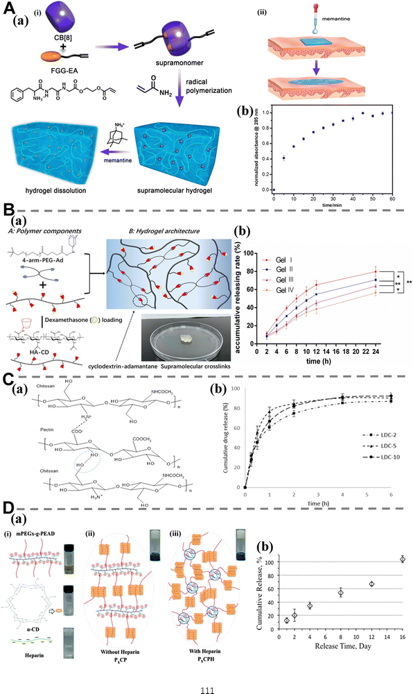 | ||
| Fig. 16 Supramolecular hydrogels with loading and delivery functions. (A) (a) Schematic depiction of (i) supramolecular hydrogel fabrication from supramonomers and its dissolution process upon memantine irrigation and (ii) its application as wound dressing materials; (b) release profile of ofloxacin from 1.4 M gel when immersed into PBS buffer observed by monitoring the absorption band of ofloxacin which peaked at 285 nm over time. Copyright 2017 American Chemical Society.70 (B) (a) Polymer components and hydrogel architecture of supramolecular hydrogel; (b) the release curves of dexamethasone from gel I, gel II, gel III and gel IV within 24 h. Copyright 2020 Elsevier.73 (C) (a) Physical interactions between chitosan (CS) and pectin (PEC) polysaccharides; (b) in vitro release of lidocaine hydrochloride (LDC) drug from 3D printed chitosan-pectin (CS-PEC) hydrogel scaffolds with different amounts of drug loading (n = 3 ± SD). Copyright 2019 Elsevier.43 (D) (a) Formation of a dually cross-linked supramolecular hydrogel based on host–guest chemistry and electrostatic interactions. (i) MPEGx-g-PEAD (MPEG-grafted poly(ethylene argininylaspartate diglyceride)), α-CD and heparin solutions are used to make the hydrogel; (ii) PxCP by mixing MPEGx-g-PEAD with α-CD based on host–guest chemistry alone. Inset: hydrogel P20CP from MPEG20-g-PEAD/α-CD (26.7 + 77.3 mg mL−1) gelled in 1 h after being subjected to magnetic stirring for 30 min; (iii) PxCPH by mixing MPEGx-g-PEAD, α-CD and heparin using host–guest chemistry as first cross-links and electrostatic interactions as secondary cross-links. Inset: hydrogel P20CPH from MPEG20-g-PEAD/α-CD/heparin (26.7 + 77.3 + 2.7 mg mL−1) gelled within 1 min after being subjected to magnetic stirring for 30 min; (b) release profile of FGF2 from P20CPH hydrogel at 37 °C. The release was nearly linear at a rate of approximately 31 ng per day with the hydrogel gradually dissociating at each time point and eventually disappearing at the end of the 16 day experiment, showing a hydrogel dissociation dependent release behavior. Copyright 2016 Royal Society of Chemistry.159 | ||
In addition to drug loading, supramolecular hydrogels also need to release drug to function at targeted sites, which is usually achieved through swelling process or degradation of hydrogels (hydrolysis of chemical bonds, structural dissociation, and stimulated dissolution).23 For instance, Lai et al.64 demonstrated that alterations in the swelling capacity can impact the release rate of drugs loaded within supramolecular hydrogel dressings (fabricated via electrostatic interactions between CS and carmellose), because the water in the dressing serves as the medium for drug molecule diffusion. Therefore, there is a positive correlation between the sustainability of the drug release and the percentage of CS in the dressing. The two drug models showed high drug release sustainability, which is due to the availability of a large number of CS amino groups in the gel process (leading to the formation of a more compact and dense cross-linked dressing with a lower swelling capacity and erosion sensitivity). Wang et al.38 loaded the model antibiotic (allicin) into casein micelles (CEs); the CEs/PVA glycerol–water (GW) hydrogel showed significant durable antibacterial properties. Moreover, the GW binary solvent system can also reduce the rate of drug delivery by restricting the swelling capacity, and can still act as wound dressing at high or low temperature. The CS–PEC hydrogel prepared by Long et al.43 contributes to the rapid release of captured lidocaine hydrochloride (LDC) (Fig. 16C(a)). The spongy structure of the hydrogel carrier promoted the absorption of solvents and controlled the release of LDC, which lasted up to 6 hours (Fig. 16C(b)). Another factor leading to rapid drug release is the degradation of CS–PEC hydrogel.
The process of wound healing necessitates intricate cellular interactions among various cell types, including keratinocytes, fibroblasts, endothelial cells, neutrophils, and macrophages. Additionally, it relies on the endogenous release of growth factors (GFs), cytokines, and chemokines. Furthermore, local administration of exogenous cells or cytokines has a favorable impact on wound healing. In addition to loading and releasing drugs, supramolecular hydrogel systems can also be used to deliver active substances (particles and growth factors). For instance, local delivery of exogenous cells or cytokines exhibits significant advantages in facilitating wound healing. In a recent work, Hu et al.260 prepared supramolecular hydrogels through electrostatic interaction and bivalent chelation with epidermal growth factor (EGF) payload. The load of EGF is distributed in the interconnected channels, and the resulting blocking network can slow down the diffusion of intercepted water. At pH 7.4, the ionic interaction between negatively charged EGF and secondary amine of CMCS also contributes to the prolonged release of EGF. Ding et al.159 incorporated fibroblast growth factor 2 into the prepared supramolecular hydrogel (Fig. 16D(a)), and the growth factor can be released at an almost stable rate continuously for up to 16 days. At each time node, when fresh salt solution is used at the top of hydrogel to replace the supernatant, the dissociation of the MPEG graft/α-CD complex occurs at the interface between gel and salt solution, leading to the gradual dissociation of hydrogel (Fig. 16D(b)). This phenomenon is combined with the stable release rate, indicating that the release kinetics is dominated by the dissociation of hydrogel.
The loading of cells into hydrogels has strict environmental requirements to ensure that the cells do not react with substances in the hydrogel skeleton structure or matrix that can cause adverse effects on the cells (damage cell structure). The fully dynamic cross-linking in the supramolecular hydrogel structure is perfectly fit for the physical loading requirements of cells, so we can use it to load cells. For example, Chavee et al.261 efficiently fabricated a supramolecular hydrogel by leveraging electrostatic and hydrophobic interactions between phospholipids and SF chains, thereby inducing the conformational transition of SF chains into beta sheet structures. Cell encapsulation experiments demonstrated that L929 and NIH/3T3 fibroblasts exhibited normal proliferation patterns when encapsulated, and the encapsulated SaOS-2 had a distinct proliferation profile.
The method of encapsulating cells in this study is comparable to the drug-loaded method of supramolecular hydrogels mentioned earlier, but in terms of the hydrogel structure, the double cross-linking mode of Chavee et al.'s supramolecular hydrogel offers more embeddable functions for hydrogels, which warrants further investigation.
In addition to the abovementioned substances, supramolecular hydrogels can also realize the loading, delivery and release of small particles (such as inorganic nonmetallic nanoparticles). This part has been introduced in more detail in Section 2.5. Moreover, a series of substance loading functions of supramolecular hydrogels are usually associated with other wound healing functions, that is, they appear as a means to achieve other functions. The relationship among these functions (anti-inflammatory function, antioxidant function, cell control, and function of promoting angiogenesis) will be discussed in detail later.
ROS plays a key role in the inflammatory response during wound healing.265–267 However, the presence of adverse factors in the wound can result in persistent infiltration of inflammatory cells and generate a substantial quantity of detrimental ROS.268 In situations where tissue's intrinsic antioxidant capacity is relatively insufficient, ROS may disrupt the DNA structure, protein integrity, and cell membrane lipids, thereby inducing cellular damage and apoptosis.269 As one of the most common antioxidant strategies, the introduction of intrinsic antioxidant activity is favored by the majority of researchers. The natural polyphenol substance TA repeatedly mentioned above is used to impart or enhance the excellent antioxidant performance of supramolecular hydrogel due to its polyphenol hydroxyls. The ability to scavenge DPPH free radicals has been used as an intuitive method to reflect the antioxidant capacity of supramolecular hydrogels. For example, Li et al.37 confirmed that the antioxidant conversion rate of double-layer hydrogel is about 32%, while the scavenging capacity of TA@double-layer hydrogel is increased to 88%. A large number of phenolic hydroxyl groups in TA enhance the antioxidant capacity of hydrogels by transferring electrons or providing hydrogen atoms. The excellent antioxidant capacity of the CS-TA@CeO2 hydrogel prepared by Teng et al.44 has also been confirmed to come from the TA and CeO2 components in hydrogel. Moreover, the reversible transition between the reduced (Ce3+) and oxidized (Ce4+) states occurring on the surface of cerium oxide nanoparticles significantly enhances their capacity to eliminate reactive oxygen species (ROS) and mitigate cellular antioxidant stress. Their test was also completed through the evaluation of DPPH free radical scavenging ability. Curcumin, a naturally occurring compound, has exhibited remarkable antioxidant and anti-inflammatory properties that could potentially enhance the process of wound healing. Despite its high value, its applications are limited because of its hydrophobicity and instability. To overcome this problem, our team developed an injectable supramolecular hydrogel with self-healing capability through host–guest and hydrophobic interactions. Based on the above, a pretreatment was performed to form the Ac-CD/Cur inclusion compound, and the anti-inflammation hydrogel with curcumin was prepared. Additionally, curcumin exerts an influence on activating antioxidant enzymes within cells. The antioxidant activity of the prepared hydrogel was predominantly attributed to curcumin, which exhibited remarkable efficacy in scavenging free radicals.14
Furthermore, supramolecular hydrogels can be loaded with antioxidant enzymes to enhance their antioxidant capacity. Notably, superoxide dismutase (SOD) and catalase (CAT) are recognized as crucial antioxidant enzymes. Upregulating the expression of SOD and CAT can mitigate oxidative stress and facilitate wound healing. Zhang et al.104 constructed a supramolecular hydrogel dressing with antioxidant properties by loading SOD through physical absorption (Fig. 17A(a)). SOD loaded by physical adsorption has been proven to have high biological activity. This kind of SOD released mildly from higher content of heparin may play a better role in wound healing (Fig. 17A(b) and (c)).
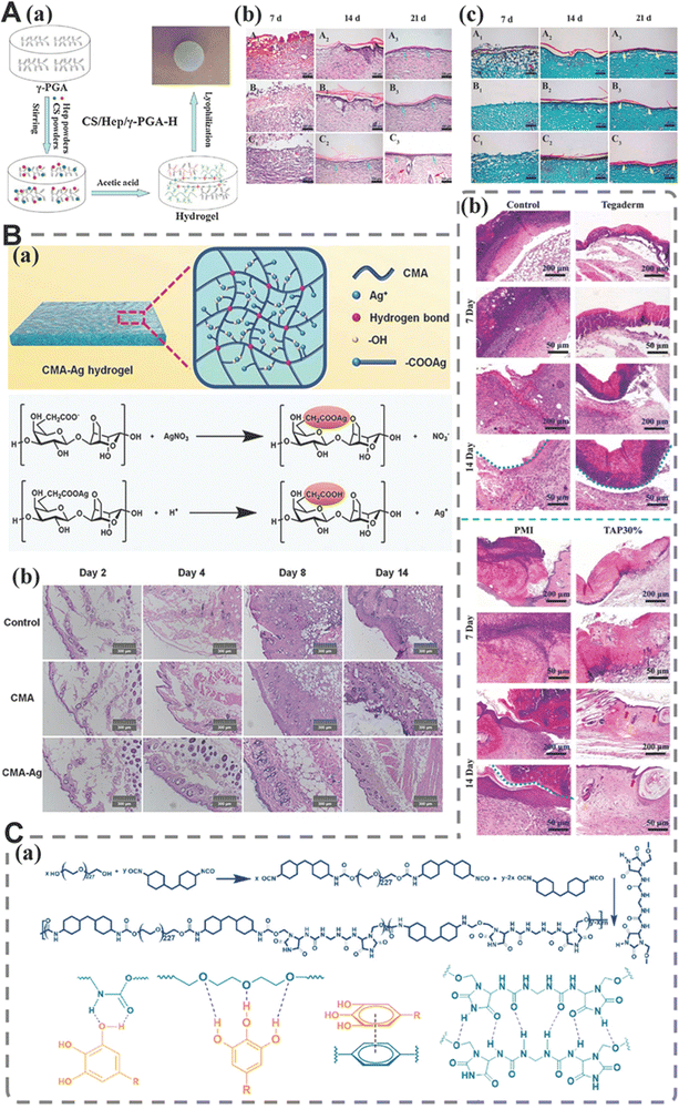 | ||
| Fig. 17 Supramolecular hydrogel dressings with anti-inflammatory and antioxidant functions. (A) (a) Preparation of CS/Hep/γ-PGA composite hydrogels; (b) tissue regeneration of the wounds treated with different dressings (H&E staining). (A1)–(A3) control; (B1)–(B3) CS/Hep/γ-PGA-H (CS/Hep/γ-PGA = 10/5/5); (C1)–(C3) SOD-CS/Hep/γ-PGA-H (CS/Hep/γ-PGA = 10/5/5) (green arrow: epidermal layer and red arrow: hair follicles); (c) collagen deposition in wounds treated with different dressings (Masson's trichrome staining). (A1)–(A3) control; (B1)–(B3) CS/Hep/γ-PGA-H (CS/Hep/γ-PGA = 10/5/5); (C1)–(C3) SOD-CS/Hep/γ-PGA-H (CS/Hep/γ-PGA = 10/5/5) (yellow arrow: epidermal layer). Copyright 2018 Elsevier.104 (B) (a) Schematics and architecture of the CMA (carboxymethyl agarose)–Ag hydrogel and the pH-responsive release of Ag+ from the CMA–Ag hydrogel; (b) micrographs of H&E stained tissue slices from different groups on days 2, 4, 8, and 14. Copyright 2020 Wiley-VCH Verlag GmbH & Co. KGaA, Weinheim.34 (C) (a) Synthetic routes of the PMI (imidazolidinyl urea reinforced polyurethane) carrier network and possible non-covalent interactions within TAP hydrogels; (b) histological evaluation of regenerated skin tissues for Tegaderm films, PMI hydrogel, TAP30% hydrogel, and control groups on day 7 and day 14. Blue dotted lines represent the residual blood crust. Red and yellow arrows represent hair follicles and sweat glands, respectively. Copyright from an open access journal of Bioactive materials.45 | ||
Group modification is also a good antioxidant strategy for hydrogels. Our team has developed a novel supramolecular gelatin (GT) hydrogel, which is composed of GT-grafted aniline tetramers and quaternized chitosan. The experiment showed that the hydrogel could effectively control the ROS concentration in wound area. Our hydrogel combines two strategies of intrinsic antioxidant components and group modification to improve the antioxidant function of hydrogel. To be more specific, QCS can scavenge reactive oxygen species (ROS) to a certain extent, thereby contributing to the antioxidant properties of the hydrogels. Additionally, aniline oligomers exhibit remarkable antioxidant activity owing to their inherent redox-active nature.200
Appropriate inflammatory reaction is an essential link in wound repair.8 However, long-term and persistent inflammation can lead to delayed wound healing and even tissue damage.264 In addition, overstimulated inflammation can cause coagulation cascade reaction, leading to microthrombosis, which further complicates the injury by causing tissue hypoxia and ischemia.34,75 Therefore, anti-inflammatory properties are also very important for supramolecular hydrogel dressings, and different anti-inflammatory strategies (loading anti-inflammatory substances, using anti-inflammatory active substances to regulate inflammatory factors) have been developed successively. For example, in the CMA Ag supramolecular hydrogel synthesized by Huang et al.34 (Fig. 17B(a)), the released Ag+ stimulates the immune function to increase the production of large amounts of white blood cells during the initial phase of wound healing. Enhanced synergistic antimicrobial activity can quickly reduce the number of white blood cells and shorten the duration of inflammation, thereby minimizing inflammation (Fig. 17B(b)).
The up-regulation of inflammatory response is the main reason for the prolongation of wound closure time for most patients. Studies have shown that effective anti-inflammatory can be achieved by down-regulating the expression of inflammatory cytokines. For example, the TAP supramolecular hydrogel developed by Yang et al.45 (Fig. 17C(a)) has demonstrated significant anti-inflammatory effects, as evidenced by its ability to scavenge free radicals and indirectly inhibit the inflammatory response. Moreover, the down-regulation of pro-inflammatory factors, such as tumor necrosis factor-alpha (TNF-α) and nitric oxide synthase 2 (Nos2), was observed (Fig. 17C(b)). Macrophages play a pivotal role in the inflammatory phase of wound healing. They can be classified into two distinct types, namely, classically activated macrophages (M1) and alternatively activated macrophages (M2). In the study of Guo et al.,270 the healed wound tissue contains more anti-inflammatory M2 macrophages after being treated with QCS/TA/Fe hydrogel, while the control group contains more pro-inflammatory M1 macrophages, which indicates that all hydrogels have good anti-inflammatory activities (Fig. 18A(a) and (b)).
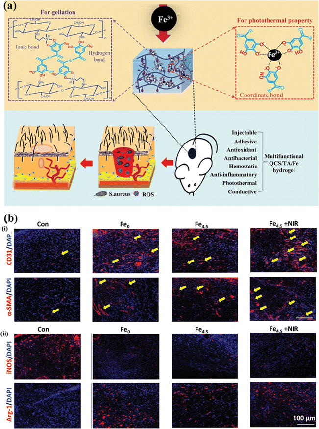 | ||
| Fig. 18 Supramolecular hydrogel dressings with anti-inflammatory and antioxidant functions. (a) Schematic and mechanism of preparation of QCS/TA/Fe hydrogel and therapeutic mechanism of QCS/TA/Fe hydrogel in S. aureus-infected wound healing; (b) (i) images of immunofluorescence staining of CD31 and α-SMA for the evaluation of angiogenesis and maturation, with the yellow arrow pointing to the vessel; (ii) images of immunofluorescence staining of iNOS and Arg-1 for the inflammation level. Copyright 2021 Wiley-VCH GmbH270 | ||
Although antioxidant properties have advanced significantly, a smart supramolecular hydrogel dressing for individualized patient care is still a bottleneck. Hydrogel dressings with precise wound responses, particularly for the various oxidative stress sensitivities, are very important for addressing the needs of different patients. Similar to this, for the same patients, the requirements for ROS at different stages of wound healing vary, necessitating the development of intelligent antioxidants. These smart designs are adaptable to changes in the wound microenvironment. Last but not least, once it is understood how precisely and intelligently antioxidation is controlled in chronic wounds, it becomes very important to treat other aspects (such as the cell control effect and angiogenesis) at various stages of healing.
Numerous studies have been conducted on the mechanisms by which supramolecular hydrogels promote the proliferation of different types of cells. In the study of Kamakshi et al.,100 the prepared supramolecular hydrogel has a more significant effect on promoting cell proliferation than the antibacterial chitosan hydrogel (one of the control groups). Their subsequent experiments revealed that cells cultured in the hydrogel were provided with a conducive environment for cell–cell interactions, as evidenced by the presence of F-actin, which indicates the existence of thin filaments within cells enabling them to interact with their surroundings. Studies have shown that natural polyphenol TA can promote the proliferation of mammalian cells. For example, the multi-cross-linked supramolecular hydrogel prepared by Yu et al.155 (Fig. 19A(a)) was verified to enhance the ability of cell proliferation and is also dependent on the concentration of TA/Fe3+. The hydrogel rich in TA is helpful in mobilizing and promoting the proliferation of 3T3 cells (Fig. 19A(b)). In addition, they demonstrated that the photothermal effect of hydrogel could induce the apoptosis of 3T3 cells to a certain extent. However, post-treatment, 3T3 cells exhibited robust proliferation and achieved nearly identical density as the control group. This observation suggests that normal cells possess a remarkable capacity for rapid self-recovery following limited photothermal damage. Mesenchymal stem cells can also be the object of cell control. The supramolecular hydrogel developed by Kartik et al.63 provides a favorable surface for cell attachment and proliferation, making it suitable for use as a scaffold for wound healing process.
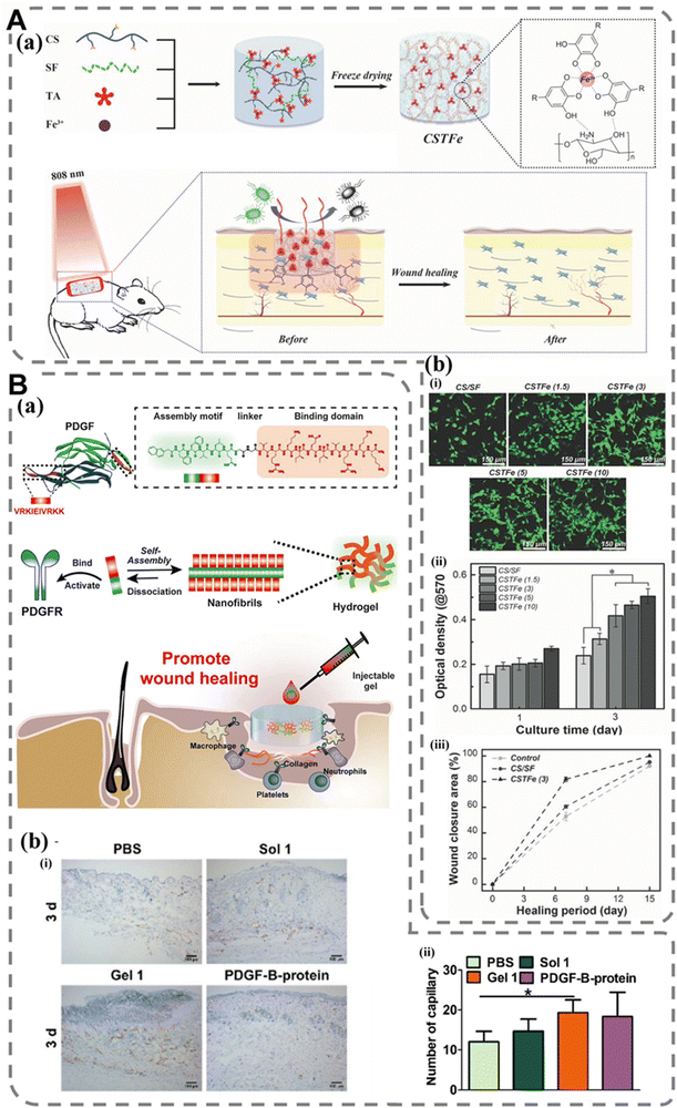 | ||
| Fig. 19 Supramolecular hydrogel dressings with cell control effects and angiogenesis promotion. (A) (a) Schematic explanation of the preparation of the hydrogels and their application as wound dressing materials; (b) in vitro cytotoxicity and cell proliferation evaluation. (i) Representative confocal laser scanning microscopy (CLSM) images of 3T3 cells after 3 days of culture on various hydrogels: CS/SF, CSTFe (1.5), (3), (5), and (10). Scale bar: 150 μm; (ii) the optical density of various hydrogels incubated with 3T3 cells for 1 and 3 days, respectively; (iii) wound recovery rate values at different healing times. Copyright 2019 Wiley-VCH Verlag GmbH & Co. KGaA, Weinheim.155 (B) (a) Conceptual illustration of a PDGF-mimicking peptide hydrogel promoting wound healing. The PDGF-mimicking peptide hydrogel contains an assembling motif, a linker, and a PDGFR binding domain. The PDGFR binding domain is residues 153–162 displayed in orange, which binds and activates the PDGF receptor. The designed peptide can self-assemble to form nanofibrils/hydrogel, which exhibits injectable properties and promotes wound healing; (b) (i) immunohistochemistry staining for CD31 of wounds in different groups on day 3. Scale bar = 100 μm; (ii) the quantification of capillary on day 3. Angiogenesis of wounds was determined using ImageJ. *p < 0.05 vs. control group. n = 3 mice per group. Copyright from an open access journal of Journal of Nanobiotechnology.219 | ||
Fibroblasts also play a key role in wound healing. Wang and his colleagues38 studied the cell proliferation of human fibroblasts. Hydrogels can regulate and provide flexibility of nutrient transport, thereby increasing cell viability. In addition, the hydrogel is not only non-toxic to cell culture, but also effective in stimulating cell proliferation. The supramolecular silk fibroin hydrogel prepared by Chavee et al.261 has been proven to support the normal growth of fibroblasts, and the cell proliferation rate of encapsulated CaSki cells increases with the increase in 1,2-dimyristoyl-sn-glycero-3-phospho-(1′-rac-glycerol)sodium salt concentration.
Although CeO2 has been shown to promote L929 cell proliferation and migration, it also exhibits dose-dependent cytotoxicity. Supramolecular hydrogels (CS-TA@CeO2 hydrogel) prepared by Teng et al.44 indicated that the introduction of TA on CeO2 can greatly reduce the cytotoxicity of large doses of CeO2, thus increasing the upper limit of CeO2 for wound healing. In a subsequent study, we found that adding rGO to QCS based supramolecular hydrogels can promote the proliferation of L929 cells. The main reason for this is that the hydrogels contain a conductive component of rGO, which can promote the proliferation of L929 cells.72 The length of the newly formed epithelium can be defined as the distance between the leading edge of migrating keratinocytes and the first hair follicle observed at the wound margin. The SF/CMCS supramolecular hydrogel prepared by Chen et al.29 can promote wound re-epithelialization by promoting keratinocyte migration, thus promoting wound healing (Fig. 20A(a) and (b)). The experimental results also demonstrate that the SF/CMCS hydrogel exhibited enhanced cell proliferation compared to the CMCS hydrogel. This can be attributed to the presence of Arg-Gly-Asp sequence in silk fibroin, which effectively facilitates cellular adhesion and proliferation.
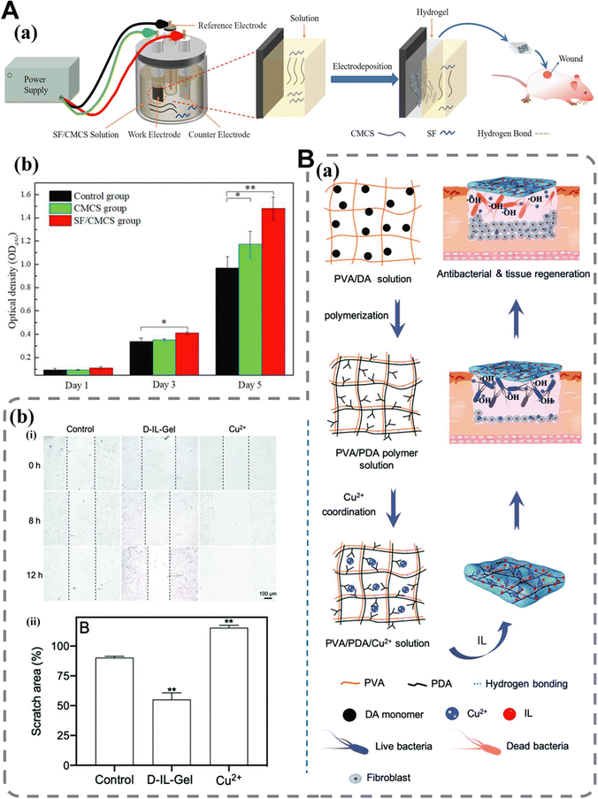 | ||
| Fig. 20 Supramolecular hydrogel with cell control effects. (A) (a) Experimental process and principle; (b) viability of HEK-293 cells after 1 day, 3 days, and 5 days of contact with leaching liquor obtained from CMCS hydrogel and SF/CMCS hydrogel. Statistical analysis was performed using an unpaired, two-tailed t-test (*p < 0.05, **p < 0.01, n = 3). Copyright from an open access journal of International Journal of Molecular Sciences.29 (B) (a) Schematic illustration of the preparation of the Cu2+ loaded gel and its capability in bacterial killing and tissue remodeling; (b) (i) the effect of D-IL-Gel (2 mM) and Cu2+ (2 mM) on the migration of MCF-7 cells. Scale bar 100 μm; (ii) the ratio of cell scratches after treatment with the control, D-IL-gel (2 mM) and Cu2+ (2 mM). **p < 0.01. Copyright 2022 Royal Society of Chemistry.198 | ||
During the process of wound healing, cells located at the wound periphery exhibit a migratory behavior towards the central region, thereby facilitating efficient closure of the wound. Consequently, augmented cellular migration plays a pivotal role in promoting effective wound repair. To evaluate the effect of composite hydrogels on cell migration, Zhai et al.158 conducted a scratch test on NIH3T3 cells. The experimental results are very consistent with previous literature which showed that RGD accelerates cell migration through the integrin avb3 receptor, in part. In the study by Gao et al.,198 the continuous release of Cu2+ from D-IL-gel (Fig. 20B(a)) facilitated cell migration, thereby enhancing wound healing. Analysis of the ratio of cell scratches following different treatments revealed a significant reduction in the scratch area after treatment with D-IL-gel (Fig. 20B(b)).
In addition to cell proliferation and migration, cell adhesion is also an important aspect of cell control in hydrogels. For example, Wei et al.203 found in their work that the ability of the hydrogel to promote cell adhesion can be regulated by adjusting the ratio of the host to guest. When the host monomer is less than or equal to the guest molecule (R ≤ 1.0), most of Ac-TU-β CD and coupling peptides are concentrated on the TU-HGMC skeleton and hindered by PAAm chains, rendering them inaccessible to cells. However, in excess (R = 2.0), the primary monomer will copolymerize with AAm and distribute within the PAAm matrix, allowing better cell proximity to Cat-RGD conjugates with host monomers and thereby enhancing cell adhesion ability. The laminin expression study by Gabriella et al.216 demonstrated that cells have the ability to construct a basement membrane. The ability of the hydrogel to hold the cells within the matrix over the stroma and making them settle over time facilitated the adhesion of human corneal epithelial cells onto the cornea.
Last but not the least, similar to the characteristics of substance delivery function, cell control effects will be more commonly used as pathways and means to achieve other wound repair functions, such as promoting angiogenesis and preventing tissue-adhesion functions. The specific links will be discussed later.
One of the common assessment methods is to monitor the expression of growth factors such as vascular endothelial growth factor (VEGF). VEGF plays a pivotal role in the initial stages of angiogenesis, orchestrating vascular disconnection (via downregulation of cell adhesion protein VCAM-1) and localized extracellular matrix degradation, while also governing the migration and proliferation of nascent endothelial cells.276,277 For example, in the study conducted by Deng et al.,40 serum levels of VEGF and TGF-β in mice from their supramolecular complex hydrogel group (prepared using fenugreek gum and cellulose through hydrogen bonds) were higher than those in the control group. The expressions of these two factors in the FCH-40% group were significantly higher than those in the control group (p < 0.05), suggesting that the application of the complex hydrogel effectively enhances neovascularization. The DN supramolecular hydrogel synthesized by our group has been proven to have good angiogenesis promoting ability.52 In our other study, the highest expression of VEGF was observed on day 7 after treatment with a conductive hydrogel dressing.92
CD31, as a marker of VEGF, is often used for immunostaining of wound neovascularization. Combinations of natural polymers with vascular promoting function that provide signals to cells have been shown to improve blood vessel formation (such as fibrin promotes angiogenic signaling and gelatin improves biosignaling).276,277 For example, in the work of Teng et al.,44 enhanced angiogenesis with well-developed capillary networks was observed in the CS and CS-TA@CeO2 groups. The multifunctional wound dressing developed by our group220 has also been experimentally demonstrated to promote the formation of blood vessels by reducing tissue edema through the absorption of wound exudate.
The function of promoting angiogenesis can also be achieved by loading vascular promoting substances. The platelet-derived growth factor (PDGF) plays a pivotal role in the process of wound healing and has been granted approval for the treatment of diabetes-associated wounds. Jian et al.219 developed a PDGF-mimicking peptide by conjugating the PDGF epitope VRKIEIVRKK with a self-assembling motif derived from β-amyloid peptide. The synthesized peptides possess the ability to self-assemble into fluorine-rich networks, resulting in remarkable stability of supramolecular hydrogels. Moreover, the hydrophilic epitopes can be exposed on nanoantibody surfaces, thereby facilitating the binding and activation of PDGF receptors. Notably, this hydrogel has demonstrated its potential in promoting collagen deposition and angiogenesis as well as facilitating skin regeneration, showcasing its exceptional therapeutic efficacy (Fig. 19B(a) and (b)).
Generally speaking, the function of supramolecular hydrogels to promote angiogenesis can be achieved through the following two common strategies: the first is to introduce components into hydrogel networks that can promote angiogenesis, and the second is to load substances which promote angiogenesis. Moreover, the reports of supramolecular hydrogels related to this function are still rare. Other methods beyond this need to be continuously explored and discovered by researchers in future research.
Postoperative adhesion occurs when fibrin strips interweave between the wound site and the surrounding tissues or organs. The incidence of postoperative adhesion in patients undergoing abdominal surgery is a staggering 90%.278 Post-operative adhesions can lead to serious complications, such as chronic pain and organ obstruction, and often require additional surgery. However, the treatment plans for postoperative adhesion such as the traditional surgery and pharmacological treatments are limited by a number of risks associated with surgery, or the rapid removal of the drug from the site of application. An increasing number of researchers have focused on barrier materials, which can physically separate the damaged area from adjacent tissues or organs to prevent the adhesion of tissue. Supramolecular hydrogels based on natural polymers such as polyphenols and polysaccharides are gradually favored by workers who study postoperative anti-adhesion materials due to their injectable nature, strong adhesion and self-healing functions.279–282 The supramolecular PVA/QUE hydrogel prepared by Song et al.51 rapidly within 10 minutes provided an effective and safe anti-adhesion barrier for drug release, significantly improving the anti-adhesion effect. In the study of Dong et al.,156 the supramolecular composite hydrogel was shown to completely cover the surface of irregular damaged tissues and effectively reduce the formation of postoperative adhesions by reducing the adhesion and growth of bacteria. The reasons are as follows: methacrylate sulfonated betaine can prevent the adhesion and invasion of fibrin during the post-operation reconstruction of the cecum and abdominal wall; moreover, the double network structure of the composite hydrogel enhances the stability of the hydrogel, and has the function of isolating and protecting the wound.
Supermonomers, which are bifunctional monomers synthesized through noncovalent methods but capable of undergoing traditional covalent polymerization, can be utilized for the construction of dynamically cross-linked and degradable supramolecular hydrogels that exhibit dissolution upon stimulation. Due to the reversible cross-linking structure of hydrogels, they quickly dissolve when they come into contact with cross-linking breaking molecules (one of the irritants). Xu and colleagues70 applied the memantine impregnated gauze to rhodamine B half-dyed hydrogels. All the experimental results showed that the prepared supramolecular hydrogels showed excellent stimulating solubility and short dissolution time. The stimulus solubility can be classified as one of the stimulus response properties, and the principle is similar to that of substance release properties driven by hydrogel degradation. For a more detailed introduction, please refer to Section 3 (Fig. 21A).
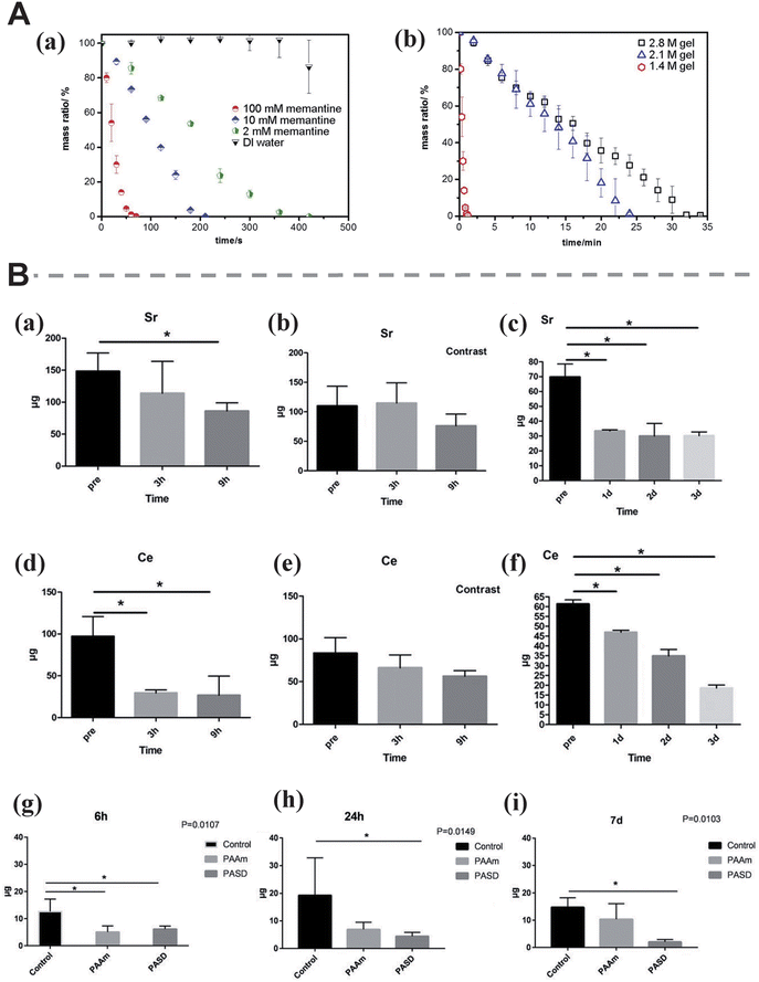 | ||
| Fig. 21 Supramolecular hydrogels with other properties that promote wound repair. (A) (a) 1.4 M hydrogel's dissolution rate upon exposure to different memantine concentrations as well as DI water; (b) dissolution rate of hydrogel with different AAm concentrations in a 100 mM memantine solution. Copyright 2017 American Chemical Society.70 (B) Decontamination effects of the PASD hydrogel on cerium and strontium solutions; decontamination effects of the PASD hydrogel on radioactive strontium-contaminated skin wounds. (a) Short-term strontium decontamination by the PASD hydrogel; (b) short-term strontium decontamination by the PAAm hydrogel; (c) decontamination of strontium by the PASD hydrogel after 3 days; (d) short-term cerium decontamination by the PASD hydrogel; (e) short-term cerium decontamination by the PAAm hydrogel; (f) decontamination of cerium by the PASD hydrogel after 3 days; (g) radioactive strontium content in the skin wound at 6 h; (h) radioactive strontium content in the skin wound at 24 h; (i) radioactive strontium content in the skin wound at 7 day. Copyright 2021 Wiley-VCH GmbH.26 | ||
People engaged in various nuclear-related activities may sustain injuries such as lacerations, puncture wounds, burns, and scalds, including the potential exposure of skin to radionuclides. Radioactive substances can readily enter the human bloodstream through these wounds and disseminate to vital organs like the liver, kidneys, and other tissues. Consequently, internal organs remain contaminated with radiation for prolonged periods leading to severe detrimental effects on human health. The supramolecular hydrogel developed by Cui et al.26 has the ability to decontaminate radionuclides from skin wounds and promote wound healing due to the presence of alginate and DTPA (diethylene triamine pentaacetic acid, commonly used for rare earth elements, plutonium, and super plutonium contamination). During the experiment, they observed the formation of granulation tissue in the skin wounds of the supramolecular hydrogel group, with macrophages and fibroblasts scattered around the center of the neovascularization. In addition, the neovascularization closes and degrades, leaving an orderly distribution of arterioles surrounded by a moderate number of macrophages and mature fusiform fibrocytes. This once again confirms that the hydrogel can effectively remove the absorbed radioactive strontium from the skin wound and promote wound healing (Fig. 21B).
Antibacterial properties are one of the requirements that almost all supramolecular hydrogel dressings must meet due to the prevalence of bacterial infections and the misuse of antibiotics. Supramolecular hydrogel dressings with drug loading and release functions, anti-inflammatory and antioxidant functions, and the ability to promote angiogenesis require supramolecular hydrogels to adhere to tissue, which enables them to remain fixed at the site of action for a long time, repeatably, or as needed. Because some of the functions mentioned above to promote skin repair are carried out by substance delivery (loading, delivering or releasing antibacterial agents, anti-inflammatory and antioxidant substances, even active factors and cells), there is a close relationship between delivery properties and them. Excessive inflammation can cause oxidative stress and an increase in ROS. Therefore, anti-inflammation and anti-oxidants usually work in tandem. Supramolecular hydrogel dressings must provide a stable activity environment for cells and control abnormal cell behavior in order to facilitate wound healing because the cell behavior permeates the entire wound healing process. In addition, vascularization, a crucial step in wound healing, should also be improved appropriately.
In a word, the functions of promoting wound repair are interrelated in the whole wound healing process, which lays a deep foundation for the research in the field of supramolecular hydrogel dressings. With the emergence of new cross-linking methods of supramolecular hydrogels and the continuous improvement of multi-network structure systems, more and more new functions will be introduced into supramolecular hydrogels in the future. They can be presented individually or in different combinations in the field of wound healing.
4.2 Application of supramolecular hydrogels in hemostasis
Hemostasis is the first step of wound healing and it is a self-repairing mechanism through which the body performs blood coagulation and platelet aggregation to prevent bleeding due to vascular injury or microvascular injury, such as vasoconstriction, platelet aggregation, coagulation, and fibrinolysis. Besides, abnormal platelet count, coagulation factor dysfunction, vascular wall function problems and other common factors can affect the normal physiological hemostasis process. Therefore, the development of wound dressings with hemostatic functions is quite important to prevent the rapid bleeding. Our group has conducted many explorations in hemostatic hydrogels and other hemostatic biomaterials,283,284 including a systematic review of hydrogels for wound healing and hemostasis.8 In these articles, we systematically elaborated the whole process of wound repair and the specific repair mechanism at each stage, and objectively analyzed the design ideas, hemostatic principles and application limitations of hemostatic hydrogels from a variety of clear perspectives.Different from our previous reviews mentioned above, this section mainly reviews the application of supramolecular hydrogels in hemostasis. On the premise of reasonable classification of hemostatic function, we analyzed the hemostatic mechanism of various hemostatic supramolecular hydrogels/hydrogel dressings. In short, the two main methods of obtaining and promoting hemostasis for supramolecular hydrogels are physical plugging and coagulation promotion. The two types of physical blocking hemostasis are achieved through hydrogel adhesion and hydrogel expansion, respectively. The three mechanisms of promoting coagulation are controlling the behavior of associated cells (such as platelets and blood cells), starting the coagulation process in vivo and interacting with coagulation factors. The more in-depth branches of the hemostatic concepts of physical plugging and coagulation promotion will be covered in detail in this section, along with assessment of earlier works. Table 6 lists a series of supramolecular hydrogels for hemostasis.
| Function of promoting hemostasis | Hemostasis mechanism | Functional components | Ref. |
|---|---|---|---|
| Forming a physical barrier | Adhere to the hemorrhaging sites and form physical barrier | HA/PVP complex | 30 |
| TA | 31,45,285 | ||
| Fully ionized isocyanoethyl methacrylate-glutamine (iLEM-GLn), poly(hexamethylene guanidine) (PHMG) | 286 | ||
| PAA and HA | 287 | ||
| Histidine, Zn2+ and sodium alginate (SA) | 288 | ||
| CS | 289 | ||
| Acrylic acid and 1-vinylimidazole | 290 | ||
| QCS, TA | 291 | ||
| Andrias davidianus skin secretion (ADS) | 292 | ||
| Catechol groups modified ε-polylysine (PL-CAT) | 293 | ||
| Poly-N-acryloyl aspartic acid (PAASP) | 249 | ||
| Procoagulant | CS can aid the establishment of interaction with the negatively charged membranes of blood cells, promote aggregation of erythrocytes, and increase platelet adhesion | CS | 39 |
| — | TA | 44 | |
| — | CS | 155 | |
| — | QCS, TA | 270 | |
| CS pro-coagulation function; better absorption ability which was beneficial for concentrating red blood cells and platelets | CS, graphene oxide (GO) | 202 | |
| Promote the blood coagulation process | Ca2+ | 61 | |
| The RGD moieties of Pept-1 enable the adhesion of platelets through α‖bβ3 receptor for further downstream activation of platelets; the release of encapsulated Ca2+ from hydrogels effectively triggers the coagulation cascade | Cell adhesive peptide conjugate (Pept-1); Ca2+ | 158 | |
| Largely initiating the extrinsic pathway of coagulation | Gelatin | 294 | |
Anionic polysaccharides such as hyaluronic acid and alginate are widely used in hemostatic hydrogel dressings. They have extremely high water absorption and can increase the concentration of platelets and coagulation factors.295,296 For example, the HC-PAA/PVP/HA complex developed by Tomoko et al.287 can form an adhesive hydrogel and bind tightly to the bleeding site, immediately achieving complete hemostasis. The addition of HA effectively prevents the accumulation of PAA/PVP complexes and results in the formation of a soft white sponge, which can instantly expand in water to form a highly bioviscous hydrogel and provides a physical barrier to stop bleeding. Histidine is a natural essential amino acid in young mammals and plays an important role in the growth and regeneration of mammalian tissues. The amine and imidazolyl groups of histidine chains are easy to protonate, forming reversible hydrogen bonds with negatively charged sodium alginate (SA) and dynamic coordination bonds with zinc ions (Zn2+). Yao et al.288 synthesized a HSZH (histidine-SA-Zn2+) supramolecular hydrogel dressing that adheres to the bleeding site by its excellent adhesion properties and further forms a barrier to prevent cardiac bleeding in vivo (Fig. 22A). Chitosan has emerged as the most commonly used natural polymer employed in hemostatic wound dressings, with chitosan-based hemostatic agents being commercially available since 2002. Diverse supramolecular hydrogel hemostatic dressings based on chitosan and its derivatives have been extensively developed.297 For example, the HBC-C hydrogel prepared by Shou et al.289 was proved to be useful as a hemostatic barrier to prevent bleeding from wounds due to its heat sensitivity and gelation within 30s after injection. In addition, hydrogen bonds, π–π accumulation and other interactions between catechol groups/amino groups and tissues in the hydrogel enable it to adhere to bleeding sites in the body, thereby reducing blood loss. In recent decades, catechol-based hydrogels have been extensively studied in medical applications298 by simulating the typical amino acids in Marine mussels.241,299 Mussel-inspired polypeptide (gamma-PGA15 and poly-lysine 16) hydrogels have previously been reported as effective hemostatic agents. TA is commonly used to form functional hydrogels through polyvalent hydrogen bonds with polyethylene glycol or DNA. For example, the PDTA hydrogel prepared by Xue et al.31 can bond to the wound site and act as a hemostatic barrier due to its acidity and stable gastric tissue adhesion properties.
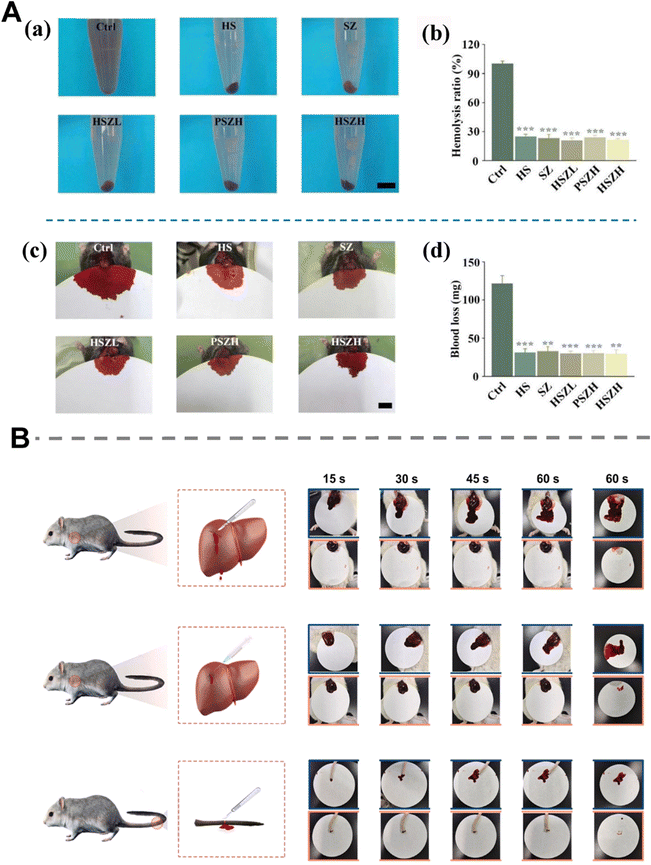 | ||
| Fig. 22 Supramolecular hydrogels that provide hemostasis by forming a physical barrier. (A) Photographs (a) and quantitative results (b) of hemolytic test using DD water and the hydrogels; images (c) and quantitative analysis (d) of blood loss from bleeding hearts treated with the hydrogels (the control group treated without any materials). Copyright 2022 Wiley-VCH GmbH.288 (B) Schematic pictures of the mouse liver incision model, mouse liver trauma model, mouse tail amputation model and the corresponding bloodstain on filter paper in the hydrogel-treated group and blank group at predetermined time intervals (15, 30, 45, and 60 s). Copyright from an open access journal of Bioactive Materials.249 | ||
Synthetic polymers are also increasingly used as the hemostatic agent in supramolecular hydrogels. Polyurethane based dressings are often hydrophobic, which can affect their hemostatic effectiveness by affecting platelet adhesion. By altering the proportion of diphenyl methyl diisocyanate, polyurethanes with varying degrees of hydrophobicity can be synthesized.300–302 In addition, modified polyurethane is increasingly used to enhance the hemostatic performance of dressings. For example, bioactive supramolecular hydrogel dressings with ideal hemostatic performance can be prepared by combining tannic acid (TA) with imidazoline-urea reinforced polyurethane (PMI).45 In another study, Yu et al.249 constructed a PAASP supramolecular hydrogel through one-pot free radical polymerization. Its excellent ability to stop bleeding in vivo is attributed to its strong adhesion, which acts as a strong physical barrier against blood loss (Fig. 22B). Similarly, Zhang et al.286 prepared a series of poly(ionized isocyanoethyl methacrylate-glutamine)/poly(hexamethylene guanidine) (iGx/PHMGy) supramolecular hydrogel dressings. Among them, robust hydrogen bonds are established between the urea groups in the P (iIEM-Gln) chain, resulting in the formation of a stable hydrogel network exhibiting exceptional tissue adhesion performance. Consequently, this hydrogel can effectively serve as a physical barrier to promptly seal bleeding wounds and achieve hemostasis.
Notably, Liu et al.292 reported a new hydrogel composed of natural substances. Upon contact with water, the Andrias davidianus skin secretion (ADS) particles self-assemble to form a hydrophobic hydrogel within seconds, which automatically binds firmly to the wet substrate. The independent hydrogel layer formed immediately and covered the wound site, exhibiting strong wet adhesion as well as cohesion and effectively stopping bleeding within 1 minute without the application of additional patches. In addition, SEM images showed that ADS powder formed a closed layer with a three-dimensional microporous network structure at the interface of wound tissue, which confirmed the mechanism of physical hemostasis.
In summary, the hemostatic strategy of physical plugging has been widely used in supramolecular hydrogel dressings with hemostatic function. The raw material selection and preparation methods of these hydrogels are similar, and the physical plugging is usually achieved through their excellent adhesion properties and additional water absorption. However, there are no supramolecular hydrogels to achieve physical hemostasis through expansion so far (which can be better applied to bleeding wounds with irregular shapes), and this direction of research may be emphasized in the future.
Supramolecular hydrogel dressings can promote the coagulation process in the human body in many different ways. Promoting the aggregation of associated cells (such as platelets and blood cells), starting the coagulation process in vivo and interacting with coagulation factors are the three parts of promoting coagulation. Promoting the aggregation of platelets or blood cells can promote blood coagulation indirectly. Hydrogel structures with strong absorptive capacity and based on natural or synthetic polymers with inherent hemostatic function can be demonstrated to achieve this hemostatic mechanism. For example, the PACG/CMCS supramolecular hydrogel prepared by Chen et al.303 showed a high blood coagulation rate in the dynamic whole blood coagulation test. This hydrogel has significant porosity, so it can absorb and coagulate blood cells, and promote the aggregation of the most prominent platelets and blood cells to trigger obvious clotting behavior, thus forming thrombus. An injectable supramolecular hydrogel developed by Feng et al.202 has been proven to have excellent hemostatic performance. They have better absorption capacity and are conducive to concentrating red blood cells and platelets, thus promoting blood coagulation and achieving rapid hemostasis. Similarly, in the QCS/TA injectable hydrogel designed by Guo et al.,291 the amino group of chitosan can undergo protonation in the bloodstream, resulting in the formation of –NH3+ groups. These positively charged groups have the ability to adsorb negatively charged platelets, induce aggregation of red blood cells, and promote thrombosis (Fig. 23A(a)). In addition, TA has the ability to cause vasoconstriction and interac with plasma proteins (Fig. 23(b)).
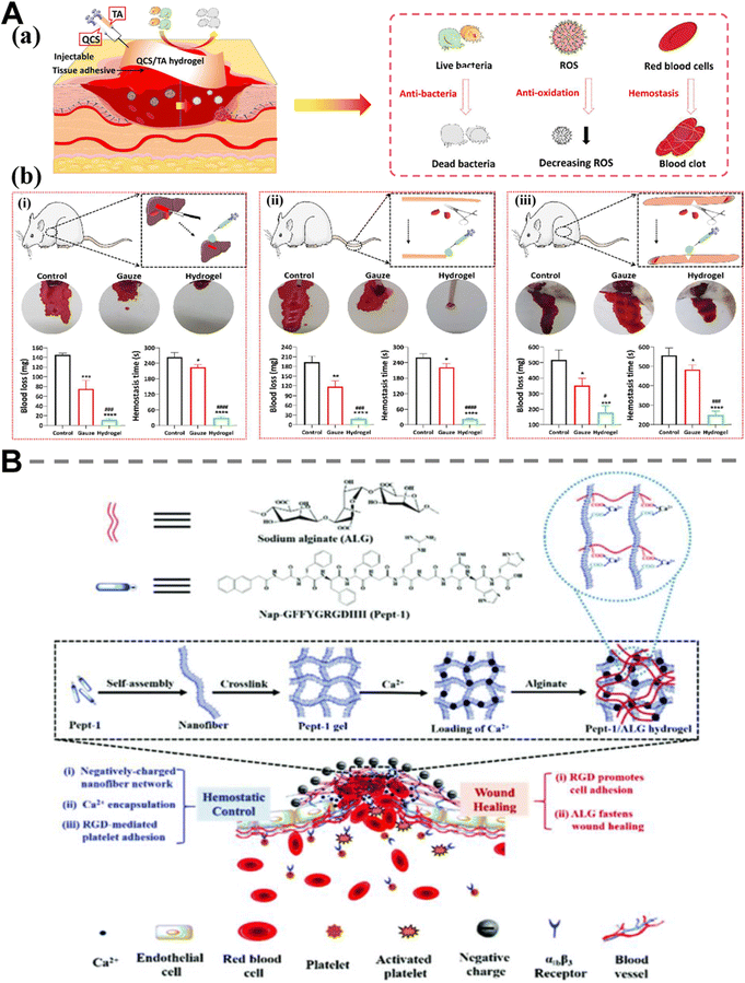 | ||
| Fig. 23 Supramolecular hydrogels with hemostatic function by promoting coagulation. (A) (a) QCS/TA hydrogels were injected into the site of injury; (b) hemostatic property of the QCS/TA2.5 hydrogel. (i) schematic diagram of the liver incision model, photographs of blood trace, and statistics of blood loss and hemostasis time without treatment (control) or with gauze and hydrogel treatments; (ii) schematic diagram of the tail amputation model, photographs of blood traces, and statistics of blood loss and hemostasis time in each group; (iii) schematic diagram of the femoral artery model, photographs of blood traces, and statistics of blood loss and hemostasis time in each group. *p < 0.05, **p < 0.01, ***p < 0.001, or ****p < 0.0001, compared with the control group; ###p < 0.001, or ####p < 0.0001, compared with the gauze group; mean ± SD, n = 3. Copyright 2022 American Chemical Society.291 (B) Schematic illustration for the design of Petp-1/ALG composite hydrogels for hemostatic control and wound healing. Copyright 2019 Royal Society of Chemistry.158 | ||
Peptide-based procoagulant supramolecular hemostatic hydrogels have also been reported. The supramolecular hydrogel (co-assembly of a cell adhesive peptide conjugate (Pept-1) and alginate (ALG)) prepared by Zhai et al.158 was proved to have excellent hemostatic control ability (Fig. 23B). The Pept-1/ALG hydrogel significantly reduced clotting time to 41 s, exhibiting a remarkable 28-fold acceleration compared to the saline control. This enhanced performance can be attributed to the rapid entrapment of blood cells facilitated by the nanofibrillar structures within the composite hydrogels. It should be noted that the RGD moieties present in Pept-1 can bind to α‖bβ3 receptors on platelets (increasing the adhesion rate of platelets to the hydrogel) and accelerate hemostasis. Furthermore, the release of Ca2+ also accelerated the coagulation process and the hemolysis test indicated that the hydrogel has minimal destructive effects on blood cells.
On the other hand, the coagulation cascade encompasses both extrinsic and intrinsic pathways. The extrinsic pathway specifically denotes the clotting pathway involving exogenous clotting factors. In the intrinsic pathway, activation of factor XII occurs upon contact of blood with a negatively charged surface, leading to downstream proteolytic activation of other coagulation factors until factor X is converted into factor Xa.296 Consequently, hemostatic agents possessing negatively charged surfaces (e.g., anionic polysaccharides) can be employed to facilitate blood clotting. Therefore, the coagulation process can be promoted by triggering endogenous and exogenous coagulation mechanisms or activating coagulation factors. For example, in the gelatin/PHEAA hydrogel prepared by Wang et al.,294 gelatin contains essential tissue growth factors that primarily initiate exogenous coagulation pathways to promote coagulation (Fig. 24A(a) and (b)). In the QCS/TA/Fe hydrogel prepared by Guo et al.,270 TA containing polyphenols can make the material negatively charged, activate the endogenous coagulation cascade and trigger coagulation factor XII, which in turn triggers the coagulation cascade. It is worth mentioning that this hydrogel also relates to hemostatic properties of physical hemostasis and the thrombocytic cell behavior mentioned earlier in this section. This cannot be ignored for the hemostatic strategy combining physical hemostasis and coagulation promotion. Zhang et al.304 verified the enhanced coagulation properties of NC supramolecular hydrogels (Fig. 24B(a)). Because clay nanosheets have a strong negative charge on their surface, they can concentrate clotting factors and shorten the clotting time (Fig. 24B(b)). In addition, substance delivery can also be introduced to promote coagulation. Zhou et al. prepared the CUR/TA hydrogel that can achieve rapid hemostasis through temperature-dependent release of tannic acid. TA modified curds may dissociate to a certain extent at low temperatures, resulting in this particular TA heat-sensitive release, which in turn drives the procoagulant process. The specific mechanism may be that TA stimulates blood coagulation through the interaction of its phenol hydroxyl group with proteins and peptides in the blood.
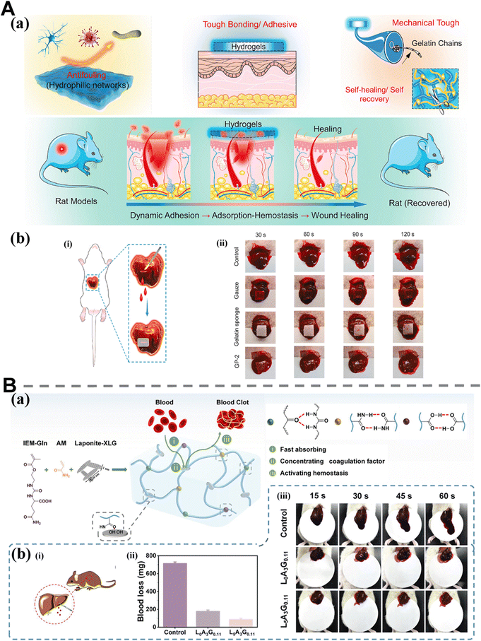 | ||
| Fig. 24 Supramolecular hydrogels with hemostatic function by promoting coagulation. (A) (a) General schematic to show integrated functions including antifouling, tough bonding, mechanically tough, and dynamic adhesion for gelatin/PHEAA hydrogels and their excellent wound healing processes (i.e., adhesion → adsorption-hemostasis → wound healing); (b) (i) schematic of the GP-2 composite hydrogel for achieving hemostasis and wound closure; (ii) digital photographs of liver bleeding treated with and without GP-2 hydrogel. Copyright 2021 Elsevier.294 (B) (a) Schematic illustration of the NC hydrogel formation. Digital photographs of the hydrogel displaying outstanding mechanical properties; (b) in vivo hemostatic performance and histocompatibility of NC hydrogels. (i) Schematic diagram of the rat liver injury model (incision depth: 5 mm and length: 5 mm); (ii) digital photographs of liver blood loss with different treatments at the preset time intervals (15, 30, 45, and 60 s); (iii) blood loss from rat liver of different treatment methods. Copyright 2021 American Chemical Society.304 | ||
The combination of physical plugging and coagulation promotion is quite competitive for hemostatic supramolecular hydrogels. Although a single hemostatic strategy is simple to realize and control, it often exposes a series of drawbacks in the process of practical application when facing various bleeding scenarios. The diversity of hemostatic methods enables hydrogels to play a more flexible role in the process of wound hemostasis.
In a word, most of today's hemostatic supramolecular hydrogels are designed based on a single hemostatic method. At present, the physical plugging hemostasis function of supramolecular hydrogels is mainly realized through tissue adhesion. In contrast, employing a variety of ways to promote coagulation (promoting the aggregation of associated cells, starting the coagulation process in vivo and interacting with coagulation factors) improves the selectivity and flexibility of the fabrication of supramolecular hydrogels to promote coagulation; however, at the same time, its realization process will be relatively complicated. The new hemostatic strategy combining physical plugging and coagulation promotion is quite competitive, because the diversity of hemostatic methods enables hydrogels to play a more flexible role in the process of wound hemostasis. In addition, supramolecular hydrogels with synergistic action of multiple hemostatic components (single-mode hemostatic or multi-mode hemostatic synergistic hemostatic) have also been reported, and these works will pave the way for the development of hemostatic supramolecular hydrogels in the future.
4.3 Application strategies and removal of supramolecular hydrogels
Injectable supramolecular hydrogels generally have shear thinning properties, which are determined by their unique properties of non-covalent cross-linking. The main difference between the preparation of injectable hydrogels and in situ polymerized hydrogels is the flexibility of gel kinetics regulation, since the molding of both is affected by varying degrees of gel kinetics. When used, a hydrogel usually preformed and then injected into the specific site.306,307 This strategy is very flexible in practice because the injection has a high degree of freedom and can be controlled by the operator. In addition, in some specific treatments, injectable properties can significantly improve patient comfort.
Unlike the two gentler application strategies mentioned above, hydrogel implants are generally pre-formed (fixed in a certain shape) blocks in vitro.308,309 The advantage of pre-formed hydrogels is that the hydrogel can be purified several times to obtain better biocompatibility and adaptive properties. In other words, some properties of the hydrogel can be modified or improved before being applied to the wound. However, the disadvantages of pre-formed hydrogels are also very obvious: (1) pre-formed hydrogels are difficult to apply to irregularly shaped wounds; (2) if the target location is a trauma in the body, the hydrogel will need to be artificially sutured or require more complex surgery to work, which will lead to discomfort for the patient and the occurrence of surgical wounds; (3) loading of active substances (such as cells, growth factors, and exosomes) is difficult to achieve in these hydrogels when post-treatment is required, and even if they are successfully loaded, the load efficiency or biological activity is far less than that of injectable hydrogels.
The chemical removal strategies of hydrogels mainly include chemical dissolution and chemical degradation. When the hydrogel is out of use or needs to be replaced, external intervention is performed on or near the surface of the hydrogel (adding a chemical solution that can react with the cross-linked components in the hydrogel, lighting, changing the temperature, pH) to finally achieve the purpose of dissolving the hydrogel or breaking the hydrogel–tissue interface cross-linking point.52,310 The chemical degradation of hydrogels can be achieved through hydrolysis, and enzymatic hydrolysis or oxidation.311 Physical removal strategies usually do not involve chemical bond breaking, but instead involve changes in the associated non-covalent interactions (non-covalent cross-linking of the hydrogel or non-covalent adhesion between the hydrogel and tissues, interfaces). Due to the purely non-covalent cross-linked network, physical removal (including physical degradation, removal of physical adhesion) is usually the method of removal for most supramolecular hydrogel dressings. For example, in our previously prepared smart supramolecular hydrogel dressings (cross-linked by metal coordination and dynamic Schiff base bonds), the artificial addition of deferoxamine mesylate (DFO) can rapidly disrupt the catechol–Fe coordinate bond, resulting in the dissolution of the hydrogel, leading to greatly reduced hydrogel tissue adhesion and easy removal.241
In the future, the removal methods for supramolecular hydrogel dressings will be further developed on the basis of today's research, and the removal concept of on-demand removal and repeatable removal will also be more introduced into the design of supramolecular hydrogel dressings to meet more complex application environments.
5 Conclusions and prospect
5.1 Conclusions
Supramolecular hydrogels based on single or variety of non-covalent interactions (including hydrogen bonds, hydrophobic interaction, electrostatic interaction, metal ligand coordination and host–guest interaction) have many characteristics that are worthy of in-depth study. Because of these reversible non-covalent interactions, supramolecular hydrogels themselves have many properties closely related to the field of wound healing, including but not limited to self-healing, tissue adhesion and sensitivity to stimulation. These unique properties provide more possibilities for the application of supramolecular hydrogel dressings. In recent years, these hydrogels have demonstrated some excellent performances which can meet the various needs of wound repair, such as antibacterial properties, substance delivery, anti-inflammatory and antioxidant functions. Researchers have further promoted the development of future supramolecular hydrogel dressings by constantly innovating and combining raw materials, supramolecular cross-linking methods and enhancement means to meet more diverse and stringent requirements in the future. In this paper, several main cross-linking methods of supramolecular hydrogels are described in detail first, including some innovative and practical high-quality research work in the whole process from preliminary discussions, material selection to hydrogel preparation and characterization, which laid a solid foundation for further research studies and innovations in the field of supramolecular hydrogel dressings. Second, we systematically classified and summarized the network structures of supramolecular hydrogels. This review mainly introduces the applications of supramolecular hydrogel dressings in wound repair and hemostasis. Specifically, we have strictly divided the functions of hydrogels into promoting wound repair and hemostasis, and systematically described hydrogel cross-linking strategies, structural characteristics and the mechanism of action for each function, aiming to present a more comprehensive overview of the applications of supramolecular hydrogel dressings in the above two aspects for readers. In addition, we also present the future prospects of supramolecular hydrogels in structure (cross-linking strategies and network design) and the above two applications (wound repair and hemostasis).5.2 Prospect
In the future, supramolecular hydrogels will play a significant role in the market with their unique dynamically non-covalent cross-linking characteristics and various biomedical applications (Fig. 25). However, although a series of excellent properties of supramolecular hydrogel dressings have gradually been recognized by researchers, the use of these materials still has limitations that cannot be ignored in many aspects, such as the cross-linking strategy (the selection conditions of cross-linking interactions and the rationality of various cross-linking interaction combinations), hydrogel network design (relationships between networks and hydrogel properties, complex relationships among multiple networks) and multiple functions (the way of function realization, the mutual interference between various functions). We believe that with the continuous adoption of new preparation methods and the reasonable integration of various strategies, supramolecular hydrogels with more novel functions and better performance will be designed and prepared in the future.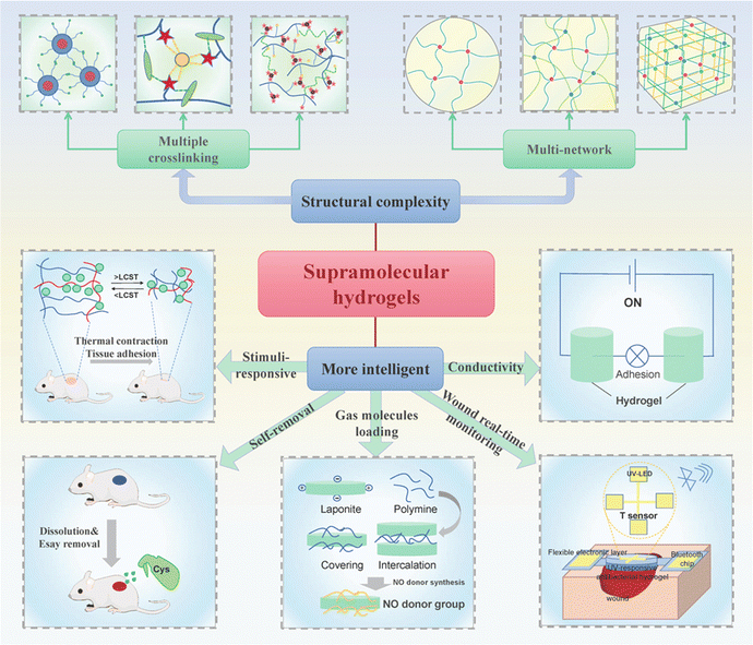 | ||
| Fig. 25 Prospects. The structure of supramolecular hydrogels will become more complex, and their functions in wound healing will be more diverse and intelligent in the future. | ||
In addition, the emergence of smart supramolecular hydrogel dressings opens up many possibilities for bleeding control in wounds.312 They can respond to external stimuli, turn on or stop hemostatic function in hydrogels to control local hemostatic behavior and avoid unnecessary downstream coagulation. Supramolecular hemostatic hydrogels that can be removed on demand also belong to this category. They can play a hemostatic role in a specific time or environment, and be removed in time later to avoid the adverse or uncontrollable effects on the wound. Although the development of super-absorbable supramolecular hydrogels places great importance on their hemostatic effect, the adverse effect of swelling of hydrogels on tissue adhesion strength is still a problem to be solved. Therefore, a more balanced choice between wet adhesion and swelling behavior should be considered in future hydrogel design.313 For more complex supramolecular hydrogel systems, the universality of their applications remains a challenge, as a variety of practical properties, including long-term stability, ease of handling and portability, need to be considered in the context of widespread use. For example, the development of hemostatic hydrogel dressings that can be stored for a long time even under extreme conditions and are easy to carry is particularly important for military applications. Finally, the wet adhesion of supramolecular hydrogels needs to be improved to provide good wet wound adhesion and hemostasis. In addition, more supramolecular hydrogel precursor materials with inherent procoagulant properties can be developed to prepare supramolecular hydrogels that promote blood coagulation, especially in the case of bleeding in patients with coagulation disorders. The introduction of analgesic properties in hemostatic hydrogel can also improve patient comfort.
Although supramolecular hydrogels have been endowed with many biomedical functions, intelligence92,314 (providing and regulating functions on demand) is still a major direction in the future. Due to the complexity of the wound repair process, numerous processes and parameters involved change at all times, making the development of wound monitoring function an important direction in research. Supramolecular hydrogel dressings that can monitor wounds in real time are rarely reported. Second, the number of studies based on the preparation of biomedical hydrogels with conductivity252,269,315–317 and stimulus response properties251,318–320 has been considerable in the last decade, and the stimulus response characteristics are the functions that most researchers hope to see in supramolecular hydrogels. Our group has made some explorations in these two aspects in recent years. For example, the supramolecular hydrogel based on host–guest interaction composed of GT-graft-aniline tetramers and quaternized chitosan has good conductivity.200 Besides, our multifunctional supramolecular hydrogels (fabricated by incorporating quaternized chitosan-graft-β-cyclodextrin, adenine, and polypyrrole nanotubes through host–guest interactions and hydrogen bonds) prepared in another work showed significant thermal response swelling behavior (hydrogel volume changes with temperature).92 The hydrogels exhibited rapid contraction, reducing their remaining areas to approximately 47.2% of their initial states. Correspondingly, during the volume-phase transition process, the maximal tensile stress was increased tenfold from its initial state, owing to the aggregation of the PNIPAm microdomains. In addition, some functional gas molecules with special functions are difficult to load into supramolecular hydrogels due to their own instability (for example, NO, CO, and H2S), so the substance delivery performance needs further exploration to meet the functional requirements such as delivering or loading gas molecules.321 Finally, components/materials that can themselves be assembled to supramolecular hydrogels and also have therapeutic functions can be developed in the future.8,283,322–324 Bioadaptive multifunctional supramolecular hydrogels with different biological functions at different stages of wound healing will also be popular.
In terms of functions, multifunctional hydrogels that combine hemostatic ability with one or more other functions are highly desirable in clinical application. For example, supramolecular hydrogels which combine hemostatic capabilities with monitoring capabilities will be more advantageous. During hemostasis, the pH and electrical properties of the wound site will change accordingly. These hydrogels monitor these physical and chemical changes in the wound, helping to determine the condition of the wound in real time. Combining hemostatic function with wound repair function is also a development trend of supramolecular hemostatic hydrogels in the future.
As a result, because of the unique cross-linking method and unprecedented advantage, supramolecular hydrogels and their application in hemostasis and wound repair have great prospects, and they deserve to be deeply studied. On the basis of published studies, it is necessary to optimize the structural design and functional allocation of supramolecular hydrogels. Clinical translational issues also require ongoing attention to better address problems in actual treatment. We believe that supramolecular hydrogels will be better developed in the future, further broadening the development path in the field of supramolecular chemistry, hydrogel materials and wound dressings.
Abbreviation
| AAm: | Acrylamide |
| Ac-CD: | Acryloyl-β-cyclodextrin |
| ACG: | N-Acryloyl-2-glycine |
| AD: | Adamantane |
| ADS: | Andrias davidianus skin secretion |
| ADxHA: | Adamantane-functionalized hyaluronic acids |
| AG: | Agarose |
| ALD-CD: | Monoaldehyde β-CD |
| ALG: | Alginate |
| AMPs: | Antimicrobial peptides |
| ANFs: | Aramid nanofibers |
| AT: | Aniline tetramer |
| BetP: | Betamethasone phosphate |
| bFGF: | Basic fibroblast growth factor |
| BSA: | Bovine serum albumin |
| C22: | Alkyl acrylate |
| CAT: | Catalase |
| CATNFC: | Cationized nanofibrillated cellulose |
| CB[6]: | Cucurbit[6]uril |
| CB[8]: | Cucurbit[8]uril |
| CCS-PCA: | Catechol-modified carboxymethyl chitosan |
| β-CD: | β-Cyclodextrin |
| CDA: | c-di-AMP |
| CEs: | Casein micelles |
| CM: | Carmellose |
| C18M: | Stearyl methacrylate |
| CMA: | Carboxymethyl agarose |
| CMCS: | Carboxymethyl chitosan |
| c-MWCNTs: | Carboxyl-functionalized multi-walled carbon nanotubes |
| CS: | Chitosan |
| CTAB: | Cetyltrimethylammonium bromide |
| CUR: | Curdlan |
| CY: | Cordycepin |
| 3D: | Three-dimensional |
| DA: | Dopamine |
| DAP: | 2,6-Diaminopurine |
| DEX: | Dexamethasone |
| Dexp: | Dexamethasone sodium phosphate |
| DMSO: | Dimethyl sulfoxide |
| DN: | Double network |
| DNSA: | 3,5-Dinitrosalicylic acid |
| DOX: | Doxycycline |
| DPCA: | Dihydrophenonthrolin-4-one-3-carboxylic acid |
| DS: | Diclofenac sodium |
| DTPA: | Diethylene triamine pentaacetic acid |
| ECM: | Extracellular matrix |
| EDA-CD: | Mono-(6-ethylenediamine-6-deoxy)-β-cyclodextrin |
| EGF: | Epidermal growth factor |
| 2-FA: | 2-Amino-2′-fluoro-2′-deoxyadenosine |
| f-BNNS: | Boron nitrogen nanosheet |
| FGG-EA: | Tripeptide Phe-Gly-Gly ester derivative |
| GA: | Gallic acid |
| GelMA: | Gelatin methacrylate |
| GO: | Graphene oxide |
| GT: | Gelatin |
| GTU: | Ureido-pyrimidinone modified gelatin |
| GW: | Glycerol–water |
| HA: | Hyaluronic acid |
| HBC: | Hydroxybutyl chitosan |
| HC: | Highly cross-linked |
| HEC: | Hydroxyethyl cellulose |
| HS: | Heparin sodium |
| Hy: | Hymexazol |
| iGx: | Poly(ionized isocyanoethyl methacrylate-glutamine) |
| iLEM-GLn: | Fully ionized isocyanoethyl methacrylate-glutamine |
| LbL: | Layer-by-layer |
| LDC: | Lidocaine hydrochloride |
| LMA: | Lauryl methacrylate |
| MC: | Methylcellulose |
| MET: | Metformin |
| MPEG-g-PEAD: | MPEG-grafted poly(ethylene argininylaspartate diglyceride) |
| MRSA: | Methicillin-resistant Staphylococcus aureus |
| NIR: | Near infrared |
| NPs: | Nanoparticles |
| OA: | Orotic acid |
| OCDPOSS: | Octa-cyclodextrin polyhedral oligomeric silsesquioxane |
| PA: | Protocatechualdehyde |
| PAA: | Poly(acrylic acid) |
| P(AAm-co-SMA): | Poly(acrylamide-co-stearyl methacrylate) |
| PAANa: | Poly(sodium acrylate) |
| PAASP: | Poly-N-acryloyl aspartic acid |
| PACG: | Poly(N-acryloyl 2-glycine) |
| PAH: | Polyallylamine |
| PAM: | Polyacrylamide |
| PANI: | Polyaniline |
| PDDA: | Poly(dimethyldiallylamide) |
| PDGF: | Platelet-derived growth factor |
| PEG: | Polyethylene glycol |
| PEGSD: | Poly(glycerol sebacate)-co-poly(ethylene glycol)-g-catechol |
| PF127-CHO: | Benzaldehyde-terminated Pluronic®F127 |
| PGA: | Polyglycolic acido |
| PHEAA: | Poly(N-hydroxyethyl acrylamide) |
| PHMG: | Poly(hexamethylene guanidine) |
| PL-CAT: | Catechol groups modified ε-polylysine |
| PMI: | Imidazolidinyl urea reinforced polyurethane |
| PNCB: | Pinocembrin |
| PNIPAM: | Poly(N-isopropylacrylamide) |
| PPP: | Poly(D,L-lactide)–poly(ethylene glycol)–poly(D,L-lactide) |
| PPVA: | Phytate polyvinyl alcohol |
| PPy: | Polypyrrole |
| PSBMA: | Poly 3-[2-(methacryloyloxy)ethyl][(dimethyl)-ammonio]-1-propanesulfonate |
| PTX: | Paclitaxel |
| PUD: | Polyurethane diol dispersion |
| PVA: | Polyvinyl alcohol |
| PVP: | Polyvinylpyrrolidone |
| QACs: | Quaternary ammonium compounds |
| QCS: | Quaternized chitosan |
| QUE: | Quercetin |
| RGD: | Tri-peptide Arg-Gly-Asp |
| rGO: | Reduced graphene oxide |
| ROS: | Reactive oxygen species |
| RSF: | Regenerated silk fibroin |
| SA-DFA: | Deferoxamine grafted alginate |
| SEM: | Scanning electron microscope |
| SF: | Silk fibroin |
| SOD: | Superoxide dismutase |
| SPU: | Sulfonate-containing polyurethane |
| TA: | Tannic acid |
| UPy: | Ureido-pyrimidinone |
| UV: | Ultraviolet light |
| VEGF: | Vascular endothelial growth factor |
Author contributions
Shaowen Zhuo: investigation, methodology, visualization, writing – original draft, and software. Yongping Liang: writing – review and editing and validation. Zhengying Wu: writing – original draft. Xin Zhao: resources, supervision, writing – review and editing, and validation. Yong Han: resources, supervision, writing – review and editing, and validation. Baolin Guo: resources, supervision, writing – review and editing, and validation.Conflicts of interest
There are no conflicts to declare.Acknowledgements
This work was jointly supported by the National Natural Science Foundation of China (grant numbers: 51971171, 52273149, 52003216 and 82272155), the Key Research and Development Program of Shaanxi (2023-YBSF-150), the Project of Supporting Young Talents in Shaanxi University Science and Technology Association (20200404), and the China Postdoctoral Science Foundation (2022M712506).References
- S. Enoch and D. J. Leaper, Surgery, 2008, 26, 31–37 Search PubMed.
- C. L. Baum and C. J. Arpey, Dermatol. Surg., 2005, 31, 674–686 CrossRef CAS.
- S. Smyth, R. McEver, A. Weyrich, C. Morrell, M. Hoffman, G. Arepally, P. French, H. Dauerman, R. Becker and D. Bhatt, J. Thromb. Haemostasis, 2009, 7, 1759–1766 CrossRef CAS PubMed.
- Y. Huang, X. Zhao, Z. Zhang, Y. Liang, Z. Yin, B. Chen, L. Bai, Y. Han and B. Guo, Chem. Mater., 2020, 32, 6595–6610 CrossRef CAS.
- Y. Yang, Y. Liang, J. Chen, X. Duan and B. Guo, Bioact. Mater., 2022, 8, 341–354 CAS.
- Y. Liang, M. Li, Y. Yang, L. Qiao, H. Xu and B. Guo, ACS Nano, 2022, 16, 3194–3207 CrossRef CAS PubMed.
- A. Maleki, J. He, S. Bochani, V. Nosrati, M.-A. Shahbazi and B. Guo, ACS Nano, 2021, 15, 18895–18930 CrossRef CAS PubMed.
- Y. Liang, J. He and B. Guo, ACS Nano, 2021, 15, 12687–12722 CrossRef CAS PubMed.
- H. Hu and F. J. Xu, Biomater. Sci., 2020, 8, 2084–2101 RSC.
- J. Zhao, X. Zhao, B. Guo and P. X. Ma, Biomacromolecules, 2014, 15, 3246–3252 CrossRef CAS PubMed.
- L. Wang, Y. Wu, T. Hu, P. X. Ma and B. Guo, Acta Biomater., 2019, 96, 175–187 CrossRef CAS PubMed.
- Y. Huang, L. Bai, Y. Yang, Z. Yin and B. Guo, J. Colloid Interface Sci., 2022, 608, 2278–2289 CrossRef CAS PubMed.
- J. He, Z. Zhang, Y. Yang, F. Ren, J. Li, S. Zhu, F. Ma, R. Wu, Y. Lv, G. He, B. Guo and D. Chu, Nano-Micro Lett., 2021, 13, 1–17 CrossRef.
- R. Yu, Y. Yang, J. He, M. Li and B. Guo, Chem. Eng. J., 2021, 417, 128278 CrossRef CAS.
- M. Li, Y. Liang, Y. Liang, G. Pan and B. Guo, Chem. Eng. J., 2022, 427, 132039 CrossRef CAS.
- X. Zhao, Y. Liang, B. Guo, Z. Yin, D. Zhu and Y. Han, Chem. Eng. J., 2021, 403, 126329 CrossRef CAS.
- Y. Ren, X. Zhao, X. Liang, P. X. Ma and B. Guo, Int. J. Biol. Macromol., 2017, 105, 1079–1087 CrossRef CAS PubMed.
- J. Qu, Y. Liang, M. Shi, B. Guo, Y. Gao and Z. Yin, Int. J. Biol. Macromol., 2019, 140, 255–264 CrossRef CAS.
- R. J. Dong, Y. Pang, Y. Su and X. Y. Zhu, Biomater. Sci., 2015, 3, 937–954 RSC.
- S. Bernhard and M. W. Tibbitt, Adv. Drug Delivery Rev., 2021, 171, 240–256 CrossRef CAS.
- T. Kakuta, Y. Takashima, M. Nakahata, M. Otsubo, H. Yamaguchi and A. Harada, Adv. Mater., 2013, 25, 2849–2853 CrossRef CAS PubMed.
- L. Voorhaar and R. Hoogenboom, Chem. Soc. Rev., 2016, 45, 4013–4031 RSC.
- J. Skopinska-Wisniewska, S. De la Flor and J. Kozlowska, Int. J. Mol. Sci., 2021, 22, 7402 CrossRef CAS.
- R. Eelkema and A. Pich, Adv. Mater., 2020, 32, 1906012 CrossRef CAS.
- B. R. Denzer, R. J. Kulchar, R. B. Huang and J. Patterson, Gels, 2021, 7, 158 CrossRef CAS PubMed.
- F.-M. Cui, Z.-J. Wu, R. Zhao, Q. Chen, Z.-Y. Liu, Y. Zhao, H.-B. Yan, G.-L. Shen, Y. Tu, D.-H. Zhou, J. Diwu, J. Hou, L. Hu and G.-J. Wang, Macromol. Biosci., 2021, 21, 2000399 CrossRef CAS.
- J. Xu, H. Wang, L. Jiang, Y. Tao, Y. Song, S. Yu, T. Wang and N. Feng, Macromol. Mater. Eng., 2021, 306, 2100286 CrossRef CAS.
- G. Xu, Y. Xiao, L. Cheng, R. Zhou, H. Xu, Y. Chai and M. Lang, J. Polym. Sci., Part B: Polym. Phys., 2017, 55, 856–865 CrossRef CAS.
- Z. Chen, X. Zhang, J. Liang, Y. Ji, Y. Zhou and H. Fang, Int. J. Mol. Sci., 2021, 22, 7610 CrossRef CAS PubMed.
- H. Yu, Q. Xiao, G. Qi, F. Chen, B. Tu, S. Zhang, Y. Li, Y. Chen, H. Yu and P. Duan, Front. Bioeng. Biotechnol., 2022, 10, 855013 CrossRef PubMed.
- W. L. Xue, R. Yang, S. Liu, Y. J. Pu, P. H. Wang, W. J. Zhang, X. Y. Tan and B. Chi, Biomater. Sci., 2022, 10, 2417–2427 RSC.
- D. Zhang, Z. Xu, H. Li, C. Fan, C. Cui, T. Wu, M. Xiao, Y. Yang, J. Yang and W. Liu, Biomater. Sci., 2020, 8, 1455–1463 RSC.
- Z. Wang, Y. Zhang, Y. Yin, J. Liu, P. Li, Y. Zhao, D. Bai, H. Zhao, X. Han and Q. Chen, Adv. Mater., 2022, 34, 2108300 CrossRef CAS.
- W.-C. Huang, R. Ying, W. Wang, Y. Guo, Y. He, X. Mo, C. Xue and X. Mao, Adv. Funct. Mater., 2020, 30, 2000644 CrossRef CAS.
- F. Gao, Y. Zhang, Y. Li, B. Xu, Z. Cao and W. Liu, ACS Appl. Mater. Interfaces, 2016, 8, 8956–8966 CrossRef CAS.
- Y. Q. Guo, X. L. An and Z. J. Fan, J. Mech. Behav. Biomed. Mater., 2021, 118, 104452 CrossRef CAS PubMed.
- Y. Li, R. Z. Fu, C. H. Zhu and D. D. Fan, Colloids Surf., B, 2021, 205, 111869 CrossRef CAS PubMed.
- J. Wang, X. Liu, Y. Wang, M. An and Y. Fan, Int. J. Biol. Macromol., 2022, 211, 678–688 CrossRef CAS.
- X. Jin, Q. Fu, Z. Gu, Z. Zhang and H. Lv, Int. J. Biol. Macromol., 2021, 184, 787–796 CrossRef CAS.
- Y. Deng, X. Yang, X. Zhang, H. Cao, L. Mao, M. Yuan and W. Liao, Int. J. Biol. Macromol., 2020, 160, 1242–1251 CrossRef CAS.
- S. Bi, J. Pang, L. Huang, M. Sun, X. Cheng and X. Chen, Int. J. Biol. Macromol., 2020, 146, 99–109 CrossRef CAS PubMed.
- Y. Huang, F. Shi, L. Wang, Y. Yang, B. M. Khan, K. L. Cheong and Y. Liu, Int. J. Biol. Macromol., 2019, 132, 729–737 CrossRef CAS.
- J. Long, A. E. Etxeberria, A. V. Nand, C. R. Bunt, S. Ray and A. Seyfoddin, Mater. Sci. Eng.: C, 2019, 104, 109873 CrossRef CAS.
- M. Teng, Z. Li, X. Wu, Z. Zhang, Z. Lu, K. Wu and J. Guo, Colloids Surf., B, 2022, 214, 112479 CrossRef CAS PubMed.
- Y. X. Yang, X. D. Zhao, J. Yu, X. X. Chen, R. Y. Wang, M. Y. Zhang, Q. Zhang, Y. F. Zhang, S. Wang and Y. L. Cheng, Bioact. Mater., 2021, 6, 3962–3975 CAS.
- F. Gao, Z. Y. Xu, Q. F. Liang, H. F. Li, L. Q. Peng, M. M. Wu, X. L. Zhao, X. Cui, C. S. Ruan and W. G. Liu, Adv. Sci., 2019, 6, 1900867 CrossRef.
- S. Cao, X. Tong, K. Dai and Q. Xu, J. Mater. Chem. A, 2019, 7, 8204–8209 RSC.
- H. Deng, Z. Yu, S. Chen, L. Fei, Q. Sha, N. Zhou, Z. Chen and C. Xu, Carbohydr. Polym., 2020, 230, 115565 CrossRef CAS.
- J. Luo, J. Yang, X. Zheng, X. Ke, Y. Chen, H. Tan and J. Li, Adv. Healthcare Mater., 2020, 9, 1901423 CrossRef CAS.
- C. Chen, X. Yang, S.-J. Li, C. Zhang, Y.-N. Ma, Y.-X. Ma, P. Gao, S.-Z. Gao and X.-J. Huang, Green Chem., 2021, 23, 1794–1804 RSC.
- X. Song, Z. Zhang, Z. Shen, J. Zheng, X. Liu, Y. Ni, J. Quan, X. Li, G. Hu and Y. Zhang, ACS Appl. Mater. Interfaces, 2021, 13, 56881–56891 CrossRef CAS.
- X. Zhao, Y. P. Liang, Y. Huang, J. H. He, Y. Han and B. L. Guo, Adv. Funct. Mater., 2020, 30, 1910748 CrossRef CAS.
- M. M. Zhang, R. L. Zhang, H. Chen, X. F. Zhang, Y. L. Zhang, H. N. Liu, C. Li, Y. H. Chen, Q. Zeng and G. Z. Huang, ACS Appl. Mater. Interfaces, 2023, 6486–6498, DOI:10.1021/acsami.2c19997,.
- C. Xu, L. Cao, C. Cao, H. Chen, H. Zhang, Y. Li and Q. Huang, Chem. Eng. J., 2023, 452, 139195 CrossRef CAS.
- Y. Y. Yang, G. Y. Feng, J. F. Wang, R. T. Zhang, S. L. Zhong, J. Wang and X. J. Cui, Int. J. Biol. Macromol., 2023, 227, 1038–1047 CrossRef CAS PubMed.
- C. Y. Qi, Z. X. Dong, Y. K. Huang, J. B. Xu and C. H. Lei, ACS Appl. Mater. Interfaces, 2022, 30385–30397, DOI:10.1021/acsami.2c06395,.
- J. Hu, R. Chen, Z. L. Li, F. Z. Wu, Y. H. Yang, Y. Yang, X. K. Li and J. Xiao, Appl. Mater. Today, 2022, 29, 101586 CrossRef.
- W. J. Yang, R. Zhang, X. Guo, R. X. Ma, Z. Y. Liu, T. Wang and L. H. Wang, J. Mater. Chem. A, 2022, 10, 23649–23665 RSC.
- M. Criado-Gonzalez, D. Wagner, J. R. Fores, C. Blanck, M. Schmutz, A. Chaumont, M. Rabineau, J. B. Schlenoff, G. Fleith, J. Combet, P. Schaaf, L. Jierry and F. Boulmedais, Chem. Mater., 2020, 32, 1946–1956 CrossRef CAS.
- X. Hu, L. Yan, Y. Wang and M. Xu, Chem. Eng. J., 2020, 388, 124189 CrossRef CAS.
- Y. Zhou, H. Li, J. Liu, Y. Xu, Y. Wang, H. Ren and X. Li, Polym. Adv. Technol., 2019, 30, 143–152 CrossRef CAS.
- Y. Zhu, Q. Luo, H. Zhang, Q. Cai, X. Li, Z. Shen and W. Zhu, Biomater. Sci., 2020, 8, 1394–1404 RSC.
- K. Ravishankar, M. Venkatesan, R. P. Desingh, A. Mahalingam, B. Sadhasivam, R. Subramaniyam and R. Dhamodharan, Mater. Sci. Eng.: C, 2019, 102, 447–457 CrossRef CAS PubMed.
- W.-F. Lai, C. Hu, G. Deng, K.-H. Lui, X. Wang, T.-H. Tsoi, S. Wang and W.-T. Wong, Int. J. Pharm., 2019, 566, 101–110 CrossRef CAS.
- Y. L. Fang, S. S. Huang, X. Gong, J. A. King, Y. Q. Wang, J. C. Zhang, X. Y. Yang, Q. Wang, Y. B. Zhang, G. X. Zhai and L. Ye, Acta Biomater., 2023, 158, 239–251 CrossRef CAS.
- T. Sangfai, V. Tantishaiyakul, N. Hirun and L. Li, AAPS PharmSciTech, 2017, 18, 605–616 CrossRef CAS PubMed.
- J. Cheng, D. Amin, J. Latona, E. Heber-Katz and P. B. Messersmith, ACS Nano, 2019, 13, 5493–5501 CrossRef CAS.
- L. Meng, C. Shao, C. Cui, F. Xu, J. Lei and J. Yang, ACS Appl. Mater. Interfaces, 2020, 12, 1628–1639 CrossRef CAS.
- Q. F. Yang, P. Wang, C. Z. Zhao, W. Q. Wang, J. F. Yang and Q. Liu, Macromol. Rapid Commun., 2017, 38, 1600741 CrossRef.
- W. Xu, Q. Song, J.-F. Xu, M. J. Serpe and X. Zhang, ACS Appl. Mater. Interfaces, 2017, 9, 11368–11372 CrossRef CAS.
- D. Y. Zhu, X. J. Chen, Z. P. Hong, L. Y. Zhang, L. Zhang, J. W. Guo, M. Z. Rong and M. Q. Zhang, ACS Appl. Mater. Interfaces, 2020, 12, 22534–22542 CrossRef CAS.
- B. Zhang, J. He, M. Shi, Y. Liang and B. Guo, Chem. Eng. J., 2020, 400, 125994 CrossRef CAS.
- B. H. Yu, A. Y. Zhan, Q. Liu, H. Ye, X. Q. Huang, Y. Shu, Y. Yang and H. Z. Liu, Int. J. Pharm., 2020, 578, 119075 CrossRef CAS.
- Y. Wu, B. Guo and P. X. Ma, ACS Macro Lett., 2014, 3, 1145–1150 CrossRef CAS PubMed.
- Z. Li, G. Li, J. Xu, C. Li, S. Han, C. Zhang, P. Wu, Y. Lin, C. Wang, J. Zhang and X. Li, Adv. Mater., 2022, 34, 2109178 CrossRef CAS.
- X. Song, Z. Zhang, J. Zhu, Y. Wen, F. Zhao, L. Lei, P.-T. Nhan, B. C. Khoo and J. Li, Biomacromolecules, 2020, 21, 1516–1527 CrossRef CAS PubMed.
- Y. Zhou, Y. Zhang, Z. Dai, F. Jiang, J. Tian and W. Zhang, Biomater. Sci., 2020, 8, 3359–3369 RSC.
- B. Pourbadiei, S. Y. Adlsadabad, N. Rahbariasr and A. Pourjavadi, Carbohydr. Polym., 2023, 313, 120667 CrossRef CAS PubMed.
- Y. Rong, R. Liu, P. Jin, C. Liu, X. Wang, L. Fang, L. Chen, W. Wu and C. Yang, J. Mater. Chem. A, 2023, 11, 5895–5901 RSC.
- S. H. Jeong, S. A. Cheong, T. Y. Kim, H. Choi and S. K. Hahn, ACS Appl. Mater. Interfaces, 2023, 16471–16481, DOI:10.1021/acsami.3c00191,.
- X. F. Yang, B. G. Yang, Y. R. Deng, X. Xie, Y. W. Qi, G. Q. Yan, X. Peng, P. C. Zhao and L. M. Bian, Adv. Mater., 2023, 2300636, DOI:10.1002/adma.202300636.
- P. Ren, L. Yang, D. Wei, M. Liang, L. Xu, T. Zhang, W. Hu, Z. Zhang and Q. Zhang, Int. J. Biol. Macromol., 2023, 242, 124885 CrossRef CAS.
- Q. Wang, X.-F. Wang, W.-Q. Sun, R.-L. Lin, M.-F. Ye and J.-X. Liu, ACS Appl. Mater. Interfaces, 2023, 15, 2479–2485 CrossRef CAS PubMed.
- X. Ke, S. Tang, H. Wang, Y. Cai, Z. Dong, M. Li, J. Yang, X. Xu, J. Luo and J. Li, ACS Appl. Mater. Interfaces, 2022, 14, 53546–53557 CrossRef CAS.
- L. Shi, Y. Zhao, Q. Xie, C. Fan, J. Hilborn, J. Dai and D. A. Ossipov, Adv. Healthcare Mater., 2018, 7, 1700973 CrossRef PubMed.
- J. Cao, P. Wu, Q. Cheng, C. He, Y. Chen and J. Zhou, ACS Appl. Mater. Interfaces, 2021, 13, 24095–24105 CrossRef CAS.
- W. C. Mai, Q. P. Yu, C. P. Han, F. Y. Kang and B. H. Li, Adv. Funct. Mater., 2020, 30, 1909912 CrossRef CAS.
- Z. L. Li, R. Yu and B. L. Guo, ACS Appl. Bio Mater., 2021, 4, 5926–5943 CrossRef CAS PubMed.
- L. L. Cai, S. Liu, J. W. Guo and Y. G. Jia, Acta Biomater., 2020, 113, 84–100 CrossRef CAS PubMed.
- H. J. Zhang, H. S. Xia and Y. Zhao, ACS Macro Lett., 2012, 1, 1233–1236 CrossRef CAS.
- M. Zhao, Z. Tang, X. Zhang, Z. Li, H. Xiao, M. Zhang, K. Liu, Y. Ni, L. Huang, L. Chen and H. Wu, Compos. Sci. Technol., 2020, 198, 108294 CrossRef CAS.
- R. Yu, M. Li, Z. Li, G. Pan, Y. Liang and B. Guo, Adv. Healthcare Mater., 2022, 11, 2102749 CrossRef CAS.
- J. Wang, F. Tang, Y. Wang, Q. P. Lu, S. Q. Liu and L. D. Li, ACS Appl. Mater. Interfaces, 2020, 12, 1558–1566 CrossRef CAS.
- D. W. Zhao, M. Feng, L. Zhang, B. He, X. Y. Chen and J. Sun, Carbohydr. Polym., 2021, 256, 117580 CrossRef CAS.
- J. Chen, R. N. Dong, J. Ge, B. L. Guo and P. X. Ma, ACS Appl. Mater. Interfaces, 2015, 7, 28273–28285 CrossRef CAS.
- Z. X. Deng, Y. Guo, X. Zhao, L. C. Li, R. N. Dong, B. L. Guo and P. X. Ma, Acta Biomater., 2016, 46, 234–244 CrossRef CAS PubMed.
- C. N. Zhu, T. W. Bai, H. Wang, J. Ling, F. H. Huang, W. Hong, Q. Zheng and Z. L. Wu, Adv. Mater., 2021, 33, 2102023 CrossRef CAS PubMed.
- J. S. Guo, W. Sun, J. P. Kim, X. L. Lu, Q. Y. Li, M. Lin, O. Mrowczynski, E. B. Rizk, J. G. Cheng, G. Y. Qian and J. Yang, Acta Biomater., 2018, 72, 35–44 CrossRef CAS.
- J. S. Guo, X. G. Tian, D. H. Xie, K. Rahn, E. Gerhard, M. L. Kuzma, D. F. Zhou, C. Dong, X. C. Bai, Z. H. Lu and J. Yang, Adv. Funct. Mater., 2020, 30, 2002438 CrossRef CAS.
- K. Bankoti, A. P. Rameshbabu, S. Datta, P. P. Maity, P. Goswami, P. Datta, S. K. Ghosh, A. Mitra and S. Dhara, Mater. Sci. Eng.: C, 2017, 81, 133–143 CrossRef CAS PubMed.
- J. Zhao, G. X. Luo, J. Wu and H. S. Xia, ACS Appl. Mater. Interfaces, 2013, 5, 2040–2046 CrossRef CAS.
- R. Shukla, S. K. Kashaw, A. P. Jain and S. Lodhi, Int. J. Biol. Macromol., 2016, 91, 1110–1119 CrossRef CAS.
- S. Uman, A. Dhand and J. A. Burdick, J. Appl. Polym. Sci., 2020, 137, 48668 CrossRef CAS.
- L. Zhang, Y. Ma, X. Pan, S. Chen, H. Zhuang and S. Wang, Carbohydr. Polym., 2018, 180, 168–174 CrossRef CAS PubMed.
- L. Liu, H. C. Shi, H. Yu, R. T. Zhou, J. H. Yin and S. F. Luan, Biomater. Sci., 2019, 7, 5035–5043 RSC.
- Z. M. Wang, C. Li, J. L. Xu, K. F. Wang, X. Lu, H. P. Zhang, S. X. Qu, G. M. Zhen and F. Z. Ren, Chem. Mater., 2015, 27, 848–856 CrossRef CAS.
- X. X. Shi, P. Yang, X. F. Peng, C. H. Huang, Q. Y. Qian, B. Wang, J. L. He, X. H. Liu, Y. W. Li and T. R. Kuang, Polymer, 2019, 170, 65–75 CrossRef CAS.
- X. Q. Zhang, Z. Li, P. Yang, G. G. Duan, X. H. Liu, Z. P. Gu and Y. W. Li, Mater. Horiz., 2021, 8, 145–167 RSC.
- X. Lin and M. W. Grinstaff, Isr. J. Chem., 2013, 53, 498–510 CrossRef CAS.
- A. Hildebrandt, D. Miesel and H. Lang, Coord. Chem. Rev., 2018, 371, 56–66 CrossRef CAS.
- T. Rossow and S. Seiffert, Adv. Polym. Sci., 2015, 268, 1–46 CrossRef CAS.
- M. Sahin, S. Schlogl, G. Kalinka, J. P. Wang, B. Kaynak, I. Muhlbacher, W. Ziegler, W. Kern and H. Grutzmacher, Polymer, 2018, 141, 221–231 CrossRef CAS.
- D. C. Tuncaboylu, M. Sahin, A. Argun, W. Oppermann and O. Okay, Macromolecules, 2012, 45, 1991–2000 CrossRef CAS.
- R. Fredrick, A. Podder, A. Viswanathan and S. Bhuniya, J. Appl. Polym. Sci., 2019, 136, 47665 CrossRef.
- T. Maeda, Bioengineering, 2019, 6, 107 CrossRef CAS PubMed.
- J. Qu, X. Zhao, Y. Liang, T. Zhang, P. X. Ma and B. Guo, Biomaterials, 2018, 183, 185–199 CrossRef CAS PubMed.
- S. Talebian, M. Mehrali, N. Taebnia, C. P. Pennisi, F. B. Kadumudi, J. Foroughi, M. Hasany, M. Nikkhah, M. Akbari, G. Orive and A. Dolatshahi-Pirouz, Adv. Sci., 2019, 6, 1801664 CrossRef.
- D. L. Zhao, Q. Tang, Q. Zhou, K. Peng, H. Y. Yang and X. Y. Zhang, Soft Matter, 2018, 14, 7420–7428 RSC.
- J. M. Leferovich, K. Bedelbaeva, S. Samulewicz, X. M. Zhang, D. Zwas, E. B. Lankford and E. Heber-Katz, Proc. Natl. Acad. Sci. U. S. A., 2001, 98, 9830–9835 CrossRef CAS PubMed.
- M. Mihajlovic, M. Staropoli, M. S. Appavou, H. M. Wyss, W. Pyckhout-Hintzen and R. P. Sijbesma, Macromolecules, 2017, 50, 3333–3346 CrossRef CAS PubMed.
- Y. Deng, I. Hussain, M. M. Kang, K. W. Li, F. Yao, S. L. Liu and G. D. Fu, Chem. Eng. J., 2018, 353, 900–910 CrossRef CAS.
- S. M. Mantooth, B. G. Munoz-Robles and M. J. Webber, Macromol. Biosci., 2019, 19, 1800281 CrossRef.
- G. Sinawang, M. Osaki, Y. Takashima, H. Yamaguchi and A. Harada, Polym. J., 2020, 52, 839–859 CrossRef CAS.
- J. Szejtli, Chem. Rev., 1998, 98, 1743–1753 CrossRef CAS PubMed.
- J. Hoque, N. Sangaj and S. Varghese, Macromol. Biosci., 2019, 19, 1800259 CrossRef PubMed.
- Y. X. Yan, Y. Zhang, Y. Chen and Y. Liu, ACS Appl. Polym. Mater., 2020, 2, 5641–5645 CrossRef CAS.
- R. Yu, Y. T. Yang, J. H. He, M. Li and B. L. Guo, Chem. Eng. J., 2021, 417, 128278 CrossRef CAS.
- W. E. Hennink and C. F. van Nostrum, Adv. Drug Delivery Rev., 2012, 64, 223–236 CrossRef.
- K. Wei, X. Chen, R. Li, Q. Feng and L. Bian, Chem. Mater., 2017, 29, 8604–8610 CrossRef CAS.
- M. M. Zhang, D. H. Xu, X. Z. Yan, J. Z. Chen, S. Y. Dong, B. Zheng and F. H. Huang, Angew. Chem., Int. Ed., 2012, 51, 7011–7015 CrossRef CAS PubMed.
- L. Gomez-Tatay and J. M. Hernandez-Andreu, Crit. Rev. Environ. Sci. Technol., 2019, 49, 1587–1621 CrossRef.
- Y. Yuan, T. Q. Nie, Y. F. Fang, X. R. You, H. Huang and J. Wu, J. Mater. Chem. B, 2022, 10, 2077–2096 RSC.
- G. H. Fang, X. W. Yang, S. M. Chen, Q. X. Wang, A. W. Zhang and B. Tang, Coord. Chem. Rev., 2022, 454, 214352 CrossRef CAS.
- L. Y. Shi, P. H. Ding, Y. Z. Wang, Y. Zhang, D. Ossipov and J. Hilborn, Macromol. Rapid Commun., 2019, 40, 1800837 CrossRef.
- Z. X. Deng, Y. Guo, X. Zhao, P. X. Ma and B. L. Guo, Chem. Mater., 2018, 30, 1729–1742 CrossRef CAS.
- D. Mozhdehi, S. Ayala, O. R. Cromwell and Z. B. Guan, J. Am. Chem. Soc., 2014, 136, 16128–16131 CrossRef CAS PubMed.
- S. C. Grindy, R. Learsch, D. Mozhdehi, J. Cheng, D. G. Barrett, Z. B. Guan, P. B. Messersmith and N. Holten-Andersen, Nat. Mater., 2015, 14, 1210–1216 CrossRef CAS.
- E. Degtyar, M. J. Harrington, Y. Politi and P. Fratzl, Angew. Chem., Int. Ed., 2014, 53, 12026–12044 CrossRef CAS PubMed.
- H. Chang, C. X. Li, R. L. Huang, R. X. Su, W. Qi and Z. M. He, J. Mater. Chem. B, 2019, 7, 2899–2910 RSC.
- B. Chen, Y. Liang, L. Bai, M. Xu, J. Zhang, B. Guo and Z. Yin, Chem. Eng. J., 2020, 396, 125335 CrossRef CAS.
- K. Yan, F. Xu, C. Wang, Y. Li, Y. Chen, X. Li, Z. Lu and D. Wang, Biomater. Sci., 2020, 8, 3193–3201 RSC.
- X. Wu, H. Sun, Z. Qin, P. Che, X. Yi, Q. Yu, H. Zhang, X. Sun, F. Yao and J. Li, Int. J. Biol. Macromol., 2020, 149, 707–716 CrossRef CAS.
- S. Zhang, B. Xu, X. Lu, L. Wang, Y. Li, N. Ma, H. Wei, X. Zhang and G. Wang, J. Mater. Chem. C, 2020, 8, 6763–6770 RSC.
- X. Zhang, Y. H. Tang, P. Y. Wang, Y. Y. Wang, T. T. Wu, T. Li, S. Huang, J. Zhang, H. L. Wang, S. M. Ma, L. L. Wang and W. L. Xu, New J. Chem., 2022, 46, 13838–13855 RSC.
- L. Shi, P. Ding, Y. Wang, Y. Zhang, D. Ossipov and J. Hilborn, Macromol. Rapid Commun., 2019, 40, 1800837 CrossRef PubMed.
- H. Geng, P. Zhang, Q. Peng, J. Cui, J. Hao and H. Zeng, Acc. Chem. Res., 2022, 55, 1171–1182 CrossRef CAS PubMed.
- D. A. Dougherty, Science, 1996, 271, 163–168 CrossRef CAS PubMed.
- B. Qi, X. Guo, Y. Gao, D. Li, J. Luo, H. Li, S. A. Eghtesadi, C. He, C. Duan and T. Liu, J. Am. Chem. Soc., 2017, 139, 12020–12026 CrossRef CAS PubMed.
- Z. Ni, H. Yu, L. Wang, X. Liu, D. Shen, X. Chen, J. Liu, N. Wang, Y. Huang and Y. Sheng, Adv. Healthcare Mater., 2022, 11, 2101421 CrossRef CAS PubMed.
- S. A. Munim and Z. A. Raza, J. Porous Mater., 2019, 26, 881–901 CrossRef CAS.
- Z. Tang, H. He, L. Zhu, Z. Liu, J. Yang, G. Qin, J. Wu, Y. Tang, D. Zhang, Q. Chen and J. Zheng, Adv. Sci., 2022, 9, 2102557 CrossRef CAS PubMed.
- J. De Las Rivas and C. Fontanillo, Briefings Funct. Genomics, 2012, 11, 489–496 CrossRef CAS.
- N. Davari, N. Bakhtiary, M. Khajehmohammadi, S. Sarkari, H. Tolabi, F. Ghorbani and B. Ghalandari, Polymers, 2022, 14, 986 CrossRef CAS.
- R. Song, J. Zheng, Y. Liu, Y. Tan, Z. Yang, X. Song, S. Yang, R. Fan, Y. Zhang and Y. Wang, Int. J. Biol. Macromol., 2019, 134, 91–99 CrossRef CAS PubMed.
- Y. Yu, P. Li, C. Zhu, N. Ning, S. Zhang and G. J. Vancso, Adv. Funct. Mater., 2019, 29, 1904402 CrossRef.
- X. Dong, F. Yao, L. Jiang, L. Liang, H. Sun, S. He, M. Shi, Z. Guo, Q. Yu and M. Yao, J. Mater. Chem. B, 2022, 10, 2215–2229 RSC.
- M. Shu, S. Long, Y. Huang, D. Li, H. Li and X. Li, Soft Matter, 2019, 15, 7686–7694 RSC.
- Z. Zhai, K. Xu, L. Mei, C. Wu, J. Liu, Z. Liu, L. Wan and W. Zhong, Soft Matter, 2019, 15, 8603–8610 RSC.
- X. Ding, J. Gao, H. Awada and Y. Wang, J. Mater. Chem. B, 2016, 4, 1175–1185 RSC.
- F. Azadikhah and A. R. Karimi, React. Funct. Polym., 2022, 173, 105212 CrossRef CAS.
- Z. Qin, X. Yu, H. Wu, J. Li, H. Lv and X. Yang, Biomacromolecules, 2019, 20, 3399–3407 CrossRef CAS.
- J. Chen, R. An, L. Han, X. Wang, Y. Zhang, L. Shi and R. Ran, Mater. Sci. Eng.: C, 2019, 99, 460–467 CrossRef CAS PubMed.
- X. Deng, B. Huang, Q. Wang, W. Wu, P. Coates, F. Sefat, C. Lu, W. Zhang and X. Zhang, ACS Sustainable Chem. Eng., 2021, 9, 3070–3082 CrossRef CAS.
- H. Q. Gao, M. C. Chen, Y. Liu, D. D. Zhang, J. J. Shen, N. Ni, Z. M. Tang, Y. H. Ju, X. C. Dai, A. Zhuang, Z. Y. Wang, Q. Chen, X. Q. Fan, Z. Liu and P. Gu, Adv. Mater., 2023, 35, 2204994 CrossRef CAS.
- J. Zhao, Y.-Y. Peng, J. Wang, D. Diaz-Dussan, W. Tian, W. Duan, L. Kong, X. Hao and R. Narain, Biomacromolecules, 2022, 23, 2552–2561 CrossRef CAS.
- J. Zhang, L. Chen, B. Shen, Y. Wang, P. Peng, F. Tang and J. Feng, Mater. Sci. Eng.: C, 2020, 117, 111298 CrossRef CAS.
- S. Tang, J. Yang, L. Lin, K. Peng, Y. Chen, S. Jin and W. Yao, Chem. Eng. J., 2020, 393, 124728 CrossRef CAS.
- C. Shao, L. Meng, C. Cui and J. Yang, J. Mater. Chem. C, 2019, 7, 15208–15218 RSC.
- S. Xia, S. Song, F. Jia and G. Gao, J. Mater. Chem. B, 2019, 7, 4638–4648 RSC.
- R. Xu, S. Ma, P. Lin, B. Yu, F. Zhou and W. Liu, ACS Appl. Mater. Interfaces, 2018, 10, 7593–7601 CrossRef CAS.
- Z. Jing, Q. Zhang, Y.-Q. Liang, Z. Zhang, P. Hong and Y. Li, Polym. Int., 2019, 68, 1710–1721 CrossRef CAS.
- J. Pan, Y. Jin, S. Lai, L. Shi, W. Fan and Y. Shen, Chem. Eng. J., 2019, 370, 1228–1238 CrossRef CAS.
- M. Rodin, J. Li and D. Kuckling, Chem. Soc. Rev., 2021, 50, 8147–8177 RSC.
- H. W. Zhou, M. Zhang, J. C. Cao, B. Yan, W. Yang, X. L. Jin, A. J. Ma, W. X. Chen, X. B. Ding, G. Zhang and C. Y. Luo, Macromol. Mater. Eng., 2017, 302, 1600352 CrossRef.
- B. Wu, S. Zhao, X. Yang, L. Zhou, Y. Ma, H. Zhang, W. Li and H. Wang, ACS Nano, 2022, 16, 4126–4138 CrossRef CAS.
- Y. Wang, X. Fang, S. Li, H. Pan and J. Sun, ACS Appl. Mater. Interfaces, 2021, 15, 25082–25090 CrossRef PubMed.
- L. Q. Li, P. W. Wu, F. Yu and J. Ma, J. Mater. Chem. A, 2022, 10, 9215–9247 RSC.
- H. Xin, Gels, 2022, 8, 247 CrossRef CAS PubMed.
- Y. Tanaka, J. P. Gong and Y. Osada, Prog. Polym. Sci., 2005, 30, 1–9 CrossRef CAS.
- A. A. Aldana, S. Houben, L. Moroni, M. B. Baker and L. M. Pitet, ACS Biomater. Sci. Eng., 2021, 7, 4077–4101 CrossRef CAS.
- X. H. Wang, F. Song, D. Qian, Y. D. He, W. C. Nie, X. L. Wang and Y. Z. Wang, Chem. Eng. J., 2018, 349, 588–594 CrossRef CAS.
- J. P. Gong, Science, 2014, 344, 161–162 CrossRef CAS.
- R. E. Webber, C. Creton, H. R. Brown and J. P. Gong, Macromolecules, 2007, 40, 2919–2927 CrossRef CAS.
- H. C. Yu, C. Y. Li, M. Du, Y. H. Song, Z. L. Wu and Q. Zheng, Macromolecules, 2019, 52, 629–638 CrossRef CAS.
- L. Y. Zhao, Q. F. Zheng, Y. X. Liu, S. Wang, J. Wang and X. F. Liu, Eur. Polym. J., 2020, 124, 109474 CrossRef CAS.
- S. E. Bakarich, G. C. Pidcock, P. Balding, L. Stevens, P. Calvert and M. I. H. Panhuis, Soft Matter, 2012, 8, 9985–9988 RSC.
- M. Sabzi, N. Samadi, F. Abbasi, G. R. Mandavinia and M. Babaahmadi, Mater. Sci. Eng.: C, 2017, 74, 374–381 CrossRef CAS PubMed.
- B. L. Guo, Y. P. Liang and R. A. Dong, Nat. Protoc., 2023, 18, 3322–3354, DOI:10.1038/s41596-023-00878-9.
- W. Ha, X. B. Zhao, K. Jiang, Y. Kang, J. Chen, B. J. Li and Y. P. Shi, Chem. Commun., 2016, 52, 14384–14387 RSC.
- G. C. Qing, X. X. Zhao, N. Q. Gong, J. Chen, X. L. Li, Y. L. Gan, Y. C. Wang, Z. Zhang, Y. X. Zhang, W. S. Guo, Y. Luo and X. J. Liang, Nat. Commun., 2019, 10, 4336 CrossRef PubMed.
- Q. Chen, H. Chen, L. Zhu and J. Zheng, Macromol. Chem. Phys., 2016, 217, 1022–1036 CrossRef CAS.
- S. P. Dai, S. Wang, X. Dong, X. Z. Xu, X. T. Cao, Y. W. Chen, X. S. Zhou, J. N. Ding and N. Y. Yuan, J. Mater. Chem. C, 2019, 7, 14581–14587 RSC.
- J. Y. Sun, X. H. Zhao, W. R. K. Illeperuma, O. Chaudhuri, K. H. Oh, D. J. Mooney, J. J. Vlassak and Z. G. Suo, Nature, 2012, 489, 133–136 CrossRef CAS.
- Y. Q. Wang, Y. Zhu, J. H. Wang, X. N. Li, X. G. Wu, Y. X. Qin and W. Y. Chen, Compos. Sci. Technol., 2021, 206, 108653 CrossRef CAS.
- H. Huang, W. Gong, X. Wang, W. He, Y. Hou and J. Hu, Adv. Healthcare Mater., 2022, 2102476 CrossRef CAS PubMed.
- X.-C. Wang, H.-B. Huang, W. Gong, W.-Y. He, X. Li, Y. Xu, X.-J. Gong and J.-N. Hu, Biomacromolecules, 2022, 23, 1680–1692 CrossRef CAS.
- F. R. Diniz, R. C. A. Maia, L. R. M. de Andrade, L. N. Andrade, M. Vinicius Chaud, C. F. da Silva, C. B. Corrêa, R. L. C. de Albuquerque Junior, L. Pereira da Costa and S. R. Shin, Nanomaterials, 2020, 10, 390 CrossRef CAS PubMed.
- Y.-R. Gao, W.-X. Zhang, Y.-N. Wei, Y. Li, T. Fei, Y. Shu and J.-H. Wang, Biomater. Sci., 2022, 10, 1041–1052 RSC.
- R. Yu, M. Li, Z. Li, G. Pan, Y. Liang and B. Guo, Adv. Healthcare Mater., 2022, 2102749 CrossRef CAS PubMed.
- R. Yu, Z. Li, G. Pan and B. Guo, Sci. China: Chem., 2022, 65, 2238–2251 CrossRef CAS.
- F. Song, J. Zhang, J. Lu, Y. Cheng, Y. Tao, C. Shao and H. Wang, Int. J. Biol. Macromol., 2021, 189, 183–193 CrossRef CAS.
- W. Feng and Z. Wang, Carbohydr. Polym., 2022, 294, 119824 CrossRef CAS.
- K. Wei, X. Chen, P. Zhao, Q. Feng, B. Yang, R. Li, Z.-Y. Zhang and L. Bian, ACS Appl. Mater. Interfaces, 2019, 11, 16328–16335 CrossRef CAS PubMed.
- X. Zhang, J. Feng, W. Feng, B. Xu, K. Zhang, G. Ma, Y. Li, M. Yang and F.-J. Xu, ACS Appl. Mater. Interfaces, 2022, 14, 31737–31750 CrossRef CAS PubMed.
- J. Huang, W. Wang, J. Yu, X. Yu, Q. Zheng, F. Peng, Z. He, W. Zhao, Z. Zhang and X. Li, Colloids Surf., B, 2017, 159, 241–250 CrossRef CAS PubMed.
- J. Wang, L. Feng, Q. Yu, Y. Chen and Y. Liu, Biomacromolecules, 2021, 22, 534–539 CrossRef CAS PubMed.
- H. Zhang, K. Liu, Y. Gong, W. Zhu, J. Zhu, F. Pan, Y. Chao, Z. Xiao, Y. Liu, X. Wang, Z. Liu, Y. Yang and Q. Chen, Biomaterials, 2022, 287, 121673 CrossRef CAS.
- X. Wang, B. Wu, Y. Zhang, X. Dou, C. Zhao and C. Feng, Acta Biomater., 2022, 153, 204–215 CrossRef CAS PubMed.
- C. Zhang, H. Huang, J. M. Chen, T. T. Zuo, Q. H. Ou, G. F. Ruan, J. He and C. H. Ding, ACS Appl. Mater. Interfaces, 2023, 15, 16369–16379 CrossRef CAS PubMed.
- J. Xu, K. N. Wang, Y. Y. Li, Y. Li, B. X. Li, H. Q. Luo, H. L. Shi, X. R. Guan, T. Zhang, Y. X. Sun, F. Chen, H. C. He, J. W. Zhang, L. Cai, W. X. Song, J. Wu and X. K. Li, Chem. Eng. J., 2023, 454, 140027 CrossRef CAS.
- F. Wang, H. Su, Z. Wang, C. F. Anderson, X. Sun, H. Wang, P. Laffont, J. Hanes and H. Cui, ACS Nano, 2023, 17, 10651–10664 CrossRef CAS.
- S. Chen, Z. Li, C. Zhang, X. Wu, W. Wang, Q. Huang, W. Chen, J. Shi and D. Yuan, Small, 2023, 19, 2301063, DOI:10.1002/smll.202301063.
- K. Park, J. I. Dawson, R. O. C. Oreffo, Y.-H. Kim and J. Hong, Biomacromolecules, 2020, 21, 2096–2103 CrossRef CAS.
- D. Wang, Y. Hu, L. Zhang, H. Cai, Y. Wang and Y. Zhang, Acta Biomater., 2023, 164, 111–123 CrossRef CAS PubMed.
- Z. Y. Li, L. D. Cao, C. C. Yang, T. N. Liu, H. Zhao, X. B. Luo and Q. M. Chen, ACS Appl. Mater. Interfaces, 2022, 14, 36379–36394 CrossRef CAS.
- G. M. Fernandes-Cunha, S. H. Jeong, C. M. Logan, P. Le, D. Mundy, F. Chen, K. M. Chen, M. Kim, G.-H. Lee and K.-S. Na, Ocular Surf., 2022, 23, 148–161 CrossRef.
- Y. Hu, Z. Zhang, Y. Li, X. Ding, D. Li, C. Shen and F. J. Xu, Macromol. Rapid Commun., 2018, 39, 1800069 CrossRef PubMed.
- W. D. Wu, S. J. Jia, H. L. Xu, Z. H. Gao, Z. Y. Wang, B. T. Lu, Y. X. Ai, Y. J. Liu, R. F. Liu, T. Yang, R. J. Luo, C. P. Hu, L. B. Kong, D. G. Huang, L. Yan, Z. M. Yang, L. Zhu and D. J. Hao, ACS Nano, 2023, 17, 3818–3837 CrossRef CAS PubMed.
- K. Jian, C. Yang, T. Li, X. Wu, J. Shen, J. Wei, Z. Yang, D. Yuan, M. Zhao and J. Shi, J. Nanobiotechnol., 2022, 20, 201 CrossRef CAS PubMed.
- S. Bochani, A. Kalantari-Hesari, F. Haghi, V. Alinezhad, H. Bagheri, P. Makvandi, M.-A. Shahbazi, A. Salimi, I. Hirata and V. Mattoli, ACS Appl. Bio Mater., 2022, 5, 4435–4453 CrossRef CAS PubMed.
- T. Takei, R. Yoshihara, S. Danjo, Y. Fukuhara, C. Evans, R. Tomimatsu, Y. Ohzuno and M. Yoshida, Int. J. Biol. Macromol., 2020, 149, 140–147 CrossRef CAS PubMed.
- X. Zhao, H. Wu, B. Guo, R. Dong, Y. Qiu and P. X. Ma, Biomaterials, 2017, 122, 34–47 CrossRef CAS.
- L. P. Qiao, Y. P. Liang, J. Y. Chen, Y. Huang, S. A. Alsareii, A. M. Alamri, F. A. Harraz and B. L. Guo, Bioact. Mater., 2023, 30, 129–141 CAS.
- Y. Xu, H. Chen, Y. Fang and J. Wu, Adv. Healthcare Mater., 2022, 11, 2200494 CrossRef CAS PubMed.
- Y. Yu, R. Tian, Y. Zhao, X. Qin, L. Hu, J.-J. Zou, Y.-W. Yang and J. Tian, Adv. Healthcare Mater., 2022, 2201651, DOI:10.1002/adhm.202201651.
- J. He, Z. Li, J. Wang, T. Li, J. Chen, X. Duan and B. Guo, Composites, Part B, 2023, 266, 110985 CrossRef CAS.
- X. Zhao, B. Guo, H. Wu, Y. Liang and P. X. Ma, Nat. Commun., 2018, 9, 2784 CrossRef.
- A. Sionkowska, B. Kaczmarek, M. Gnatowska and J. Kowalonek, J. Photochem. Photobiol., B, 2015, 148, 333–339 CrossRef CAS.
- X. Zhao, P. Li, B. Guo and P. X. Ma, Acta Biomater., 2015, 26, 236–248 CrossRef CAS PubMed.
- B. Jia, G. W. Li, E. R. Cao, J. L. Luo, X. Zhao and H. Y. Huang, Mater. Today Bio, 2023, 19, 100582 CrossRef CAS.
- H. L. Tan, R. Ma, C. C. Lin, Z. W. Liu and T. T. Tang, Int. J. Mol. Sci., 2013, 14, 1854–1869 CrossRef CAS.
- K. Y. Zhang, Q. Feng, Z. W. Fang, L. Gu and L. M. Bian, Chem. Rev., 2021, 121, 11149–11193 CrossRef CAS.
- S. Saini, N. Belgacem, J. Mendes, G. Elegir and J. Bras, ACS Appl. Mater. Interfaces, 2015, 7, 18076–18085 CrossRef CAS PubMed.
- T. Ikeda, H. Hirayama, H. Yamaguchi, S. Tazuke and M. Watanabe, Antimicrob. Agents Chemother., 1986, 30, 132–136 CrossRef CAS.
- J. W. He, E. Soderling, P. K. Vallittu and L. V. J. Lassila, J. Biomater. Sci., Polym. Ed., 2013, 24, 565–573 CrossRef CAS PubMed.
- X. He, Y. F. Ding, W. J. Xie, R. Y. Sun, N. C. Hunt, J. Song, X. X. Sun, C. Peng, Q. H. Zeng, Y. N. Tan and Y. Liu, ACS Biomater. Sci. Eng., 2019, 5, 4726–4738 CrossRef CAS.
- A. Gopal, V. Kant, A. Gopalakrishnan, S. K. Tandan and D. Kumar, Eur. J. Pharmacol., 2014, 731, 8–19 CrossRef CAS PubMed.
- H. Qiu, F. Pu, Z. W. Liu, X. M. Liu, K. Dong, C. Q. Liu, J. S. Ren and X. G. Qu, Nano Res., 2020, 13, 496–502 CrossRef CAS.
- M.-C. Wu, A. R. Deokar, J.-H. Liao, P.-Y. Shih and Y.-C. Ling, ACS Nano, 2013, 7, 1281–1290 CrossRef CAS PubMed.
- Accounts of Chemical ResearchOcular Surface D. Hu, H. Li, B. Wang, Z. Ye, W. Lei, F. Jia, Q. Jin, K.-F. Ren and J. Ji, ACS Nano, 2017, 11, 9330–9339 CrossRef CAS PubMed.
- Y. Liang, Z. Li, Y. Huang, R. Yu and B. Guo, ACS Nano, 2021, 15, 7078–7093 CrossRef CAS PubMed.
- X. He, Z. Li, J. Li, D. Mishra, Y. Ren, I. Gates, J. Hu and Q. Lu, Small, 2021, 17, 2103521 CrossRef CAS.
- X. Peng, Y. Li, T. Li, Y. Li, Y. Deng, X. Xie, Y. Wang, G. Li and L. Bian, Adv. Sci., 2022, 9, 2203890 CrossRef CAS.
- L. W. Zhang, M. Liu, Y. J. Zhang and R. J. Pei, Biomacromolecules, 2020, 21, 3966–3983 CrossRef CAS PubMed.
- X. L. Ma, Q. Bian, J. Y. Hu and J. Q. Gao, J. Controlled Release, 2022, 345, 292–305 CrossRef CAS.
- J. He, M. Shi, Y. Liang and B. Guo, Chem. Eng. J., 2020, 394, 124888 CrossRef CAS.
- Y. Liang, X. Zhao, T. Hu, B. Chen, Z. Yin, P. X. Ma and B. Guo, Small, 2019, 15, 1900046 CrossRef.
- J. W. Yang, R. B. Bai, B. H. Chen and Z. G. Suo, Adv. Funct. Mater., 2020, 30, 1901693 CrossRef CAS.
- J. Yu, Y. Qin, Y. Yang, X. Zhao, Z. Zhang, Q. Zhang, Y. Su, Y. Zhang and Y. Cheng, Bioact. Mater., 2023, 19, 703–716 CAS.
- Y. Q. Liang, H. R. Xu, Z. L. Li, A. D. Zhangji and B. L. Guo, Nano-Micro Lett., 2022, 14, 185 CrossRef CAS.
- B.-L. Guo and Q.-Y. Gao, Carbohydr. Res., 2007, 342, 2416–2422 CrossRef CAS PubMed.
- R. Dong, X. Zhao, B. Guo and P. X. Ma, ACS Appl. Mater. Interfaces, 2016, 8, 17138–17150 CrossRef CAS PubMed.
- L. Zhang, L. Wang, B. Guo and P. X. Ma, Carbohydr. Polym., 2014, 103, 110–118 CrossRef CAS PubMed.
- M. J. Webber and E. T. Pashuck, Adv. Drug Delivery Rev., 2021, 172, 275–295 CrossRef CAS PubMed.
- P. Liu, W. Lin, R. Wieduwild, R. Towers, A. K. Thomas, M. Guenther, S. Butdayev, M. Wobus, M. Bornhaeuser and Y. Zhang, Chem. Mater., 2021, 33, 2756–2768 CrossRef CAS.
- Z. Y. Sun, C. J. Song, C. Wang, Y. Q. Hu and J. H. Wu, Mol. Pharm., 2020, 17, 373–391 CAS.
- L. J. Lei, Y. J. Bai, X. Y. Qin, J. Liu, W. Huang and Q. Z. Lv, Gels, 2022, 8, 301 CrossRef CAS PubMed.
- N. M. Sangeetha and U. Maitra, Chem. Soc. Rev., 2005, 34, 821–836 RSC.
- M. R. Saboktakin and R. M. Tabatabaei, Int. J. Biol. Macromol., 2015, 75, 426–436 CrossRef CAS PubMed.
- Y. Hu, Z. Y. Zhang, Y. Li, X. K. Ding, D. W. Li, C. N. Shen and F. J. Xu, Macromol. Rapid Commun., 2018, 39, 1800069 CrossRef PubMed.
- C. Laomeephol, M. Guedes, H. Ferreira, R. L. Reis, S. Kanokpanont, S. Damrongsakkul and N. M. Neves, J. Tissue Eng. Regener. Med., 2020, 14, 160–172 CrossRef CAS PubMed.
- J. He, Y. Liang, M. Shi and B. Guo, Chem. Eng. J., 2020, 385, 123464 CrossRef CAS.
- M. Li, J. Chen, M. Shi, H. Zhang, P. X. Ma and B. Guo, Chem. Eng. J., 2019, 375, 121999 CrossRef CAS.
- C. Huang, L. L. Dong, B. H. Zhao, Y. F. Lu, S. R. Huang, Z. Q. Yuan, G. X. Luo, Y. Xu and W. Qian, Clin. Transl. Med., 2022, 12, e1094 CrossRef CAS PubMed.
- C. Dunnill, T. Patton, J. Brennan, J. Barrett, M. Dryden, J. Cooke, D. Leaper and N. T. Georgopoulos, Int. Wound J., 2017, 14, 89–96 CrossRef PubMed.
- L. L. Deng, C. Z. Du, P. Y. Song, T. Y. Chen, S. L. Rui, D. G. Armstrong and W. Q. Deng, Oxid. Med. Cell. Longevity, 2021, 2021, 8852759 Search PubMed.
- W. Q. Zhang, L. Chen, Y. Xiong, A. C. Panayi, A. Abududilibaier, Y. Q. Hu, C. Y. Yu, W. Zhou, Y. Sun, M. F. Liu, H. Xue, L. C. Hu, C. C. Yan, X. D. Xie, Z. Lin, F. Q. Cao, B. B. Mi and G. H. Liu, Front. Bioeng. Biotechnol., 2021, 9, 707479 CrossRef PubMed.
- U. S. Srinivas, B. W. Q. Tan, B. A. Vellayappan and A. D. Jeyasekharan, Redox Biol., 2019, 25, 101084 CrossRef CAS PubMed.
- J. Qu, X. Zhao, Y. Liang, Y. Xu, P. X. Ma and B. Guo, Chem. Eng. J., 2019, 362, 548–560 CrossRef CAS.
- S. Guo, M. Yao, D. Zhang, Y. He, R. Chang, Y. Ren and F. Guan, Adv. Healthcare Mater., 2022, 11, 2101808 CrossRef CAS.
- M. T. Shi, R. A. Dong, J. Hu and B. L. Guo, Chem. Eng. J., 2023, 457, 141110 CrossRef CAS.
- L. Wang, T. Li, Z. H. Wang, J. D. Hou, S. T. Liu, Q. Yang, L. Yu, W. H. Guo, Y. J. Wang, B. L. Guo, W. H. Huang and Y. B. Wu, Biomaterials, 2022, 285, 121537 CrossRef CAS PubMed.
- Z. Wei, E. Volkova, M. R. Blatchley and S. Gerecht, Adv. Drug Delivery Rev., 2019, 149, 95–106 CrossRef.
- Z. Xing, C. Zhao, S. Wu, C. Zhang, H. Liu and Y. Fan, Biomaterials, 2021, 274, 120872 CrossRef CAS PubMed.
- R. Chapla and J. L. West, PRGB, 2021, 3, 012002 Search PubMed.
- H. Stratesteffen, M. Koepf, F. Kreimendahl, A. Blaeser, S. Jockenhoevel and H. Fischer, Biofabrication, 2017, 9, 045002 CrossRef.
- A. E. Haggerty, I. Maldonado-Lasuncion and M. Oudega, Biomed. Mater., 2018, 13, 044105 CrossRef.
- Z. Wan, J. He, Y. Yang, T. Chong, J. Wang, B. Guo and L. Xue, Acta Biomater., 2022, 152, 157–170 CrossRef CAS.
- Z. Li, L. Liu and Y. Chen, Acta Biomater., 2020, 110, 119–128 CrossRef CAS PubMed.
- C. Cui, T. Wu, X. Chen, Y. Liu, Y. Li, Z. Xu, C. Fan and W. Liu, Adv. Funct. Mater., 2020, 30, 2005689 CrossRef CAS.
- Y. Yang, X. Liu, Y. Li, Y. Wang, C. Bao, Y. Chen, Q. Lin and L. Zhu, Acta Biomater., 2017, 62, 199–209 CrossRef CAS PubMed.
- M. P. Diamond, S. D. Wexner, G. S. diZereg, M. Korell, O. Zmora, H. Van Goor and M. Kamar, Surg. Innov., 2010, 17, 183–188 CrossRef PubMed.
- B. Guo, R. Dong, Y. Bang and M. Li, Nat. Rev. Chem., 2021, 5, 773–791 CrossRef CAS PubMed.
- Y. T. Yang, Y. Z. Du, J. Zhang, H. L. Zhang and B. L. Guo, Adv. Fiber Mater., 2022, 4, 1027–1057 CrossRef CAS.
- H. Liu, S. Qin, J. Liu, C. Zhou, Y. Zhu, Y. Yuan, D. A. Fu, Q. Lv, Y. Song, M. Zou, Z. Wang and L. Wang, Adv. Funct. Mater., 2022, 32, 2201108 CrossRef CAS.
- M. Zhang, P. Lin, X. Song, K. Chen, Y. Yang, Y. Xu, Q. Zhang, Y. Wu, Y. Zhang and Y. Cheng, J. Polym. Sci., 2022, 60, 1511–1520 CrossRef CAS.
- T. Ito, S. Yamaguchi, D. Soga, T. Yoshimoto and Y. Koyama, Gels, 2022, 8, 462 CrossRef CAS PubMed.
- S. Yao, Y. Zhao, Y. Xu, B. Jin, M. Wang, C. Yu, Z. Guo, S. Jiang, R. Tang, X. Fang and S. Fan, Adv. Healthcare Mater., 2022, 11, 2200516 CrossRef CAS.
- Y. Shou, J. Zhang, S. Yan, P. Xia, P. Xu, G. Li, K. Zhang and J. Yin, ACS Biomater. Sci. Eng., 2020, 6, 3619–3629 CrossRef CAS PubMed.
- X. Wang, Y. Guo, J. Li, M. You, Y. Yu, J. Yang, G. Qin and Q. Chen, ACS Appl. Mater. Interfaces, 2022, 14, 36166–36177 CrossRef CAS PubMed.
- S. Guo, Y. Ren, R. Chang, Y. He, D. Zhang, F. Guan and M. Yao, ACS Appl. Mater. Interfaces, 2022, 14, 34455–34469 CrossRef CAS PubMed.
- Y. Liu, Y. Li, H. Shang, W. Zhong, Q. Wang, K. Mequanint, C. Zhu, M. Xing and H. Wei, iScience, 2022, 25, 105106 CrossRef CAS PubMed.
- Z. Man, L. Sidi, Y. Xubo, Z. Jin and H. Xin, Int. J. Biol. Macromol., 2021, 191, 714–726 CrossRef CAS PubMed.
- S. Wang, F. Wang, K. Shi, J. Yuan, W. Sun, J. Yang, Y. Chen, D. Zhang and L. Che, Composites, Part B, 2022, 241, 110010 CrossRef CAS.
- N. K. Preman, S. E. S. Priya, A. Prabhu, S. B. Shaikh, C. Vipin, R. R. Barki, Y. P. Bhandary, P. D. Rekha and R. P. Johnson, J. Mater. Chem. B, 2020, 8, 8585–8598 RSC.
- Q. Q. Tang, C. W. Chen, Y. G. Jiang, J. J. Huang, Y. Liu, N. M. Peter, G. S. Gu, X. W. Wu, Y. Zhao and J. N. Ren, J. Mater. Chem. B, 2020, 8, 5756–5764 RSC.
- I. Bano, M. Arshad, T. Yasin, M. A. Ghauri and M. Younus, Int. J. Biol. Macromol., 2017, 102, 380–383 CrossRef CAS PubMed.
- Z. Ma, G. Bao and J. Li, Adv. Mater., 2021, 33, 2007663 CrossRef CAS PubMed.
- S. Jin, S. Kim, D. S. Kim, D. Son and M. Shin, Adv. Funct. Mater., 2022, 32, 2110320 CrossRef CAS.
- T. L. Landsman, T. Touchet, S. M. Hasan, C. Smith, B. Russell, J. Rivera, D. J. Maitland and E. Cosgriff-Hernandez, Acta Biomater., 2017, 47, 91–99 CrossRef CAS PubMed.
- Y.-F. Zhao, J.-Y. Zhao, W.-Z. Hu, K. Ma, Y. Chao, P.-J. Sun, X.-B. Fu and H. Zhang, J. Mater. Chem. B, 2019, 7, 1855–1866 RSC.
- F. I. Broekema, W. van Oeveren, A. Boerendonk, P. K. Sharma and R. R. M. Bos, Bio-Med. Mater. Eng., 2016, 27, 149–159 CAS.
- X. Chen, C. Cui, Y. Liu, C. Fan, M. Xiao, D. Zhang, Z. Xu, Y. Li, J. Yang and W. Liu, Biomater. Sci., 2020, 8, 3760–3771 RSC.
- M. Zhang, J. Yu, K. Shen, R. Wang, J. Du, X. Zhao, Y. Yang, K. Xu, Q. Zhang, Y. Zhang and Y. Cheng, Chem. Mater., 2021, 33, 6453–6463 CrossRef CAS.
- S. R. Van Tomme, G. Storm and W. E. Hennink, Int. J. Pharm., 2008, 355, 1–18 CrossRef CAS PubMed.
- Z. Y. Wan, J. H. He, Y. T. Yang, T. Chong, J. X. Wang, B. L. Guo and L. Xue, Acta Biomater., 2022, 152, 157–170 CrossRef CAS PubMed.
- M. Zawani and M. B. Fauzi, Biomedicines, 2021, 9, 527 CrossRef CAS PubMed.
- H. D. N. Tran, K. D. Park, Y. C. Ching, C. Huynh and D. H. Nguyen, J. Ind. Eng. Chem., 2020, 89, 58–82 CrossRef CAS.
- Z. Sheikh, P. J. Brooks, O. Barzilay, N. Fine and M. Glogauer, Materials, 2015, 8, 5671–5701 CrossRef CAS PubMed.
- M. D. Konieczynska, J. C. Villa-Camacho, C. Ghobril, M. Perez-Viloria, K. M. Tevis, W. A. Blessing, A. Nazarian, E. K. Rodriguez and M. W. Grinstaff, Angew. Chem., Int. Ed., 2016, 55, 9984–9987 CrossRef CAS PubMed.
- M. Grosjean, L. Gangolphe and B. Nottelet, Adv. Funct. Mater., 2023, 33, 2205315 CrossRef CAS.
- M. Li, G. Pan, Y. Yang and B. Guo, Nano Today, 2023, 48, 101720 CrossRef CAS.
- R. Dong, H. Zhang and B. Guo, Natl. Sci. Rev., 2022, 9, nwac162 CrossRef CAS PubMed.
- D. Liu, H. Zhou, Y. Zhao, C. Huyan, Z. Wang, H. Torun, Z. Guo, S. Dai, B. B. Xu and F. Chen, Small, 2022, 18, 2203258 CrossRef CAS PubMed.
- B. Guo, L. Glavas and A.-C. Albertsson, Prog. Polym. Sci., 2013, 38, 1263–1286 CrossRef CAS.
- Z. Deng, Y. Guo, X. Zhao, P. X. Ma and B. Guo, Chem. Mater., 2018, 30, 1729–1742 CrossRef CAS.
- Y. P. Liang, L. P. Qiao, B. W. Qiao and B. L. Guo, Chem. Sci., 2023, 14, 3091–3116 RSC.
- Z. Deng, T. Hu, Q. Lei, J. He, P. X. Ma and B. Guo, ACS Appl. Mater. Interfaces, 2019, 11, 6796–6808 CrossRef CAS PubMed.
- Y. Liang, X. Zhao, P. X. Ma, B. Guo, Y. Du and X. Han, J. Colloid Interface Sci., 2019, 536, 224–234 CrossRef CAS PubMed.
- J. Qu, X. Zhao, P. X. Ma and B. Guo, Acta Biomater., 2017, 58, 168–180 CrossRef CAS PubMed.
- Y. T. Yang, M. Li, G. Y. Pan, J. Y. Chen and B. L. Guo, Adv. Funct. Mater., 2023, 33, 2214089 CrossRef CAS.
- R. Dong and B. Guo, Nano Today, 2021, 41, 101290 CrossRef CAS.
- B. Guo and P. X. Ma, Biomacromolecules, 2018, 19, 1764–1782 CrossRef CAS PubMed.
- R. Dong, P. X. Ma and B. Guo, Biomaterials, 2020, 229, 119584 CrossRef CAS PubMed.
| This journal is © The Royal Society of Chemistry 2024 |


