Quenched cetuximab conjugate for fast fluorescence imaging of EGFR-positive lung cancers†
Abstract
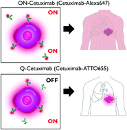
Cetuximab–dye conjugates have shown great potential for image-guided surgery of epidermal growth factor receptor (EGFR)-positive cancers in clinical trials. However, their long circulation half-life and prolonged generation of high background signals require the injection of antibody conjugates several days prior to imaging, which limits the clinical applications. Herein, we developed a cetuximab–ATTO655 conjugate (i.e., Q-Cetuximab) for fast and real-time fluorescence imaging of EGFR-positive lung cancers. The fluorescence intensity of Q-Cetuximab was quenched to just 6.9% of that of the unconjugated dye when only 2.14 ATTO655 dyes were conjugated to cetuximab. In vitro real-time cell imaging showed that EGFR-positive A549 cells emitted strong fluorescence at 10 min after Q-Cetuximab treatment in the absence of the washing step, implying target-specific activation of quenched Q-Cetuximab fluorescence upon binding with EGFR-positive cancer cells. When mice with orthotropic A549 tumors received intravenous injection of Q-Cetuximab, scattered microsized tumors in the lungs could be clearly identified from near-infrared fluorescence imaging with a tumor-to-background ratio of 4.28 at 8 h post-injection. For comparison, the cetuximab–Alexa647 conjugate (i.e., ON-Cetuximab), which does not show fluorescence quenching, was synthesized as an always-on type of probe. The ON-Cetuximab-treated mice expressed strong fluorescence throughout their body at 8 h post-injection; therefore, lung tumor sites could not be discriminated using fluorescence imaging. These results confirm the benefits of Q-Cetuximab for image-guided precision surgery of EGFR-positive lung cancers.

- This article is part of the themed collection: Biomaterials for Imaging and Sensing


 Please wait while we load your content...