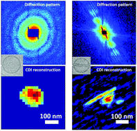
Free-electron-laser coherent diffraction images of individual drug-carrying liposome particles in solution†
Abstract
Using the excellent performances of a SACLA (RIKEN/HARIMA, Japan) X-ray free electron laser (X-FEL), coherent diffraction imaging (CDI) was used to detect individual liposome particles in water, with or without inserted doxorubicin nanorods. This was possible because of the electron density differences between the carrier, the liposome, and the drug. The result is important since liposome nanocarriers at present dominate drug delivery systems. In spite of the low cross-section of the original ingredients, the diffracted intensity of drug-free liposomes was sufficient for spatial reconstruction yielding quantitative structural information. For particles containing doxorubicin, the structural parameters of the nanorods could be extracted from CDI. Furthermore, the measurement of the electron density of the solution enclosed in each liposome provides direct evidence of the incorporation of ammonium sulphate into the nanorods. Overall, ours is an important test for extending the X-FEL analysis of individual nanoparticles to low cross-sectional systems in solution, and also for its potential use to optimize the manufacturing of drug nanocarriers.


 Please wait while we load your content...