Issue 20, 2018

In situ microscopy across scales for the characterization of crystal growth mechanisms: the case of europium oxalate

Abstract

A better understanding of how production pathway affects the final product is required in order to produce targeted syntheses, but many of the classical ex situ techniques used for studying nanoparticle growth are unsuitable as stand-alone methods for identifying and characterizing growth mechanisms. Using a combination of high resolution transmission electron microscopy (TEM), cryogenic TEM, liquid cell scanning electron microscopy, and optical microscopy we monitor europium oxalate growth over the range of nanometers to tens of micrometers and identify potential crystal growth pathways. Interpretation of the evolving crystallites reveals the significant impact of inhomogeneity in diffusion fields on diffusion limited crystal growth. We also compare the effects of stirring during crystal growth as an example of how changing processing conditions changes growth mechanisms.

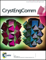
Graphical abstract: In situ microscopy across scales for the characterization of crystal growth mechanisms: the case of europium oxalate

Article information

Article type
Paper
Submitted
09 Aug 2017
Accepted
12 Apr 2018
First published
30 Apr 2018

CrystEngComm, 2018,20, 2822-2833

Author version available

In situ microscopy across scales for the characterization of crystal growth mechanisms: the case of europium oxalate

J. A. Soltis, W. C. Isley, M. Conroy, S. M. Kathmann, E. C. Buck and G. J. Lumetta, CrystEngComm, 2018, 20, 2822 DOI: 10.1039/C7CE01450C

To request permission to reproduce material from this article, please go to the Copyright Clearance Center request page.

If you are an author contributing to an RSC publication, you do not need to request permission provided correct acknowledgement is given.

If you are the author of this article, you do not need to request permission to reproduce figures and diagrams provided correct acknowledgement is given. If you want to reproduce the whole article in a third-party publication (excluding your thesis/dissertation for which permission is not required) please go to the Copyright Clearance Center request page.

Read more about how to correctly acknowledge RSC content.

Social activity

Spotlight

Advertisements