Targetable, two-photon fluorescent probes for local nitric oxide capture in the plasma membranes of live cells and brain tissues†
Abstract
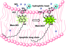
Although the plasma membrane is a major site for nitric oxide (NO) generation and action, few targetable probes that specifically sense and image NO in the plasma membrane have been reported. In this study, a membrane targetable, two-photon nitric oxide probe, Mem-NO, was developed and evaluated for bio-imaging of both exogenous and endogenous NO. By installing a quaternary ammonium compound as the hydrophilic head and a long alkyl chain as the hydrophobic tail on 4-amino-1,8-naphthalimide, we designed Mem-NO into a bi-polar structure. Due to the interaction with the phospholipid bilayer of plasma membrane, Mem-NO could specifically and stably localize in the plasma membrane. Mem-NO is almost nonfluorescent, but it displayed substantial fluorescence enhancement (16-fold) upon NO capture with sensitive (74 nM limit of detection) and fast response (within 1 min). Moreover, Mem-NO displayed strong two-photon excitation fluorescence activity (δ = 177 GM at 810 nm) and low cytotoxicity. It was found that Mem-NO is capable of two-photon imaging of endogenous NO in live neurons and human umbilical vein endothelial cells (HUVECs) and exogenous NO in mouse brain tissues. Therefore, Mem-NO qualifies as an essential and unique analytical tool for monitoring NO for future physiological, pathological, and pharmacological studies.


 Please wait while we load your content...