Association of cultured myotubes and fasting plasma metabolite profiles with mitochondrial dysfunction in type 2 diabetes subjects
Abstract
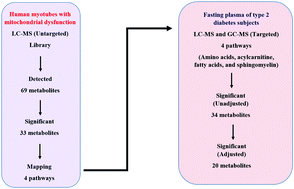
Accumulating evidence implicates mitochondrial dysfunction-induced insulin resistance in skeletal muscle as the root cause for the greatest hallmarks of type 2 diabetes (T2D). However, the identification of specific metabolite-based markers linked to mitochondrial dysfunction in T2D has not been adequately addressed. Therefore, we sought to identify the markers-based metabolomics for mitochondrial dysfunction associated with T2D. First, a cellular disease model was established using human myotubes treated with antimycin A, an oxidative phosphorylation inhibitor. Non-targeted metabolomic profiling of intracellular-defined metabolites on the cultured myotubes with mitochondrial dysfunction was then determined. Further, a targeted MS-based metabolic profiling of fasting blood plasma from normal (n = 32) and T2D (n = 37) subjects in a cross-sectional study was verified. Multinomial logical regression analyses for defining the top 5% of the metabolites within a 95% group were employed to determine the differentiating metabolites. The myotubes with mitochondrial dysfunction exhibited insulin resistance, oxidative stress and inflammation with impaired insulin signalling activities. Four metabolic pathways were found to be strongly associated with mitochondrial dysfunction in the cultured myotubes. Metabolites derived from these pathways were validated in an independent pilot investigation of the fasting blood plasma of healthy and diseased subjects. Targeted metabolic analysis of the fasting blood plasma with specific baseline adjustment revealed 245 significant features based on orthogonal partial least square discriminant analysis (PLS-DA) with a p-value < 0.05. Among these features, 20 significant metabolites comprised primarily of branched chain and aromatic amino acids, glutamine, aminobutyric acid, hydroxyisobutyric acid, pyroglutamic acid, acylcarnitine species (acetylcarnitine, propionylcarnitine, dodecenoylcarnitine, tetradecenoylcarnitine hexadecadienoylcarnitine and oleylcarnitine), free fatty acids (palmitate, arachidonate, stearate and linoleate) and sphingomyelin (d18:2/16:0) were identified as predictive markers for mitochondrial dysfunction in T2D subjects. The current study illustrates how cellular metabolites provide potential signatures associated with the biochemical changes in the dysregulated body metabolism of diseased subjects. Our finding yields additional insights into the identification of robust biomarkers for T2D associated with mitochondrial dysfunction in cultured myotubes.


 Please wait while we load your content...