Detection of SiO2 nanoparticles in lung tissue by ToF-SIMS imaging and fluorescence microscopy
Abstract
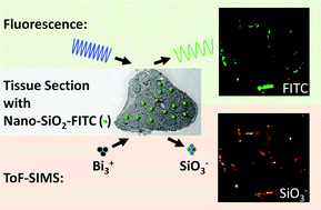
The direct detection of nanoparticles in tissues at high spatial resolution is a current goal in nanotoxicology. Time-of-Flight Secondary Ion Mass Spectrometry (ToF-SIMS) is widely used for the direct detection of inorganic and organic substances with high spatial resolution but its capability to detect nanoparticles in tissue sections is still insufficiently explored. To estimate the applicability of this technique for nanotoxicological questions, comparative studies with established techniques on the detection of nanoparticles can offer additional insights. Here, we compare ToF-SIMS imaging data with sub-micrometer spatial resolution to fluorescence microscopy imaging data to explore the usefulness of ToF-SIMS for the detection of nanoparticles in tissues. SiO2 nanoparticles with a mean diameter of 25 nm, core-labelled with fluorescein isothiocyanate, were intratracheally instilled into rat lungs. Subsequently, imaging of lung cryosections was performed with ToF-SIMS and fluorescence microscopy. Nanoparticles were successfully detected with ToF-SIMS in 3D microanalysis mode based on the lateral distribution of SiO3− (m/z 75.96), which was co-localized with the distribution pattern that was obtained from nanoparticle fluorescence. In addition, the lateral distribution of protein (CN−, m/z 26.00) and phosphate based signals (PO3−, m/z 78.96) originating from the tissue material could be related to the SiO3− lateral distribution. In conclusion, ToF-SIMS is suitable to directly detect and laterally resolve SiO2 nanomaterials in biological tissue at sufficient intensity levels. At the same time, information about the chemical environment of the nanoparticles in the lung tissue sections is obtained.


 Please wait while we load your content...