Catalase-functionalized SiO2 nanoparticles mediate growth of gold nanoparticles for plasmonic biosensing of attomolar microRNA with the naked eye†
Abstract
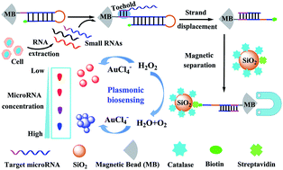
MicroRNAs (miRNAs) have been considered as promising biomarkers for cellular events and disease diagnosis. However, current methods for miRNA detection often require sophisticated instruments that may limit the applications in a resource-constrained setting. Herein, we developed a plasmonic biosensor for the colorimetric detection of miRNA with high specificity based on hairpin capture probe-coated magnetic beads (MBs) and catalase (CAT)/streptavidin (SA)-functionalized SiO2 nanoparticles. One end of the hairpin capture probe was labeled with a biotin group which was located close to the surface of MBs and failed to bind to CAT/SA-SiO2 nanoparticles in the absence of target. However, the toehold-mediated strand displacement between target miRNA and hairpin capture probe stretched the biotin group far away from the surface of MBs, enabling the capture and separation of CAT/SA-SiO2 nanoparticles. CAT can rapidly consume hydrogen peroxide (H2O2) which was then used to mediate the growth of gold nanoparticles (AuNPs). A blue aggregated AuNP solution and a red non-aggregated AuNP solution were obtained under low and high concentrations of H2O2, respectively. A single SiO2 nanoparticle was decorated with a large number of CAT molecules to offer an amplified colorimetric signal. As low as 5 amol miRNA was detected with the naked eye. The biosensor can also distinguish target miRNA from homologous sequences even with single nucleotide variation due to the long stem-containing hairpin capture probe. Furthermore, the practical application in real samples was demonstrated by detecting the small RNA samples extracted from cancer cells. Thus, we expect this simple biosensor may work as a routine tool for on-site detection of miRNA in resource-limited settings for clinical early diagnosis.


 Please wait while we load your content...