Activated T cells exhibit increased uptake of silicon phthalocyanine Pc 4 and increased susceptibility to Pc 4-photodynamic therapy-mediated cell death†
Abstract
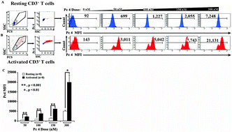
Photodynamic therapy (PDT) is an emerging treatment for malignant and inflammatory dermal disorders. Photoirradiation of the silicon phthalocyanine (Pc) 4 photosensitizer with red light generates singlet oxygen and other reactive oxygen species to induce cell death. We previously reported that Pc 4-PDT elicited cell death in lymphoid-derived (Jurkat) and epithelial-derived (A431) cell lines in vitro, and furthermore that Jurkat cells were more sensitive than A431 cells to treatment. In this study, we examined the effectiveness of Pc 4-PDT on primary human CD3+ T cells in vitro. Fluorometric analyses of lysed T cells confirmed the dose-dependent uptake of Pc 4 in non-stimulated and stimulated T cells. Flow cytometric analyses measuring annexin V and propidium iodide (PI) demonstrated a dose-dependent increase of T cell apoptosis (6.6–59.9%) at Pc 4 doses ranging from 0–300 nM. Following T cell stimulation through the T cell receptor using a combination of anti-CD3 and anti-CD28 antibodies, activated T cells exhibited increased susceptibility to Pc 4-PDT-induced apoptosis (10.6–81.2%) as determined by Pc 4 fluorescence in each cell, in both non-stimulated and stimulated T cells, Pc 4 uptake increased with Pc 4 dose up to 300 nM as assessed by flow cytometry. The mean fluorescence intensity (MFI) of Pc 4 uptake measured in stimulated T cells was significantly increased over the uptake of resting T cells at each dose of Pc 4 tested (50, 100, 150 and 300 nM, p < 0.001 between 50 and 150 nM, n = 8). Treg uptake was diminished relative to other T cells. Cutaneous T cell lymphoma (CTCL) T cells appeared to take up somewhat more Pc 4 than normal resting T cells at 100 and 150 nm Pc 4. Confocal imaging revealed that Pc 4 localized in cytoplasmic organelles, with approximately half of the Pc 4 co-localized with mitochondria in T cells. Thus, Pc 4-PDT exerts an enhanced apoptotic effect on activated CD3+ T cells that may be exploited in targeting T cell-mediated skin diseases, such as cutaneous T cell lymphoma (CTCL) or psoriasis.


 Please wait while we load your content...