Hybrid nanomaterials YVO4:Eu/Fe3O4 for optical imaging and hyperthermia in cancer cells†
Abstract
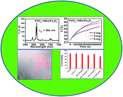
YVO4:xEu nanoparticles having a spherical shape at different concentrations of Eu (x = 0.02, 0.05, 0.07 and 0.10 at%) are prepared by a polyol method at 120 °C and their luminescence properties at different annealing temperatures (as-prepared, 500 and 900 °C) are studied. In the luminescence study, the typical emission peaks of Eu3+ ion at 590 and 612 nm are observed for all samples. The intensity of the emission peak increases with the increase of annealing temperature due to the decrease of the contribution from the non-radiative process arising from the surface dangling bond/OH. The crystallite size calculated from the X-ray diffraction is found to increase with annealing from 500 to 900 °C but the as-prepared sample has a larger crystallite size than the 500 °C annealed sample. This can be explained by the incorporation of C in the interstitial sites of the lattice after heating the as-prepared sample at 500 °C. The C residue remains after decomposition of ethylene glycol at 500 °C. Luminescence decays for the 5D0 level of Eu3+ are studied under 395 nm (direct excitation) and 270, 320 nm (indirect excitation). The energy transfer process from V–O to Eu3+ is studied from decay curves. The quantum yields for as-prepared, 500 and 900 °C annealed samples of 5 at% Eu3+ doped YVO4 nanoparticles are 1, 14 and 46%, respectively. We have also synthesized YVO4:10Eu/Fe3O4 hybrid nanocomposites and determined their intracellular localization as well as hyperthermia efficacy in tumor cells. These materials will have potential application in the diagnosis as well as therapy of cancer cells.


 Please wait while we load your content...