Lactoferrin conjugated iron oxide nanoparticles for targeting brain glioma cells in magnetic particle imaging
Abstract
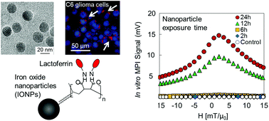
Magnetic Particle Imaging (MPI) is a new real-time imaging modality, which promises high tracer mass sensitivity and spatial resolution directly generated from iron oxide nanoparticles. In this study, monodisperse iron oxide nanoparticles with median core diameters ranging from 14 to 26 nm were synthesized and their surface was conjugated with lactoferrin to convert them into brain glioma targeting agents. The conjugation was confirmed with the increase of the hydrodynamic diameters, change of zeta potential, and Bradford assay. Magnetic particle spectrometry (MPS), performed to evaluate the MPI performance of these nanoparticles, showed no change in signal after lactoferrin conjugation to nanoparticles for all core diameters, suggesting that the MPI signal is dominated by Néel relaxation and thus independent of hydrodynamic size difference or presence of coating molecules before and after conjugations. For this range of core sizes (14–26 nm), both MPS signal intensity and spatial resolution improved with increasing core diameter of nanoparticles. The lactoferrin conjugated iron oxide nanoparticles (Lf-IONPs) showed specific cellular internalization into C6 cells with a 5-fold increase in MPS signal compared to IONPs without lactoferrin, both after 24 h incubation. These results suggest that Lf-IONPs can be used as tracers for targeted brain glioma imaging using MPI.


 Please wait while we load your content...