Mapping electrogenerated chemiluminescence reactivity in space: mechanistic insight into model systems used in immunoassays†
Abstract
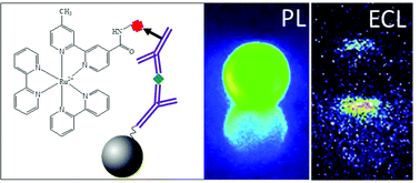
The remarkable characteristics of electrogenerated chemiluminescence (ECL) as a readout method are successfully exploited in numerous microbead-based immunoassays. However there is still a lack of understanding of the extremely high sensitivity of such ECL bioassays. Here the mechanisms of the reaction of the Ru(bpy)32+ luminophore with two efficient co-reactants (TPrA or DBAE) were investigated by mapping the ECL reactivity at the level of single Ru(bpy)32+-functionalized beads. Micrometric non-conductive beads were decorated with the ruthenium label via a sandwich immunoassay or via a peptide bond. Mapping the ECL reactivity on one bead demonstrates the generation of the excited state at a micrometric distance from the electrode by reaction of surface-confined Ru(bpy)32+ with diffusing TPrA radicals. The signature of the TPA˙+ lifetime is obtained from the ECL profile. Unlike the reaction with Ru(bpy)32+ in solution, DBAE generates very low ECL intensity in the bead-based format suggesting more unstable radical intermediates. The 3D imaging approach provides insights into the ECL mechanistic route operating in bioassays and on the optical effects that focus the ECL emission.


 Please wait while we load your content...