Response of single leukemic cells to peptidase inhibitor therapy across time and dose using a microfluidic device†
Abstract
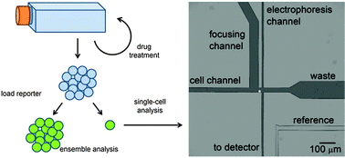
Single-cell methodologies are revealing cellular heterogeneity in numerous biological processes and pathologies. For example, cancer cells are characterized by substantial heterogeneity in basal signaling and in response to perturbations, such as drug treatment. In this work, we examined the response of 678 individual U937 (human acute myeloid leukemia) cells to an aminopeptidase-inhibiting chemotherapeutic drug (Tosedostat) over the course of 95 days. Using a fluorescent reporter peptide and a microfluidic device, we quantified the rate of reporter degradation as a function of dose. While the single-cell measurements reflected ensemble results, they added a layer of detail by revealing unique degradation patterns and outliers within the larger population. Regression modeling of the data allowed us to quantitatively explore the relationships between reporter loading, incubation time, and drug dose on peptidase activity in individual cells. Incubation time was negatively correlated with the number of peptide fragment peaks observed, while peak area (which was proportional to reporter loading) was positively correlated with both the number of fragment peaks observed and the degradation rate. Notably, a statistically significant change in the number of peaks observed was identified as dose increased from 2 to 4 μM. Similarly, a significant difference in degradation rate as a function of reporter loading was observed for doses ≥2 μM compared to the 1 μM dose. These results suggest that additional enzymes may become inhibited at doses >1 μM and >2 μM, demonstrating the utility of single-cell data to yield novel biological hypotheses.


 Please wait while we load your content...