Issue 20, 2016

Super-resolution imaging for monitoring cytoskeleton dynamics

Abstract

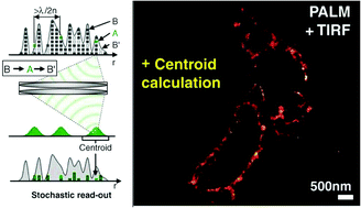
The cytoskeleton is a key cellular structure that is important in the control of cellular movement, structure, and sensing. To successfully image the individual cytoskeleton components, high resolution and super-resolution fluorescence imaging methods are needed. This review covers the three basic cytoskeletal elements and the relative benefits and drawbacks of fixed versus live cell imaging before moving on to recent studies using high resolution and super-resolution techniques. The techniques covered include the near-diffraction limited imaging methods of confocal microscopy and TIRF microscopy and the super-resolution fluorescence imaging methods of STORM, PALM, and STED.

Graphical abstract: Super-resolution imaging for monitoring cytoskeleton dynamics

Article information

Article type
Critical Review
Submitted
29 Mar 2016
Accepted
10 Aug 2016
First published
10 Aug 2016

Analyst, 2016,141, 5674-5688

Super-resolution imaging for monitoring cytoskeleton dynamics

S. A. Finkenstaedt-Quinn, T. A. Qiu, K. Shin and C. L. Haynes, Analyst, 2016, 141, 5674 DOI: 10.1039/C6AN00731G

To request permission to reproduce material from this article, please go to the Copyright Clearance Center request page.

If you are an author contributing to an RSC publication, you do not need to request permission provided correct acknowledgement is given.

If you are the author of this article, you do not need to request permission to reproduce figures and diagrams provided correct acknowledgement is given. If you want to reproduce the whole article in a third-party publication (excluding your thesis/dissertation for which permission is not required) please go to the Copyright Clearance Center request page.

Read more about how to correctly acknowledge RSC content.

Social activity

Spotlight

Advertisements