Micellar refolding of coiled-coil honeybee silk proteins†
Abstract
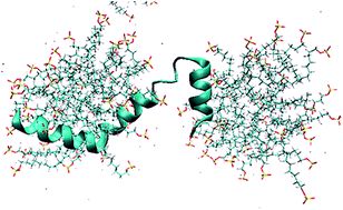
There is growing interest in materials generated from coiled coil proteins for medical applications. In this study we describe controlled micellar refolding of coiled coil honeybee silk proteins using the detergent SDS. Circular dichroism and dynamic light scattering experiments demonstrate that micellar SDS promotes folding of randomly coiled honeybee silk proteins into isolated α-helices, and that removal of detergent micelles, or addition of salt, leads to coiled coil formation. Comparative molecular dynamics simulations of protein helices, with and without SDS, have allowed us to characterize detergent–protein interactions and propose a mechanism of protein folding. In the presence of micellar detergent, hydrophobic residues are associated with the detergent tail groups within the micelles whereas hydrophilic residues are paired with the detergent head-groups on the micelle's surface. These detergent–protein interactions prevent residue–residue interactions and allow the protein to fold according to the natural tendency of individual residues. From this condition, when hydrophobic residue–micellar interactions are reduced by lowering detergent levels to below the critical micelle concentration (CMC) or by using salt to increase detergent packing in micelles and thereby excluding the protein from the interior, the proteins fold into coiled coils. We propose that under low SDS conditions, hydrophobic–monomeric SDS tail-group and hydrophilic–monomeric head-group interactions (low SDS conditions) or hydrophilic–micellar SDS head-group interactions (high salt conditions) stabilize a transient α-helix intermediate in coiled coil folding. The folding pathway constitutes a new kind of micellar refolding, which may be profitably employed to refold other proteins rich in coiled coils.


 Please wait while we load your content...