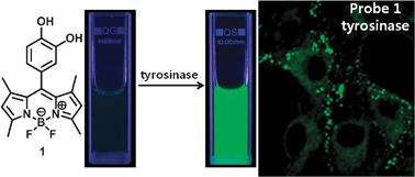
Visualization of tyrosinase activity in melanoma cells by a BODIPY-based fluorescent probe†
Abstract
We have presented a

* Corresponding authors
a
Department of Chemistry, 126 Jukjeon-dong, Yongin-si, Gyeonggi-do, Korea
E-mail:
youngmi@dankook.ac.kr
Fax: +82 31-8005-3148
Tel: +82 31-8005-3156
b
Molecular Imaging & Therapy Branch, National Cancer Center, 323 Ilsan-ro, Goyang-si, Gyeonggi-do 410-769, Korea
E-mail:
ydchoi@ncc.re.kr
Fax: +82 31-920-2529
Tel: +82 31-920-2512
We have presented a

 Please wait while we load your content...
Something went wrong. Try again?
T. Kim, J. Park, S. Park, Y. Choi and Y. Kim, Chem. Commun., 2011, 47, 12640 DOI: 10.1039/C1CC15061H
To request permission to reproduce material from this article, please go to the Copyright Clearance Center request page.
If you are an author contributing to an RSC publication, you do not need to request permission provided correct acknowledgement is given.
If you are the author of this article, you do not need to request permission to reproduce figures and diagrams provided correct acknowledgement is given. If you want to reproduce the whole article in a third-party publication (excluding your thesis/dissertation for which permission is not required) please go to the Copyright Clearance Center request page.
Read more about how to correctly acknowledge RSC content.
 Fetching data from CrossRef.
This may take some time to load.
Loading related content
