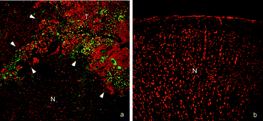
Fluorescence-guided resection (FGR) and photodynamic therapy (PDT) have previously been investigated separately with the objectives, respectively, of increasing the extent of brain tumour resection and of selectively destroying residual tumour post-resection. Both techniques have demonstrated trends towards improved survival, pre-clinically and clinically. We hypothesize that combining these techniques will further delay tumour re-growth. In order to demonstrate technical feasibility, we here evaluate fluorescence imaging and PDT treatment techniques in a specific intracranial tumour model. The model was the VX2 carcinoma grown by injection of tumour cells into the normal rabbit brain. An operating microscope was used for white light imaging and a custom-built fluorescence imaging system with co-axial excitation and detection was used for FGR. PDT treatment light was applied by intracranially-implanted light emitting diodes (LED). The fluorescent photosensitizer used for both FGR and PDT was ALA-induced PpIX. For PDT, ALA (100 mg kg−1) and low light doses (15 and 30 J) were administered over extended periods, which we refer to as metronomic PDT (mPDT). Eighteen tumour bearing rabbits were divided equally into three groups: controls (no resection); FGR; and FGR followed by mPDT. Histological whole brain sections (H&E stain) showed primary and recurrent tumours. No bacteriological infections were found by Gram staining. Selective tumour cell death through mPDT-induced apoptosis was demonstrated by TUNEL stain. These results demonstrate that the combined treatment is technically feasible and this model is a candidate to evaluate it. Further optimization of mPDT treatment parameters (drug/light dose rates) is required to improve survival.

You have access to this article
 Please wait while we load your content...
Something went wrong. Try again?


 Please wait while we load your content...