
A ‘turn-on’ fluorescent probe for lysosomal phosphatase: a comparative study for labeling of cancer cells†

Abstract
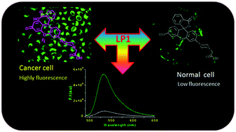
We have described the ability of a newly synthesized fluorescent probe (LP1) to detect phosphatase activity in lysosomes in cancer cells. Probe LP1 displayed a 33-fold fluorescence intensity enhancement at λem 532 nm in the presence of phosphatase in HEPES buffer (pH 4.5). The quantum yield of probe LP1 was increased by ∼21-fold upon exposure to phosphatase at acidic pH. The probe LP1 is highly chemoselective toward phosphatase (ALP/ACP) and is insensitive to interference by ubiquitous biological analytes. The high cell adhesion property and cell viability of LP1 indicate that LP1 is biocompatible and nontoxic; these two characteristic features make it a suitable candidate for phosphatase tracking in living cells. LP1 dose-dependent fluorescence images in living cells suggested that the expression of phosphatase in cancer cells (HeLa) is 2-fold higher as compared to the normal NIH-3T3 cells. The colocalization images confirmed that LP1 was exclusively localized in lysosomes. We envision that LP1 could be a potential tool in clinical diagnosis for discriminating cancer cells from normal cells depending on the expression of phosphatase in lysosomes.
 Please wait while we load your content...

