Issue 14, 2018

Visualization of mercury(ii) accumulation in vivo using bioluminescence imaging with a highly selective probe

Abstract

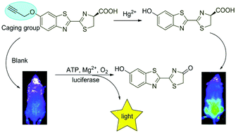
Mercury is a highly toxic environmental pollutant that negatively affects human health. Thus, an in vivo method for noninvasive imaging of mercury(II) and visualization of its accumulation within living systems would be advantageous. Herein, we describe a reaction-based bioluminescent probe for detection of mercury(II) in vitro and accumulation in vivo. The application of this probe would help to shed light on the intricate contributions of mercury(II) to various physiological and pathological processes.

Graphical abstract: Visualization of mercury(ii) accumulation in vivo using bioluminescence imaging with a highly selective probe

Supplementary files

Article information

Article type
Communication
Submitted
14 Feb 2018
Accepted
13 Mar 2018
First published
13 Mar 2018

Org. Biomol. Chem., 2018,16, 2388-2392

Visualization of mercury(II) accumulation in vivo using bioluminescence imaging with a highly selective probe

B. Ke, H. Chen, L. Ma, S. Zingales, D. Gong, D. Hu, L. Du and M. Li, Org. Biomol. Chem., 2018, 16, 2388 DOI: 10.1039/C8OB00398J

To request permission to reproduce material from this article, please go to the Copyright Clearance Center request page.

If you are an author contributing to an RSC publication, you do not need to request permission provided correct acknowledgement is given.

If you are the author of this article, you do not need to request permission to reproduce figures and diagrams provided correct acknowledgement is given. If you want to reproduce the whole article in a third-party publication (excluding your thesis/dissertation for which permission is not required) please go to the Copyright Clearance Center request page.

Read more about how to correctly acknowledge RSC content.

Social activity

Spotlight

Advertisements