Two-color dark-field (TCDF) microscopy for metal nanoparticle imaging inside cells†

Abstract
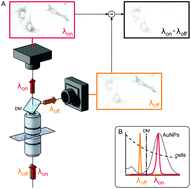
Noble metal nanoparticles (NPs) supporting localized surface plasmon resonances are widely used in the context of biotechnology as optical and absorption contrast agents with great potential applicability to both diagnostics and less invasive therapies. In this framework, it is crucial to have access to simple and reliable microscopy techniques to monitor the NPs that have internalized into cells. While dark field (DF) microscopy takes advantage of the enhanced NP scattering at their plasmon resonance, its use in cells is limited by the large scattering background from the internal cell compartments. Here, we report on a novel two-color dark field microscopy that addresses these limitations by significantly reducing the cell scattering contribution. We first present the technique and demonstrate its enhanced contrast, specificity and reliability for NP detection compared to a standard optical dark field. We then demonstrate its potential suitability in two different settings, namely wide-field parallel screening of circulating cells in microfluidic chips and high-resolution tracking of internalized NPs in cells. These proof of principle experiments show a promising capability of this approach with possible extension to other kinds of targeted systems like bacteria and vesicles.
 Please wait while we load your content...

