Metallome of cerebrovascular endothelial cells infected with Toxoplasma gondii using μ-XRF imaging and inductively coupled plasma mass spectrometry†
Abstract
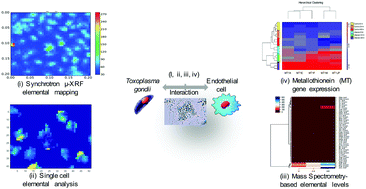
In this study, we measured the levels of elements in human brain microvascular endothelial cells (ECs) infected with T. gondii. ECs were infected with tachyzoites of the RH strain, and at 6, 24, and 48 hours post infection (hpi), the intracellular concentrations of elements were determined using a synchrotron–microfocus X-ray fluorescence microscopy (μ-XRF) system. This method enabled the quantification of the concentrations of Zn and Ca in infected and uninfected (control) ECs at sub-micron spatial resolution. T. gondii-hosting ECs contained less Zn than uninfected cells only at 48 hpi (p < 0.01). The level of Ca was not significantly different between infected and control cells (p > 0.05). Inductively Coupled Plasma Mass Spectrometry (ICP-MS) analysis revealed infection-specific metallome profiles characterized by significant increases in the intracellular levels of Zn, Fe, Mn and Cu at 48 hpi (p < 0.01), and significant reductions in the extracellular concentrations of Co, Cu, Mo, V, and Ag at 24 hpi (p < 0.05) compared with control cells. Zn constituted the largest part (74%) of the total metal composition (metallome) of the parasite. Gene expression analysis showed infection-specific upregulation in the expression of five genes, MT1JP, MT1M, MT1E, MT1F, and MT1X, belonging to the metallothionein gene family. These results point to a possible correlation between T. gondii infection and increased expression of MT1 isoforms and altered intracellular levels of elements, especially Zn and Fe. Taken together, a combined μ-XRF and ICP-MS approach is promising for studies of the role of elements in mediating host–parasite interaction.


 Please wait while we load your content...