
Combination of micro X-ray fluorescence spectroscopy and time-of-flight secondary ion mass spectrometry imaging for the marker-free detection of CeO2 nanoparticles in tissue sections†

Abstract
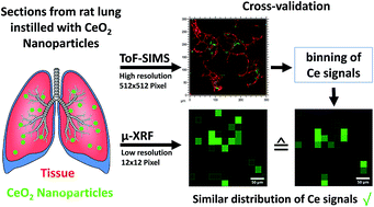
The description of nanoparticle distributions in tissue and associated effects is an important goal of nanotoxicology. Three-dimensional time-of-flight-secondary ion mass spectrometry (3D ToF-SIMS) is a promising label-free method to analyse tissue sections for nanoparticle distribution and to simultaneously detect organic and inorganic molecule species at high spatial resolution. As sample regions accessible to ToF-SIMS 3D imaging are limited to several hundred square micrometers, the proper selection of nanoparticle containing regions is mandatory. Micro X-ray fluorescence spectrometry (μ-XRF) provides a non-destructive, fast elemental analysis of large tissue sections with lateral resolution in the micrometer range. The latter technique was, therefore, used to screen samples as well as to validate elemental distribution images from ToF-SIMS 3D analysis. Specifically, cryo-sections from lungs inhomogeneously laden with CeO2 nanoparticles (10–200 nm) were subjected to μ-XRF and reflected light microscopy to pre-select lung tissue areas containing CeO2 NPs. The lateral resolution (670 nm) of ToF-SIMS imaging analyses in these areas exceeds the lateral resolution of μ-XRF imaging by a factor of 42 and reveals the lateral distribution of CeO2 NPs within the tissue structure. In addition, simultaneously acquired further secondary ions at a mass resolution of R = 4000 shed light on the sample composition. In summary, the investigation shows that μ-XRF is highly suitable to identify relevant tissue regions for subsequent high resolution ToF-SIMS 3D microanalysis of nanoparticle-laden tissue sections.
- This article is part of the themed collection: Atomic spectrometry for the analysis of biological samples
 Please wait while we load your content...

